
Introduction
Shrimp aquaculture in Vietnam has continuously developed during the last few years, which is an economically important part of the aquaculture industry. According to the Directorate of Fisheries,1 the production of shrimp was reported to increase from 1.06 million metric tons in 2022 to 1.12 million metric tons in 2023. However, with intensive and super-intensive cultivation technology, shrimp farming is facing several challenges with regard to disease outbreaks and water quality degradation. In the past decades, the use of antibiotics and other chemicals has been commonly applied to control bacterial diseases and enhance the growth of cultured species.2 Reverter et al.3 reported that antibiotic overuse may result in other problems such as environmental effects, food safety, and bacterial multidrug resistance. As a result, many alternative findings have been developed to reduce the use of antibiotics in shrimp culture. In the recent past, probiotics have been considered one of the most promising ways to control disease and improve growth, survival, and water quality.4
Lactobacillus are lactic acid producing bacteria that have been commonly used as probiotic candidates in the aquaculture industry.5 They could be found in fermented foods and in the gastrointestinal tracts of fish, prawns, and shrimp.6 The species of the genus Lactobacillus are gram-positive, non-motile, and non-sporulation bacteria that can produce lactic acid. Normally, Lactobacillus species could be administered to aquatic animals as a feed additive. They are capable of inhibiting pathogenic bacteria, improving the intestinal microbial balance, producing digestive enzymes, and promoting growth performance and disease resistance.5 Several studies have reported that dietary supplementation of L. plantarum could improve growth performance, immune parameters, and disease resistance in fish species such as rainbow trout, common carp, and Nile tilapia.7–9 In a recent study, Zheng et al.10 demonstrated that dietary administration of L. plantarum improved the composition and diversity of beneficial bacteria in the gut of whiteleg shrimp. Moreover, the whiteleg shrimp fed L. plantarum were isolated from kefir, which could enhance their survival, growth, and immune response.11 Although L. plantarum has been widely used as a probiotic in whiteleg shrimp culture, most of them are non-shrimp sourced, which could not show the most effective results. This is because the selected strain of probiotic is one of the crucial factors for evaluating probiotic effectiveness.12
The objective of this study is to screen and isolate potential probiotic L. plantarum from the intestine of healthy whiteleg shrimp through in vitro assays. The isolated strain was screened based on morphology, biochemical characteristics, antimicrobial activity, and extracellular enzyme production. In addition, an in vivo experiment was also conducted to assess the effects of L. plantarum CMT1 on the growth performance and survival of whiteleg shrimp when administered in feed.
Materials and Methods
Isolation and screening of Lactobacillus spp.
A total of 120 whiteleg shrimp (8-10 g) were collected from six different extensive and semi-intensive ponds in Dam Doi district, Ca Mau province, Vietnam. Shrimp samples were rinsed with 70% ethanol. Each whole gut of shrimp was dissected out and homogenized in an Eppendorf tube with 1.5 ml of sterile saline solution (0.85% NaCl). Then, 100 μL of homogenate was aseptically spread onto De Man–Rogosa–Sharpe (MRS) agar plates, and incubated at 37°C for 24–72 hours. After incubation, each colony was purified on new MRS plates by the streaking method. All isolates were determined Gram-stain, catalase, oxidase, motility, and spore-forming tests based on the methods of Mohamad et al.12 The isolated strains were stored at -80oC in Eppendorf tubes containing MRS broth with 20% glycerol for further study.
The genomic DNA of Lactobacillus isolates was extracted using a genomic DNA purification kit following the manufacturer’s instructions. The 16s rDNA gene was amplified using 27F (5’-AGAGTTTGATCCTGG CTCAG-3’) and 1492R (5’-GGTTACCTTGTTACGACTT-3’) primers. Sequences identified were compared directly with sequences obtained from Gen Bank using BLAST at the NCBI.
Antimicrobial activity of the isolated strains
The antimicrobial activity of Lactobacillus isolates against pathogenic bacteria, Vibrio parahaemolyticus was performed using the agar disk diffusion method described by Noordiana et al.13 Vibrio parahaemolyticus was stored in the lab of aquatic probiotic, College of Aquaculture and Fisheries, Can Tho University. In brief, the fresh cultures of isolated Lactobacillus and V. parahaemolyticus were cultured in MRS broth and TSB broth, respectively. After incubation at 37oC for 24 h, the bacteria were collected by centrifugation at 10000 rpm for 4 min and then diluted with sterile saline solution to obtain an absorbance of 0.8 at 600 nm using a spectrophotometer (Shimadzu UV-1900i, Malaysia), which was equivalent to a density of 107 CFU mL-1 based on the standards bacterial growth curves (data not shown). Then, 100 μL of pathogen solution was spread over MHA plates. Four wells of 6.0 mm in diameter were made and filled with 100 µl of Lactobacillus isolates. The plates were incubated at 37oC for 24 h, and the inhibition zone diameter was measured in millimeters by caliper. The antimicrobial activity test was conducted in two replicates.
Screening of extracellular enzyme activity of the isolated strains
Three isolated strains with high antibacterial activity were selected and cultured in MRS broth at 37oC for 24 h. After incubation, cell-free supernatant (CFS) was harvested to measure extracellular α-amylase, protease, and leu-aminopeptidase activity. Amylase activity was determined based on the method of Bernfeld et al. (1955) with 2% starch solution as the substrate. The activity of protease was measured according to the method of Lowry et al.,14 using the casein hydrolysis method. All enzyme activities were expressed as U mg protein-1. The concentration of soluble protein was determined following the method of Lowry et al.,14 using bovine serum albumin as a standard.
Effect of Lactobacillus plantarum CMT1 on growth and survival of whiteleg shrimp
Juveniles whiteleg shrimp (0.90 ± 0.05g) were obtained from a local hatchery in Can Tho city, Vietnam, and acclimatized to a 4000-L composite tank for 5 days prior to the experiment. After acclimatizing, shrimp were randomly distributed to 12 composite tanks (500 L capacity) in recirculating aquaculture systems. Each tank contained 100 shrimp in 400 L of water at 15 ppt salinity. Shrimp were fed with different experimental diets four times a day (6:00, 12:00, 17:00, and 20:00). Dietary experiments included a commercial diet (Grobest Landfound Co. Ltd., Vietnam) without L. plantarum CMT1 (control), T1, T2, and T3 containing 106, 107 and 108 CFU kg-1 L. plantarum CMT1, respectively. The experimental diets were prepared as described by Zheng et al.10 The bacteria solution was sprayed and mixed homogeneously to a commercial feed (Grobest Landfound Co. Ltd., Vietnam) to reach the desired concentration. The bacterial density in the diets were then checked using MRS plate count method. The amount of uneaten feed was collected after 1 h of feeding and dried in an oven at 80℃. During a 56-day feeding experiment, water quality, including temperature and pH, was checked daily using a Hana HI98128 pH/temperature tester. Dissolved oxygen (DO), total ammonia nitrogen (TAN), and nitrite (NO2--N) were weekly determined following APHA.15 At the end of the experiment, growth parameters such as initial weight, final weight, weight gain (WG), daily weight gain (DWG), specific growth rate (SGR), feed conversion ratio (FCR), and survival rate (%) were calculated based on a method previously described by Tu et al.16 In this experiment, three shrimp from each tank were randomly sampled every week for the determination of intestinal bacterial counts. Briefly, the intestine of shrimp was weighed and homogenized in sterile normal saline (0.85%). An aliquot of 100 µL from appropriated dilutions was spread on MRS agar and TCBS agar plates to determine total Vibrio sp. and Lactobacillus sp., respectively.
Statistical analysis
All experimental data were analyzed by one-way analysis of variance (ANOVA) and a Tukey multiple range test using SPSS 22.0 software. Statistically significant level was set at p < 0.05. Values were presented as mean ± standard deviation (SD).
Results
Isolation and characterization of Lactobacillus candidates
A total of 20 Lactobacillus isolates (CMT1-CMT20) were obtained from the intestine of healthy whiteleg shrimp. All isolates were gram-positive microorganisms with a rod morphology. The isolated strains showed negative results for catalase, oxidase, motility, and spore-forming tests (Table 1). During the preliminary screening, ten of 20 isolates exhibited antimicrobial activity against V. parahaemolyticus. Among these 10 isolates, only 3 isolates, CMT1, CMT6, and CMT20 were selected for further study due to their strong antimicrobial performance. The isolated bacterial strain was first identified as Lactobacillus sp. based on its morphology, biochemical characteristics, antibacterial activity, and extracellular enzyme activity. PCR analysis using 16S rDNA gene sequences showed 99% identity with Lactobacillus plantarum for CMT1.
Extracellular enzyme activities
The highest amylase activity (U mg protein-1) was recorded in strain CMT1, and there was a significant difference as compared with the strains CMT6 and CMT20 (Figure 1A). Similar results were observed in protease activity. Specifically, the value in strain CMT1 was significantly higher than that in strains CMT6 and CMT20 (Figure 1B).
_amylase_and_(b)_protease_of_three_*lactobacillus*_isol.jpeg)
Effects of Lactobacillus plantarum CMT1 on growth performance and survival of whiteleg shrimp
The effect of L. plantarum CMT1 on water quality in shrimp tanks is presented in Table 2. Water temperature, pH, DO, and alkalinity in each tank were within a suitable range for shrimp growth and survival, and there was no significant difference among all groups. Although TAN and NO2--N values in the control group were slightly higher than those of the other groups, no significant difference was found.
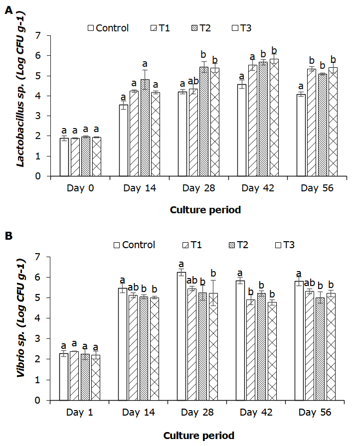
During the experimental period, the total Lactobacillus count in the intestine of shrimp significantly increased from day 28 of the experimental period (Figure 2A). Significant differences in Lactobacillus count were found among T2, T3, and the control groups at days 28, 42, and 56 (p < 0.05). As for Vibrio counts, the levels were significantly decreased (p < 0.05) by increasing the dose of L. plantarum CMT1 supplementation. To be specific, the number of Vibrio in the T2 and T3 groups was lower than that in the control group (Figure 2B).
_and_*vibrio*_sp._(b)_in_the_intestine_of_whiteleg.png)
After a 56-day feeding period, the final weight, WG, DWG, and SGR were significantly enhanced in the T3 group (108 CFU kg diet-1) compared to the control, T1, and T2 groups (Table 3). However, there were no significant differences in final weight, WG, and DWG between the control and T1 groups or between T1 and T2 groups (p > 0.05). Additionally, the survival rates of shrimp fed T2 and T3 diets were significantly higher than those of the control group. Conversely, the lowest FCR value (1.24±0.72) was observed in the T3 group, and there was a significant difference as compared with the control group (p > 0.05).
Discussion
Probiotics are one of the potential solutions to support the development of sustainable shrimp farming practices. Therefore, the use of probiotics in shrimp culture has been increasing from day to day. In fact, the application of probiotics in sufficient quantities helps to enhance the growth performance, feed conversion efficiency, immune responses, disease resistance, and intestinal health of aquatic organisms, including shrimp species.17,18 According to Mohamad et al.,12 most Lactobacillus species were commonly detected in the intestinal tracts of shellfish, crabs, and shrimp. Therefore, the digestive gut of a healthy shrimp could be the best source for the isolation of potential probiotic strains. In the present study, 20 Lactobacillus isolates were identified based on morphological and biochemical tests. Due to the strong antimicrobial activity against V. parahamolyticus and extracellular enzyme activity, the isolated strain CMT1 was selected and confirmed as L. plantarum using 16S rDNA gene sequencing, which was further used for evaluation on a laboratory scale in feed application.
The present result showed that the application of feed supplemented with L. plantarum CMT1 significantly enhanced the growth performance of the whiteleg shrimp, wherein growth parameters in the T3 group (108 CFU kg-1) were significantly higher than those in the other groups. Similar results were reported in other species, including rainbow trout (Oncorhynchus mykiss) fed L. plantarum at 2 × 10⁷ CFU g-1 feed for 72 days,7 Nile tilapia (Oreochromis niloticus) fed L. plantarum at 108 CFU g diet-1 for 28 days,9 and giant prawn (Macrobrachium rosenbergii) fed L. plantarum at 109 CFU g diet-1 for 90 days.19 As reported by Xie et al.,20 increased activity of digestive enzymes could improve nutrient absorption ability, hence promoting growth performance. Based on the results of this study, L. plantarum strain CMT1 isolated from shrimp could produce digestive enzymes such as protease and amylase. In addition, probiotics have been demonstrated to increase epithelial cell microvilli in the intestine, which provide a great surface area for the absorption of available nutrients.21 The dietary supplementation of L. plantarum could significantly improve the survival rate of whiteleg shrimp, and similar results were observed in blue swimming crab (Portunus pelagicus),22 bighead catfish (Clarias macrocephalus),23 and olive flounder (Paralichthys olivaceus).24 Moreover, shrimp fed diet supplemented with L. plantarum CMT1 at a dose of 108 CFU kg-1 significantly decreased FCR compared to the control diet. This result is similar to the finding of Dash et al.,19 who reported lower FCR in freshwater prawn M. rosenbergii fed with L. plantarum. Total Lactobacillus counts in the intestines of shrimp fed T2 and T3 diets were significantly higher than those in the other diets from day 14 to the end of the experiment, but no significant difference was observed in the T2 and T3 groups. In contrast with the number of Vibrio counts, the levels of the T2 and T3 groups significantly decreased compared to the control and T1 groups. These findings suggested that L. plantarum CMT1 could inhibit the growth of pathogenic bacteria in the gut of shrimp. Similarly, Kongnum and Hongpattarakere25 reported that the administration of L. plantarum MRO3.12 in feed effectively inhibited non-fermenting Vibrios in the guts of shrimp. This could be explained by the fact that lactic acid bacteria are capable of producing antimicrobial substances and competing for nutrients and space from harmful bacteria.26 Furthermore, the high density of beneficial bacteria in the gastrointestinal tracts of shrimp could reduce and eliminate the colonization of pathogenic bacteria.27
In conclusion, the present study isolated L. plantarum CMT1 from the intestines of shrimp collected from extensive shrimp farming, which could be used as a potential probiotic candidate. In an in vivo experiment, the diet supplementation of L. plantarum CMT1 had a positive effect on the survival and growth performance of shrimp, and the suggested dose of the probiotic strain CMT1 is 108 CFU kg diet-1.
Acknowledgments
The authors would like to thank Ms. Le Thi Van Anh and Ms. Truong Thi Kim Chi for their assistance during the experiment. Phan Thi Cam Tu was funded by the Postdoctoral Scholarship Programme of Vingroup Innovation Foundation (VINIF) under the code VINIF.2023.STS.45.
Authors’ Contribution
Conceptualization: Thi Cam Tu Phan (Equal), Thi Thanh Hien Tran (Equal). Methodology: Thi Cam Tu Phan (Equal), Truong Giang Huynh (Equal), Thi Thanh Hien Tran (Equal). Formal Analysis: Thi Cam Tu Phan (Equal), Thi Thu Nguyen (Equal). Writing – original draft: Thi Cam Tu Phan (Lead). Writing – review & editing: Thi Cam Tu Phan (Equal), Thi Kim Lien Nguyen (Equal), Truong Giang Huynh (Equal), Thi Thanh Hien Tran (Equal). Funding acquisition: Thi Cam Tu Phan (Lead).
Ethical Conduct Approval – IACUC
The experiment complied with the regulations of Can Tho University and Vietnam’s policy for animals used in research.