

1. Introduction
The giant salamander, known as Wawayu, is a second-grade animal under state protection in China and is classified as Amphibia, Vrodtle, Cryptobranchidae, and Andrias. IUCN (The International Union for the Conservation of Nature) has classified this animal as a critically endangered species and has listed this species in Appendix I of CITES (Convention on International Trade in Endangered Species of Wild Fauna and Flora).1,2 The Chinese giant salamander is found mostly in the Pearl, Yellow, and Yangtze rivers, in the middle and upper stages of deep valley streams. Consequently, this species represents a typical amphibian that occupies the transition zone between aquatic and terrestrial habitats. Of the 28 amphibian species in China, the Chinese giant salamander is the largest, making it extremely significant for research, especially regarding the history of biological evolution.3,4
Acinetobacter is a short and rod-shaped Gram-negative bacterium belonging to Moraxella5 that widely exists in nature and exists in soil, water, and spoiled food.5,6 In addition, Acinetobacter is a zoonotic pathogen that can cause infections in mammals, birds, amphibians, and fish.7,8 Acinetobacter is responsible for global epidemics that endanger wildlife and domestic livestock populations, ultimately leading to large-scale morbidity and mortality.9
Since the second half of 2022, infectious diseases have been increasingly detected in the Chinese giant salamander at around three years of age. These bacterial diseases are associated with a high mortality rate in diseased salamanders, and the tendency for infection grows annually. In this study, we detected the pathogens responsible for disease in samples of infected Chinese giant salamanders and performed antibiotic susceptibility measurements and drug sensitivity analysis.
2. Materials and Methods
2.1. Experimental Materials
Typical diseased Chinese giant salamander specimens (weighing 40-60 g) were collected from the designated A. davidianus aquaculture facility in Zhangjiajie. They were sent to the Laboratory of Fish Diseases, Yangtze River Fisheries Research Institute at the Chinese Academy of Fishery Sciences to isolate and identify pathogens.
2.2. Isolation and Purification
The surfaces of the diseased salamander were decontaminated with 75% alcohol in a biological safety cabinet. Then, each salamander was dissected, and an inoculation loop was used to remove a portion of the liver, which was subsequently inoculated onto a Brain-Heart Infusion (BHI; HopeBio, Qingdao, China) agar plate and incubated for 24 h at 28 °C. Dominant single colonies were chosen after culture and placed onto brand-new BHI agar plates. Purified single colonies were obtained after 24 hours of growth under the same conditions. Single colonies were then selected and implanted into 5 mL of BHI liquid medium, then cultured for approximately 24 hours at 28°C with rotation at 200 rpm. Subsequently, we mixed each bacterial solution with an equal volume of 50% glycerol. We transferred this mixture into a microcentrifuge tube (1.5 mL) kept at -80°C in an extremely low-temperature refrigerator to await future analysis. The isolated strain was designated DN-2.
2.3. Pathogenic identification
2.3.1. Identification of Bacterial Morphology
The isolated strains were cultured for 24 hours at 28°C on a BHI solid substrate while the colony morphology was examined. The bacterial suspension was prepared using phosphate-buffered (PBS, Procell, Wuhan, China) as the solvent and subjected to Gram staining. Individual colonies were fixed, dried, dehydrated, sprayed with gold and then analyzed by scanning electron microscopy (SEM) analysis.
2.3.2. Molecular Biological Identification
A single prepared colony was selected from the biosafety cabinet and transferred to a 1.5mL microcentrifuge tube. Then, bacterial genomic DNA was extracted with a Bacterial DNA Kit (Tiangen, Beijing, China) following the manufacturer’s recommendations. Next, the 16S rRNA of DN-2 was amplified by PCR utilizing generic primers (27 the force F: 5-AGAGTTTGATCATGGCTCAG-3, 1492the rectangle R: 5-TACGGTTACCTTGTTACGACTT-3). A total amount of 25 μL was used for the PCR reactions, which included 12.5 μL of PCR Mix, 1 μL of primers (10 μmol/L for the upstream and downstream), 1 μL of template DNA, and 9.5 μL of diluted water. Pre-denaturation at 94°C for 5 minutes, denaturation at 94°C for 30 seconds, annealing at 55°C for 1 minute, and denaturation at 72°C for 30 seconds (30 cycles) were the reaction conditions.
A final elongation step was performed at 72°C for 5 minutes. Subsequently, the amplified products were visualized by electrophoresis on a 1% agarose gel; amplicons were then selected and sequenced. Sequencing outcomes were then uploaded into the NCBI database (National Center for Biotechnology Information, http://blast.ncbi.nlm.nih.gov), and the Blast search tool was used to make sequence homology comparisons. Subsequently, we used 16S rRNA gene sequences from different sources of Acinetobacter to construct a tree of phylogenetic relationships with the MEGA 11.0 program by applying the Neighbor-Joining approach (http://www.megasoftware.net/previousVersions.ph); 1000 bootstrap analysis was used for confidence detection.
2.4. Antibiotic Susceptibility Testing
Strain DN-2 was cultured, and sterile PBS was used to control the concentration of bacteria in the solution. we coated 100 mL of bacterial liquid onto the surface of the BHI agar culture in a biological safety cabinet. Following the bacterial inoculum had been completely absorbed into the agar medium, the pre-arranged antibiotic tablet was carefully placed on the surface of the agar plate. After 24 hours of growth at 28°C in a constant temperature incubator, the plates were inverted, and bacteriostatic circles were measured (in mm). The sensitivity of this method was determined according to the drug sensitivity kit manufacturer’s recommendations.
2.5 Biochemical Identification of Bacteria
Strain 2 and the isolates from the regression infection experiment were cultured on BHI plates for analysis with the Bacterial identification system BIOLOG (Hayward, CA, USA); this procedure was performed by the manufacturer’s instructions. Single colonies grown on plates were picked with an uncontaminated inoculation ring, placed inside with IF-A reagent, and mixed, and 1 μL of the mixture was placed into each well of a 96-well plate. Then, we added reagents from the system for automated microbiological identification; this allowed us to determine the types of bacteria present.
2.6 Experimental Regression Infection
After being isolated, the DN-2 strain was cultivated at 28°C with 200 rpm shaking in a liquid BHI medium. Following the bacterial solution’s optical density (OD) value reaching 0.5, the cultures were centrifuged for two minutes at 4000 r/min, and the supernatant was disposed of. After that, the bacterial pellet was cleaned three more times using sterile PBS. Next, sterile PBS was added to the bacterial solution to dilute it.
We kept stocks of Chinese giant salamanders in the laboratory for in vivo experiments. Five groups of 30 animals each were randomly selected from the animals. Each animal in these test groups was injected intraperitoneally with aseptic PBS, 1×10⁴, 1×10⁵, 1×10⁶, and 1×10⁷ CFU/g of the bacterial solution. Subsequently, the Chinese giant salamander was monitored for infections; we also recorded morbidity and mortality. We performed autopsies on all Chinese giant salamander showing apparent signs of morbidity; then, we re-isolated and re-identified the pathogenic bacteria responsible for infection. This experiment was repeated three times in total. Following that, the DN-2 strain’s median lethal dosage (LD50) was determined using Reed-Muench’s approach.10
3. Results
3.1 Clinical Symptoms of Disease
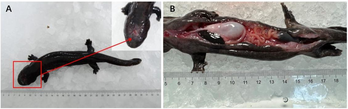
The diseased giant salamander moved slowly and exhibited slightly enlarged limbs. On the top of the head, we observed enormous irregular blisters. We also observed ulcerated signs on the body, along with a few small white patches. The surrounding skin tissue exhibited signs of inflammation and lesions.

3.2. Histopathological Changes
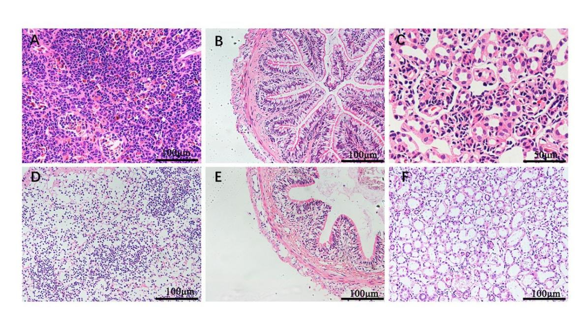
For histological analysis, the intestine, kidney, and spleen of Chinese giant salamanders that were sick and those that weren’t were removed. The spleens of healthy Chinese giant salamanders were structurally intact with tightly packed lymphocytes (Figure 2A). The structures of the spleens from diseased Chinese giant salamander appeared scattered and filamentous, and t cells were loosely arranged (Figure 2D). Kidney sections of healthy giant salamanders showed clear boundaries for the glomeruli, tubules, and other structures with normal gaps (Figure 2C). However, in the kidneys of diseased salamanders, we observed edema and congestion of the glomeruli, the enlargement of gaps, swelling, and necrosis of the renal tubular cells (Figure 2F). Healthy intestinal tissue appeared tightly packed, with good structural integrity and the absence of loose muscular tissue (Figure 2B). The intestinal epithelium of diseased animals showed obvious rupture, loosely packed cells, and atrophy of the muscle layer; in addition, the serosa layer was loose (Figure 2E).

3.3. Morphological Characterization of the Isolated Bacterium
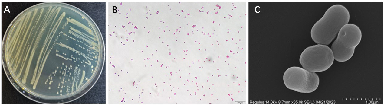
After 24 hours of culture on BHI plates, the DN-2 strain produced pale yellow colonies with diameters ranging from 1.2 to 1.6 mm; these colonies exhibited tidy and shiny edges. Gram staining revealed that the DN-2 strain was a form of Gram-negative bacteria. The bacteria obtained were short and rod-shaped and grew in a single manner, as determined by scanning electron microscopy (Figure 3)
__dn-2_gram_staining_(b)__morphology_of_th.png)
3.4. Molecular Biological Identification Analysis
The sequencing results arising from the PCR amplicons were compared in the NCBI database by using the BLAST tool. Analysis showed that the bacterial strain recovered from the livers of testing Chinese giant salamander was Acinetobacter lwoffii. The similarity between DN-2 and A. lwoffii was 99%. Figure 4 shows a 16S rRNA phylogenetic tree for the near-source standard strain. A. lwoffii is positioned on the same branch.

3.5. Antibiotic Susceptibility Analysis
In total, fourteen types of antibacterial drugs were selected, and the sensitivity of the DN-2 strain to each experimental drug was analyzed with a drug sensitivity kit in accordance with the manufacturer’s instructions. Drug susceptibility tests revealed that the DN-2 strain was highly sensitive to 11 of the 14 drugs, including erythromycin, gentamicin, neomycin, streptomycin, midecamycin, ciprofloxacin, piperacillin, florfenicol, doxycycline, carbenicillin, and sulfanilamide, and moderately sensitive to 3 antibiotics, including norfloxacin, vancomycin, and furazolidone (Table 1).
3.6. Physiological and biochemical identification of bacteria
Strain DN-2 was identified with a BIOLOG automatic microbial identification system. Analysis showed that the DN-2 strain was A. lwoffii. The parameters are:PROB = 0. 988; SIM = 0. 822; DIST = 5. 155. Key physiological and biochemical indicators are shown in Table 2.
3.7. Regression infection experiment
The experimental infection of Chinese giant salamander over a 14-day period led to different degrees of symptoms or death; there were no deaths in the control group (Figure 5). Bacteria were isolated from the infected group and then re-identified to confirm the pathogenicity of the bacteria. the median fatal dosage (LD50) of A. lwoffii DN-2 for A. davidianus was determined to be 4.63×10⁴ CFU/g in Chinese giant salamander.

4. Discussion
By using standard physicochemical assays on DN-2 in our investigation, we were able to determine that the strain was a single developing Gram-negative short rod. The isolated strain’s 16S rDNA sequence matched A. lwoffii by 99%, and it was found clustered on the same branch as A. lwoffii. We also carried out physiological and biochemical identification at the same time. The two most crucial values in the Biolog identification results were SIM and DIST. The degree of matching between the identification result and the matching data bar in the database is indicated by the DIST value. A good match is found when DIST is less than 5.0. The identification result is more dependable the closer the SIM value is to 1.0012. The DIST value of 5. 155 and the SIM value of 0. 822 in the identification of the bacteria in this investigation show that the identification result was correct. In the end, our investigation verified that Ivonella was the primary culprit causing the lesions in A. davidianus.
Acinetobacter is a ubiquitous zoonosis bacterium; previous studies reported the isolation of Acinetobacter junnii, Acinetobacte bauman and Acinetobacter xiamenensis from Schizothorax prenanti, Silurus asotus, Crucian carp and Ctenopharyngodon idella.11–14 The most common pathogenic bacteria isolated from Chinese giant salamander are Aeromonas hydrophila, Aeromonas sobria, Aeromonas veronii, Pseudomonas fluorescens, Edwardsiella tarda and Pseudomonas putida.15–18 The isolation of A. lwoffii from Chinese giant salamander has not been reported previously.
The diseased Chinese giant salamander monitored in the present study showed limb enlargement, sluggish movement, different degrees of ulceration and inflammation on the body surface, along with signs of bleeding. We also identified that the bacteria primarily targeted the liver, spleen, kidney, and intestine; similar damage has been reported for Oncorhynchus mykiss, Cyprinus carpio and the Schizothorax genus when infected by A. lwoffii. Furthermore, this pathogenic bacterium has been clinically associated with pneumonia, meningitis, peritonitis and gastroenteritis19–21 and has also been reported to cause respiratory infections in Lama glama and piglets,22,23 and arthritis in racing pigeons.24 Collectively, these results indicate that A. lwoffii exerts different effects on target organs in amphibians and fishes when compared to those reported in terrestrial animals.
Reference recommendations for the management of A. lwoffii infection were provided by means of antibiotic susceptibility testing. The DN-2 isolate was highly sensitive to erythromycin, doxycycline, neomycin, streptomycin, medicamycin, ciprofloxacin, piperacillin, florfenicol, gentamicin, carbenicillin, and sulfamethoxazole. Previous research showed that A. lwoffii isolated from Cygnus olor exhibited sensitivity to Amikacin, gatifloxacin, doxycycline, kanamycin, ciprofloxacin, cefotaxime, gentamicin and Ofloxacin, but was resistant to amoxicillin, ampicillin and cefixime25 Resistance is not as widespread as in humans and farmed animals,26,27 thus indicating that resistance to these antibiotics is not natural; rather, it is the result of selection after heavy use.
“The lowest dose or concentration that can cause 50% mortality of experimental animals” is known as the semi-lethal concentration. The fundamental idea behind this figure is that when 50% of experimental animals pass away due to poisonings at varying concentrations, people hypothesize about the concentration value.28 A. lwoffii’s LD50 varied depending on the aquatic animal. The hybrid sturgeon’s LD50 was discovered to be 1.22×10² CFU/g31. The present investigation revealed that the LD50 of A. davidianus was 4.63×10⁴ CFU/g, a much higher value than that of the former. Intraperitoneal injection of DN-2 cultures into A revealed clinical signs resembling spontaneous infection*. davidianus*.
A. lwoffii, as an opportunistic pathogen, exhibits a good rate of survival in a host under normal circumstances but can transform into a harmful form of bacteria when its site of aggregation changes, the body’s resistance weakens, or an imbalance of the flora develops.29,30 Under conditions of artificial domestication, an excessively high breeding density can cause Chinese giant salamanders to bite each other; nutrition is compromised, and breeding patterns are not standardized. Furthermore, management strategies often include the use of a range of antibiotics. Collectively, these factors can lead to serious reductions in immunity, thus resulting in dysbacteriosis and eventually leading to A. lwoffii infections.
Chinese giant salamanders are commonly raised in flowing water that varies in temperature and water quality. It is suggested that the prevention and control of bacterial diseases caused by giant salamanders should be carried out, and the daily monitoring of bacterial resistance should be carried out so that farmers can have effective varieties of antibiotics. In the case of an outbreak, the right drugs should be used in the first instance to control the disease. In the breeding process, doing a good job of drug sensitivity tests, targeted medication, and medication records is very important. A combination of narrow-spectrum antibiotics, a combination of drugs, and a rotation of drugs should be used as much as possible.31 Meanwhile, it is necessary to investigate further the risk factors of A. lwoffii infection in Chinese giant salamanders.
5. Conclusions
This investigation discovered a Chinese giant salamander strain known as DN-2. Testing for artificial infections revealed that DN-2 is extremely harmful to Chinese giant salamanders and that this isolate was extremely sensitive to eleven different medications, including streptomycin, erythromycin, gentamicin, and neomycin. When combined, our findings offer a guide for identifying and managing A. lwoffii infections in Chinese giant salamanders.
Acknowledgments
We are very grateful to the Yangtze River Fisheries Research Institute, Chinese Academy of Fishery Sciences, for helping us with the experiments in this paper.
Authors’ Contribution
Conceptualization: Pan Mao (Equal), Ying Wei (Equal). Data curation: Pan Mao (Equal). Formal Analysis: Pan Mao (Equal). Investigation: Pan Mao (Equal), Yixing Xie (Equal). Methodology: Pan Mao (Equal), Yixing Xie (Equal). Resources: Pan Mao (Equal), Yixing Xie (Equal). Validation: Pan Mao (Equal), Ying Wei (Equal). Writing – original draft: Pan Mao (Lead). Writing – review & editing: Pan Mao (Equal), Yixing Xie (Equal). Software: Yixing Xie (Equal), Cheng Wang (Equal). Supervision: Cheng Wang (Equal), Zhiyong Deng (Equal), Huayan Yuan (Equal), Mingzhu Tian (Equal). Funding acquisition: Ying Wei (Lead), Ying Wei (Lead). Project administration: Ying Wei (Lead).
Competing of Interest – COPE
No competing interests were disclosed.
Ethical Conduct Approval – IACUC
In the present study, all experimental procedures were conducted according to guidelines of the appropriate Animal Experimental Ethical Inspection of Laboratory Animal Centre, Yangtze River Fisheries Research Institute, Chinese Academy of Fishery Sciences (ID Number: YFI2022-zhouyong-022).
Informed Consent Statement
All authors and institutions have confirmed this manuscript for publication.
Data Availability Statement
All are available upon reasonable request.