


Introduction
Streptococcosis is an infectious disease caused by members of the genus Streptococcus that can seriously cause huge losses to the food sector. It has been listed as a secondary infectious disease by the Ministry of Agriculture and Rural Affairs of China (Affiche No. 256) in 2020. Streptococcal pathogens in fish to date including S. iniae,1 S. parauberis, ,2 S. agalactiae,3 S. ictalurid,4 and S. dysgalactiae.5,6 According to reports, S. dysgalactiae can lead to significant diseases in several types of farmed fish, including marine fish such as king fish (Seriola lalandi)7 and grey mullet (Mugil cephalus),8 and freshwater-farmed fish such as Nile tilapia (Oreochromis niloticus)9 and sturgeon (Acipenser schrenckii and Acipenser gueldenstaedti).10,11 Furthermore, as an emerging pathogen, it has significantly impacted the economy within the aquaculture sector.5,12 In addition, S. dysgalactiae is thought to be the primary cause of bovine mastitis,13 ovine suppurative polyarthritis,14 and neonatal mortality in puppies.15 However, rapid and sensitive detection of S. dysgalactiae is crucial for timely treatment. Moreover, to detect S. dysgalactiae in outdoor settings where laboratory testing instruments are unavailable, inspectors prefer a quick method that does not require complex equipment.16 Therefore, it is certainly worth developing a fast, sensitive, and simple method to detect S. dysgalactiae to better control its transmission and enable early diagnosis in fish.
Isothermal amplification of nucleic acid sequences has emerged as a pivotal method in molecular biology detection, witnessing rapid advancements over the past decade. Key techniques in this field include nucleic acid sequence-based amplification (NASBA), loop-mediated isothermal amplification (LAMP), and recombinase-aided amplification (RAA).17 As a new isothermal amplification technology, the RAA needs lower requirements on the temperature conditions of amplification, and the reaction can be finished in 30-minute water bath at a simple constant temperature (37-42 °C). The amplified products cannot only be displayed on a portable instrument with blue light,18 but also can be detected at the endpoint combined with a lateral flow dipstick (LFD),19,20 which can be used for on-site testing. LFD is an endpoint detection technique that can be used to observe amplification results directly with the naked eye. The RAA-LFD method, which combines RAA amplification and LFD detection technology, has good specificity and sensitivity, easy-to-read results, low equipment, and experimental operator requirements, and is suitable for rapid detection in non-laboratory environments. As an efficient detection method, the RAA-LFD has been widely used in multiple fields, including early detection and screening of pathogenic microorganisms, rapid quarantine of entry-exit food, and food safety.21–23
The immunogenic secreted protein (ISP) gene sequence is highly conserved, and the encoded protein has strong immunogenicity, which is the dominant antigen of S. dysgalactiae.24,25 In this study, the ISP gene of S. dysgalactiae was considered as the target gene, and RAA and LFD were combined to establish an RAA-LFD method for rapid detection of S. dysgalactiae isolated from channel catfish (Ictalurus punctatus), and its reliability was verified by sample detection, which provided a basis for establishing a method for fast detection of S. dysgalactiae in the field.
Materials and methods
Source of strains and DNA extraction
In this study, S. dysgalactiae (WJ001), S. agalactiae (GY104),26 S. iniae (Ab130920),27 Staphylococcus aureus,28 Vibrio mimicus (SCCF01),29 Aeromonas hydrophila (YYL),30 and Edwardsiella ictalurid (LW102)31 were isolated from fish and preserved in our laboratory. These strains were cultured on brain heart infusion (BHI, BD corporation, USA) agar plates and cultured at 28 °C for 48 h. The cultured bacteria were transferred to fresh BHI broth (1:100) and cultured until the OD600 value reached 0.6-0.8. These culture suspensions were used for the preparation of DNA templates.
Total DNA was extracted from each strain using the bacterial DNA extraction kit (TIANGEN, Beijing, China). The DNA concentration was determined by a spectrophotometer (DU730, Beckman Coulter, USA). The purified DNA extractions were stored at −20 °C until needed.
S. dysgalactiae WJ001 sequencing analysis
Genomic libraries were prepared using Nextera XT Kit (Illumina, Essex, UK), and 150 base pairs (bp) paired-end sequencing was performed using HiSeq. 4000 (Illumina) and the P6-C4 Reagent Kit from PacBio. Read quality was assessed using FastQC (bioinformatics.bbsrc.ac.uk) and filtered using Trimmomatic.32 Trimmed reads were submitted to the Center for Genomic Epidemiology for online assembly by Spades (Nurk et al., 2013) and a subsequent assessment was done using Quast.33
Using the CVTree3 Web Server (http://tlife.fudan.edu.cn/cvtree/cvtree/), a phylogenetic tree based on the whole genome was constructed by the synthetic vector method, with the K-tuple length set to 12. The other S. dysgalactiae of genomic sequences that were used for phylogenetic analysis were downloaded from the National Center for Biotechnology Information (NCBI) (https://www.ncbi.nlm. nih.gov/).
Primers and probe design
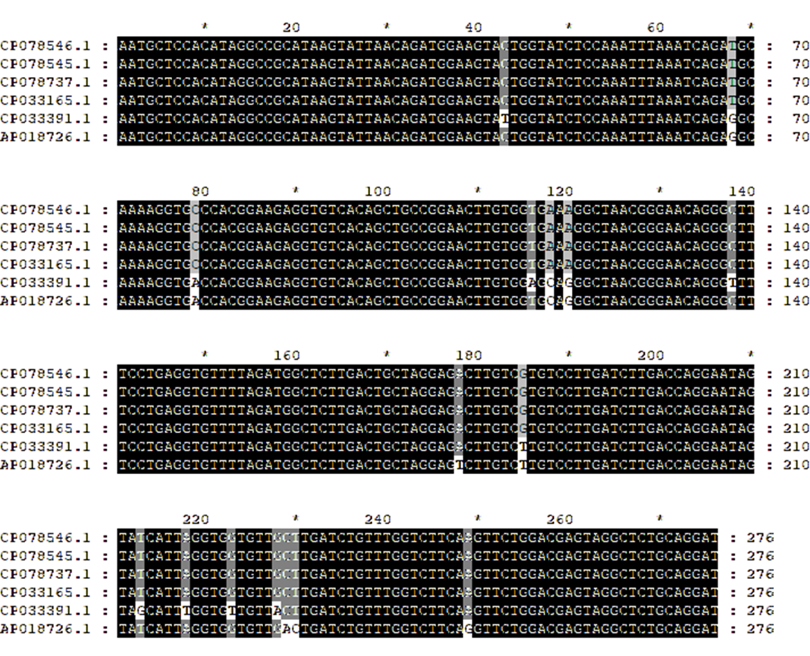
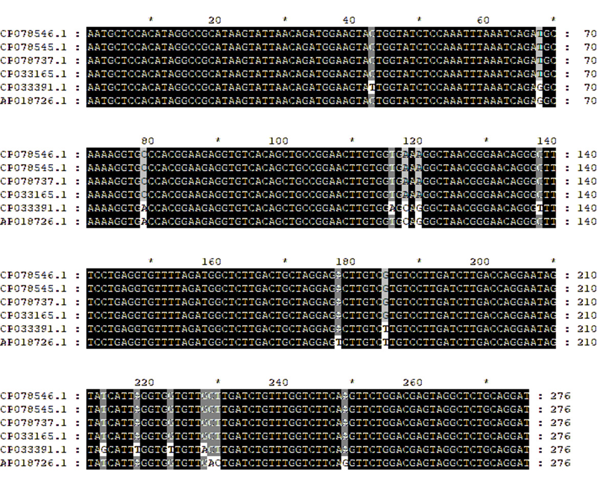
The primers for the ISP gene of S. dysgalactiae were designed specifically based on the instruction manual by lesunbio® kits (RAA). The ISP gene sequence can be obtained in GenBank (Accession Numbers: AP018726.1, CP033391.1, CP033165.1, CP078737.1, CP078545.1, CP078546.1). Homology analysis of ISP gene sequences and determination of conserved regions was performed by DNAMAN alignment (Fig. 1). The amplification reaction was performed as per the instructions of the kit. RAA products were detected by 1% agarose gel electrophoresis after the reaction to determine the best primer pair. According to the instructional manual provided by lesunbio® kits (RAA-LFD), the best primer pair and probe were synthesized and modified. All primers and probes were synthesized by Sangon Biotech (Shanghai, China).
RAA-LF assay
RAA reaction was finished using lesunbio® kits (RAA-LFD), with primers and probe as described above. Reaction mixtures containing 25 μL 1×rehydration buffer, 15.2 μL ddH2O, 2.1 μL forward primer (10 μM), 2.1 μL reverse primer (10 μM), 0.6 μL probe (10 μM), 2 μL template DNA, and 47 μL reaction premix were transferred to the reaction unit, the freeze-dried powder was fully dissolved by hand flicking and centrifuged briefly. Next, 3 μL magnesium acetate (280 mM) was added to the reaction tube cover, carefully closed the tube cover, and briefly centrifuged to make the starter into the premix, then hand flick evenly and briefly centrifuged. After fully mixing, centrifuge and put into a water bath for 20 min at 39 °C.
The outcome of RAA-LFD reactions was analyzed by RAA and LFD-strips in this study. 10 μL RAA products were added to the sample pad, and the LFD-strips were vertically inserted into the 100 μL buffer solution in the EP tube. The determination of the LFD result is a test band. The control band is clearly visible with red lines within 5-10 minutes, indicating that the RAA amplicon is detected (positive). However, displaying only the control band (red line) means that no RAA amplifier is detected (negative). In any case, the control band must be visible at all times to confirm that the strips are functioning properly.34 The working principle of the RAA-LFD for the detection of S. dysgalactiae is shown in Fig. 2.

Optimization of RAA-LFD amplification reaction system and conditions
To determine the optimal temperature for this assay, we performed RAA-LFD with 2 μL of genomic DNA at various temperatures ranging from 29 to 49 °C. The experiment to determine the optimum reaction time was carried out at 39 °C, and the reaction was terminated after adding MgAc at 5 min, 10 min, 15 min, 20 min, 25 min,and 30 min, respectively.
Analytical sensitivity and specificity of RAA-LFD
To assess the sensitivity of the RAA-LFD assay, genomic DNA of the S. dysgalactiae was extracted by the kit and accomplished with 10-fold serial dilutions (1.002×10³ ng/μL, 1.002×10² ng/μL, 1.002×101 ng/μL, 1.002×100 ng/μL, 1.002×10² pg/μL, 1.002×101 pg/μL, 1.002×100 pg/μL) for RAA-LFD assay. Conventional PCR was used as a control to more valuable assessment the sensitivity of the RAA-LFD assay, and ddH2O was used as a negative control.
To determine the analytical specificity of the RAA-LFD assay developed in this study, the genomic DNA of a set of pathogens including S. dysgalactiae, S. agalactis, S. iniae, S. aureus, V. mimicus, A. hydrophila, and E. ictalurid were used as templates for the RAA reactions. Different RAA products were directly analyzed by LFD-strips.
Evaluation of the RAA-LFD assay using experimental samples
To evaluate the performance of the RAA-LFD assays for the detection of active S. dysgalactiae infection, 60 healthy Zebrafish were used for challenge experiments. The temporarily reared Zebrafish were divided into two groups, with 50 Zebrafish used as an experimental group and 10 Zebrafish used as a negative control group. Each fish in the experimental group was challenged with 50 μl of bacterial suspensions in 0.9% NaCl solution at a concentration of approximately 1.0×10⁶ CFU/ml by intraperitoneal injection, and the control group was intraperitoneally injected with 50 μl 0.9% NaCl. All Zebrafish were checked daily for clinical signs for 7 days. DNA extracted from splanchnic tissue obtained from experimentally infected Zebrafish using the Genomic DNA extraction kit (TIANGEN, Beijing, China). RAA-LFD and conventional PCR were used to detect, and the coincidence rates of the two detection methods were compared. The receiver operating characteristic (ROC) curve analysis was conducted with Statistical Package for Social Sciences (SPSS version 26.0) to evaluate the diagnostic value of RAA-LFD. All animal infections were conducted following protocols approved by the Institutional Animal Care and Use Committee of Sichuan Agricultural University.
Statistical analysis
Three measurements were made for each experiment, and all experiments were made in triplicate. Statistical analyses were performed with the GraphPad Software package (GraphPad Software, La Jolla, CA, USA). Statistical significance was determined with a Student’s t-test or two-way ANOVA for multiple comparisons. At p<0.05, the difference was considered significant.
Results
Genome sequence comparisons
To determine the phylogenetic relationship between S. dysgalactiae WJ001 and other S. dysgalactiae, which were based on whole genome sequences, the CVTree3 was used based on the synthetic vector method.35 The results showed that the phylogenetic relationship between S. dysgalactiae and its subtypes from different regions was relatively close. Besides, there was no obvious trend between different regions (Fig. 3).

Screening and synthesis of ISP gene-specific RAA-LFD primers
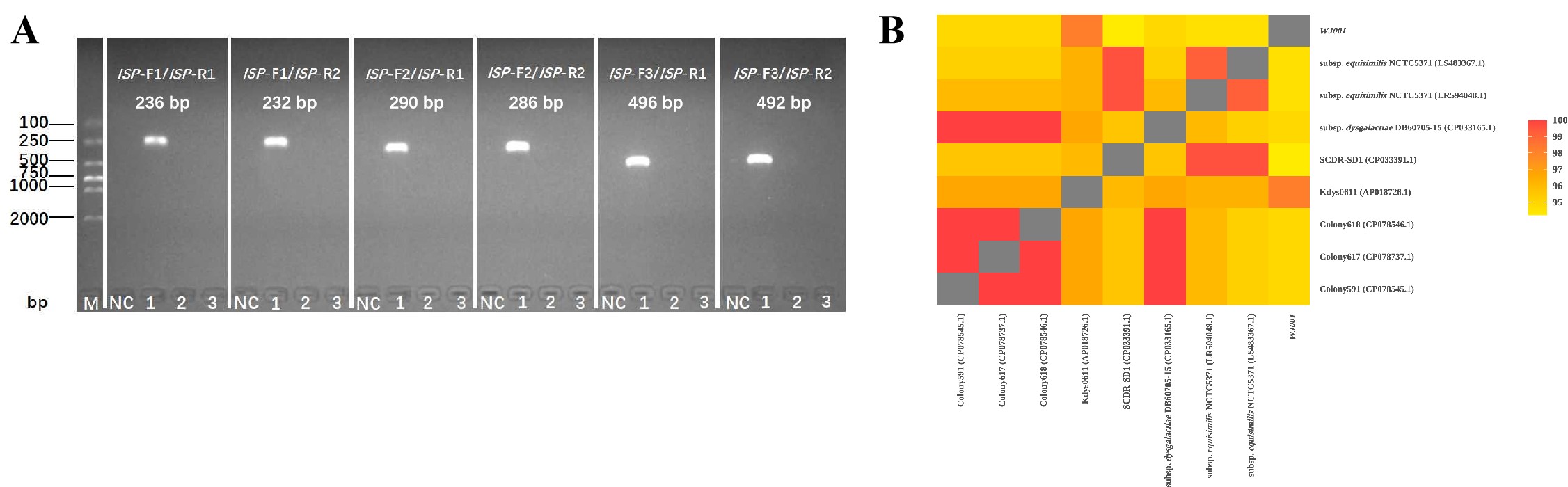
Six pairs of ISP gene-specific primers were designed based on the RAA primer design principle, with three forward and two reverse candidate primers displayed in Table 1. The primers were screened by lesunbio® kits (RAA). The results showed that all 6 pairs of primers were able to amplify the specific band (Fig. 4A). ISP-F2/ISP-R2 primers were randomly selected for establishing RAA-LFD. The sequencing results of the amplified products of the ISP-F2/ISP-R2 primers were compared with the corresponding sequences in GenBank. The analysis of the sequence homology was 94.2%-100% (Fig. 4B), indicating the ISP-F2/ISP-R2 primers had high specificity. Biotin and FAM were modified at the 5’ end of the reverse primer and probe respectively, C3 spacer was modified at the 3’ end of the probe, besides, base T was replaced with dSpacer (Table 1) for subsequent LFD-strips detection of amplification products.

Optimization of the RAA-LFD conditions
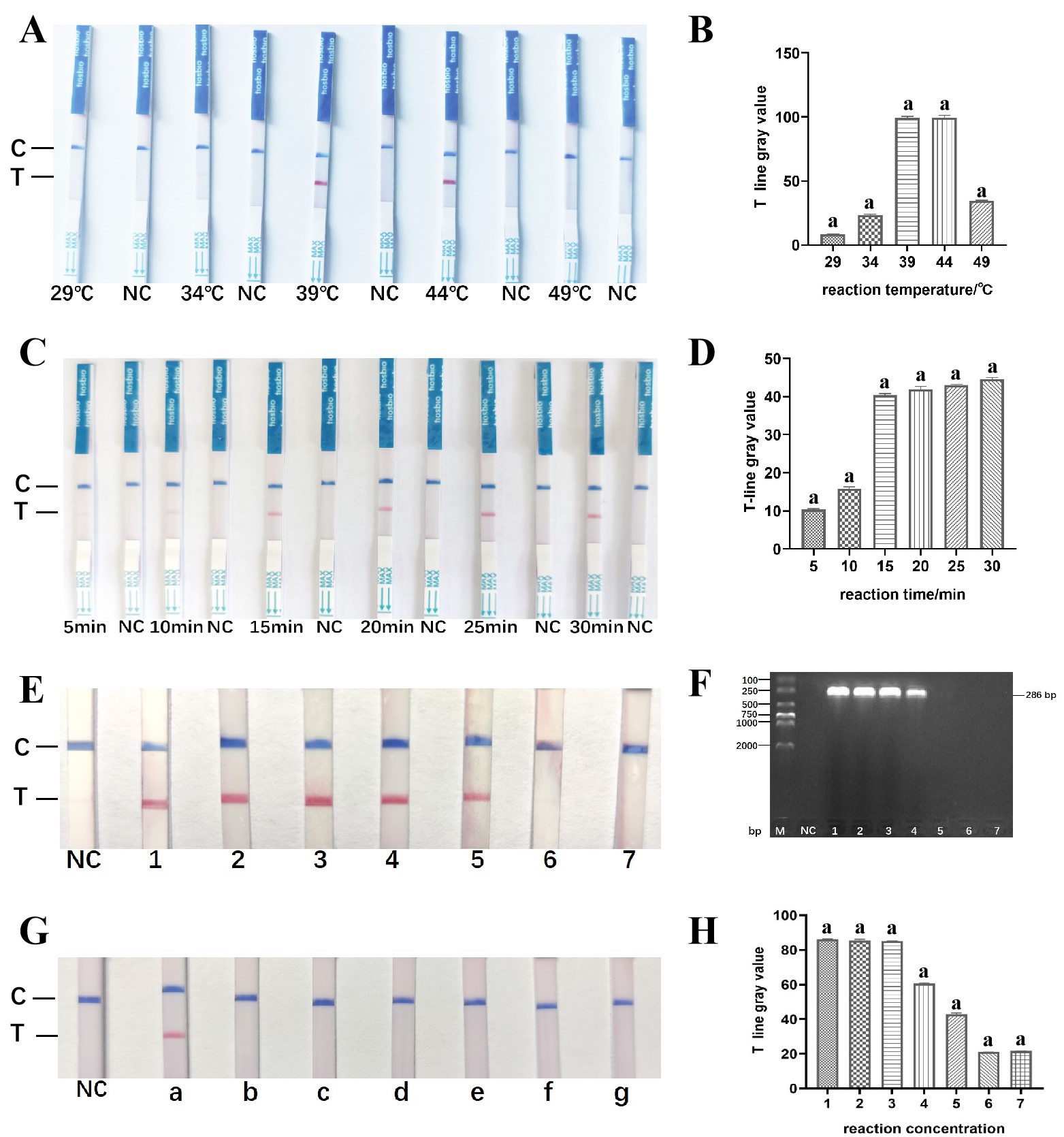
RAA amplification products can be detected by LFD-strip in the temperature range of 39 - 44 °C. However, the results showed that with the increase of temperature, the brightness on the T-line of the LFD-strip increased first and then decreased, when the temperature was lower than 39 °C or higher than 44 °C, the brightness on the T-line was not obvious (Fig. 5A), there was no significant difference in the gray value of T-line analyzed with ImageJ software (P > 0.05) (Fig. 5B). In order to make the detection temperature closer to the ambient temperature, 39 °C was chosen as the optimal reaction temperature.

39 °C was determined as the reaction temperature, then the optimal temperature for this assay was determined. The reaction time was 5 min, 10 min, 15 min, 20 min, 25 min, and 30 min, respectively. The T-line of the LFD-strip began to show a weak bright band after 5 min of amplification reaction and gradually brightened with the increase of the reaction time (Fig. 5C). When the reaction time was 15, 20, 25, and 30 min, respectively, there was no significant difference in the T-ray gray value (P > 0.05) (Fig. 5D). Therefore, 15 min was chosen as the best reaction time to ensure the rapidity of the experiment.
Sensitivity and specificity tests of RAA-LFD
The RAA-LFD assay’s sensitivity was tested by employing 10-fold serial dilutions of S. dysgalactiae genomic DNA, and the amplified products were identified using conventional PCR and LFD strips. The conventional PCR results showed positive signals from 1.002×10³ ng/μL to 1.002×100 ng/μL of DNA (Fig. 5H), which indicated that the lowest detection limit was 1.002×100 ng/μL. While the concentration of S. dysgalactiae genomic DNA decreased, the brightness of the T-line band gradually became weaker. When the genomic DNA concentration was lower than 1.002×10² pg/μL, there was no positive band in the T-line (Fig. 5E), and there was no significant difference in the gray value of the T-line analyzed with ImageJ software (P > 0.05) (Fig. 5F). Therefore, the detection limit of S. dysgalactiae by the RAA-LFD was 1.002×10² pg/μL.
The specificity analysis of RAA was carried out using the genomic DNA of S. dysgalactiae, S. agalactis, S. iniae, S. aureus, V. mimicus, A. hydrophila, and E. ictalurid. S. dysgalactiae have positive bands on the T line of the LFD-strips, while the other 6 pathogens have no positive bands on the T line of the LFD-strips after amplification (Fig. 5G), indicating that the ISP gene S. dysgalactiae amplification primers designed with conserved sequences are highly specific.
Performance of the RAA-LFD on experimental samples
The performance of the S. dysgalactiae RAA-LFD was evaluated using 50 Zebrafish in the experimental group and compared with that of conventional PCR. In the experiment, On the first day after the infection, Zebrafish began to show abnormal symptoms, including anorexia, slow and weak swimming, and directionless swimming in the water. The next day, the affected Zebrafish began to die. No mortality or significant changes were observed in the control group. Artificially infected Zebrafish had the same clinical symptoms of the disease as naturally infected channel catfish. In these experimental samples, 41 positive samples and 9 negative samples were detected by the RAA-LFD method, with a positive rate of 82.0% and a negative rate of 18.0%; 40 positive samples and 10 negative samples were detected by conventional PCR, with a positive rate of 80.0% and a negative rate of 20.0%. The two methods had a positive coincidence rate of 97.6%, a negative coincidence rate of 90.0%, and the total coincidence rate of 98.0% (Fig. 6A). Compared with conventional PCR, the AUC of RAA-LFD was 0.950, the sensitivity was 100% and the specificity was 90% (Fig. 6B). Through the test results of actual samples, RAA-LFD method has higher detection accuracy and better practical application effect.

In another word, the RAA-LFD method established in our study is more suitable for the field detection of S. dysgalactiae.
Discussion
With the stable development of China’s aquaculture business, streptococcosis has become a more dangerous bacterial disease.36 S. dysgalactiae, as a newly emerged Gram-positive streptococcal pathogen in aquaculture, has caused significant economic losses to the aquaculture industry.5,12 To reduce the economic losses caused by S. dysgalactiae, a convenient, quick, accurate, and sensitive assay for detecting infection must be developed.
To develop a diagnostic technology capable of detecting pathogens in outdoor settings or environments lacking laboratory equipment, isothermal amplification technology was optimized to replace the PCR method, which relies on precision thermocouples. It has become a technology with far-reaching influence, wide use, and rapid nucleic acid amplification about isothermal nucleic acid amplification, which has been widely used in the detection of common infectious pathogens.37,38 At present, there are many isothermal amplification techniques for nucleic acid. Different isothermal amplification techniques are designed according to different reaction principles. The common methods are: nucleic acid sequence-based amplification (NASBA), loop-mediated isothermal amplification (LAMP), and recombinase-aided amplification (RAA). Compared with other methods, RAA has the following advantages: (1) NASBA needs to be preheated, but RAA does not need it. It can be maintained at 37 °C throughout the whole process; (2) LAMP needs to design 4-6 pairs of primers, while RAA-LFD only needs to design 1 pair of primers and probes; (3) RAA-LFD takes a short time, and the reaction can be completed in 20-30 min; (4) RAA reaction temperature is lower than that of other amplification methods.39,40
Generally speaking, the false positives were more probable to cause amplification results because of undesirable amplification or the presence of primer dimers.41 The less possibility of false positives or sensitivity can be avoided to a greater extent by designing and creating RAA primer/probe combinations.42 In addition, it was found that probes play a crucial role in RAA. On the contrary, the changes in primers did not have a significant effect.43 In our study, we designed primers and probes for the ISP sequence of S. dysgalactiae to reduce the probability of false positives and to improve the feasibility of the success of the RAA-LFD assay in our study.
Up to now, the practicability of isothermal amplification used in outdoor settings has been limited due to the useful, cheap, and specific signal transduction.44 Optimizing reaction conditions is critical for obtaining accurate and reliable RAA results. One of the factors that influences amplification is the RAA’s reaction temperature. In this study, 39 °C was determined to be the optimal reaction temperature. We can learn that the RAA reaction can be carried out in a water bath with a temperature slightly higher than the ambient temperature by these results.45 In addition, the RAA-positive amplification can be visually confirmed within 15 min, whereas the conventional PCR analysis typically takes approximately 2–3 h to complete. It can be seen that the entire detection time is significantly shortened. There is no expensive instrument in the presentation of LFD-strip results, which can be observed directly with the naked eye at room temperature. The RAA-LFD is appropriate for on-site detection, which can be accomplished for nucleic acid detection in remote areas with low cost and no instrument. Nevertheless, the RAA-LFD method has its shortcomings. The most significant issue is the propensity for false positives to arise when operations are improperly conducted. Additionally, there are stringent requirements regarding the design of primers and probes, which can incur certain costs during selection. Furthermore, while nucleic acid test strips offer rapid testing advantages, they also present challenges related to quantification that may limit the broader application of this methodology.
In our study, the detection method of RAA-LFD established had high specificity and had no cross-reactivity with the nucleic acids of other common aquatic pathogens. The minimum detection limit was 1.002×10² pg/μL, which was higher sensitivity than conventional PCR. Our research results are consistent with those of other previous studies, and RAA-LFD method is more sensitive and specific compared to conventional PCR.46–49 Moreover, it is very feasible to screen S. dysgalactiae in the preliminary clinical study. To verify the effectiveness of the RAA-LFD method constructed in this study, we tested 50 experimental samples infected by S. dysgalactiae disease materials and compared them with conventional PCR. The results showed that the positive and negative rates of the RAA-LFD method were 82.0% and 18.0%, respectively; the positive and negative rates of the conventional PCR method were 80.0% and 20.0%, respectively. The positive and negative coincidence rates of the two methods were 97.6% and 90.0%, respectively, with a total coincidence rate of 98.0%. Compared with conventional PCR, the AUC of RAA-LFD was 0.950, with sensitivity and specificity of 100% and 90%, respectively. The results of this study demonstrated that the RAA-LFD method is effective for diagnosing S. dysgalactiae infection in fish.
In our study, we successfully established the RAA-LFD method, which can not only be used for the fast and visual detection of S. dysgalactiae, but also has the remarkable advantages of high sensitivity and good specificity. Furthermore, the above method has potential clinical application, as the reaction can be finished at 39 °C for 15 minutes and can be directly observed with the naked eye. In conclusion, the RAA-LFD method in our study is extremely meaningful to rapidly detect S. dysgalactiae in the field.
Conclusions
In this study, we established an ISP gene sequence that was highly conserved of the S. dysgalactiae, and the effectiveness of this method in detecting S. dysgalactiae specific nucleic acids was verified. The products amplified by RAA, coupled with LFD, can be interpreted by the naked eye. The RAA-LFD method in our study can be performed at 39 °C for 15 minutes, positively detect as low as 1.002×10² pg/μL of DNA, and has the remarkable advantages of high sensitivity and good specificity. This method is rapid and does not require specialized equipment or professional personnel. Overall, we provide an accurate and effective tool for the detection of S. dysgalactiae, especially in resource-limited areas.
Acknowledgments
This research was supported by the Sichuan Natural Science Foundation (24NSFSC0381), Sichuan Innovation Team Project of Agricultural Industry Technology System (SCCXTD-15).
Authors’ Contribution
Methodology: Jiao Wang (Equal), Kun Peng (Equal), Keyu Zhou (Equal), Ping Ouyang (Equal), Weimin Lai (Equal). Formal Analysis: Jiao Wang (Equal), Wei Fan (Equal), Keyu Zhou (Equal), Ping Ouyang (Equal), Hongrui Guo (Equal). Investigation: Jiao Wang (Equal), Wei Fan (Equal), Keyu Zhou (Equal), Xiaoli Huang (Equal), Ping Ouyang (Equal), Hongrui Guo (Equal), Weimin Lai (Equal). Writing – original draft: Jiao Wang (Lead). Data curation: Yang Feng (Equal), Kun Peng (Equal), Defang Chen (Equal). Writing – review & editing: Yang Feng (Equal), Yi Geng (Equal). Funding acquisition: Wei Fan (Equal), Xiaoli Huang (Equal), Yi Geng (Equal). Resources: Defang Chen (Equal), Yi Geng (Equal). Conceptualization: Yi Geng (Lead). Supervision: Yi Geng (Lead). Project administration: Yi Geng (Lead).
Ethics approval and consent to participate
All animal handling procedures were approved by the Animal Care and Use Committee of Sichuan Agricultural University (Chengdu, China) and followed the university’s guidelines for animal experiments under permit number 2020303116.
Availability of data and material
All data generated or analyzed during this study are included in this published article.
Competing interests
The authors declare no competing financial interests.