


Introduction
Microalgae are incredibly adaptable photosynthetic organisms that can thrive in a wide range of aquatic environments, from the open ocean to wastewater treatment systems.1,2 Species like Chlorella, Nannochloropsis, Tetraselmis, and Chaetoceros are among the most commonly cultured in controlled environments because of their wide-ranging uses in aquaculture and various other industries.3,4 These microalgae play a key role in biotechnology, with applications ranging from health foods and animal feeds to natural colorants, biofuels, and even pharmaceuticals.5–7 In aquaculture, microalgae are vital to larval nutrition. They are either fed directly to organisms like penaeid shrimp and mollusks or used to nourish live prey, such as copepods and rotifers, which are then fed to fish larvae.8,9 Moreover, studies have shown that combining different microalgal species results in better nutritional value and improved growth in aquatic animals compared to feeding them a single species.10,11 Microalgae also play a key role in enriching zooplankton, making them a more nutritious feed for fish and their larvae.12,13
Among the many microalgae species, Nannochloropsis sp. stands out for its ability to produce a range of valuable pigments, most notably chlorophyll a, violaxanthin, vaucheriaxanthin, and beta-carotene, making it a key player in commercial pigment production.14,15 Given their potential as a rich source of pigments with many uses in aquaculture and various industries, microalgae have attracted a lot of research attention.1,16 Among the pigments they produce, carotenoids and chlorophylls stand out for their versatility and wide range of applications.16,17 Moreover, to boost the efficiency of microalgae production, researchers are focusing on two main strategies. One involves centralizing production at specialized mass culture facilities that use methods like heterotrophic growth or photobioreactors, while the other focuses on improving post-harvest processing techniques, such as spray drying or concentrating the algae to make the biomass easier and more affordable to distribute to hatcheries.18–20
AMPEP is selected for this study because it offers a unique, natural alternative to synthetic biostimulants. Made from the brown seaweed Ascophyllum nodosum, AMPEP is rich in bioactive compounds like polysaccharides, amino acids, and essential micronutrients that are known to support growth and stress resistance in marine organisms.21 It has already been successfully used in macroalgal cultivation and aquaculture, where it has been shown to improve the performance of various species. These qualities make it a promising candidate for use in microalgae, especially with species like Nannochloropsis sp., which are widely used in hatcheries. Additionally, AMPEP’s marine origin and natural composition distinguish it as a more sustainable and aquaculture-friendly alternative to many land-based or synthetic growth enhancers.22
Beyond the usual ways of growing microalgae, recent studies have started exploring Acadian Marine Plant Extract Powder (AMPEP) as a new way to boost both growth and pigment production. AMPEP comes from the brown seaweed Ascophyllum nodosum and is well-known for helping marine life and crops grow better and handle stress more effectively.2,23 Although AMPEP is widely used in macroalgae cultivation and agriculture, its potential for boosting microalgal growth, especially in microalgae Nannochloropsis sp., is still not well understood. Early research shows promise, suggesting that AMPEP might help microalgae grow better and withstand stress. However, more studies are needed to determine how it affects microalgae, including the best concentrations to use and how to apply it effectively.2,24 This is surprising, given how important Nannochloropsis sp. is in aquaculture and biotechnology. There is currently very limited data on how AMPEP might influence its growth, pigment production, or overall performance. Exploring this could lead to new and more efficient methods for improving microalgae cultivation. This study aims to fill that gap by testing how different concentrations of AMPEP affect the cell density, growth rate, and pigment content of Nannochloropsis sp., with the goal of uncovering its potential as a natural growth enhancer in microalgal production systems.
Materials and Methods
Experimental Site and Duration of the Study
The research was conducted at the Marine Integrated Laboratory (MIL) located in the College of Fisheries Mindanao State University-Tawi-Tawi College of Technology and Oceanography (MU-TCTO) Sanga Sanga, Bongao, Tawi-Tawi, Philippines for 21 days of culture.
Culture Condition of Nannochloropsis sp.
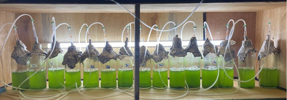
Improvised glass bottles with 1L volume were used in Nannochloropsis sp. experiment. Freshwater was used in the study and filtered through a filter paper before being transferred to 15 Improvised glass bottles containing 1L each. Starter green microalgae of Nannochloropsis sp. with initial cell density of 2.94 x 10⁵ cells mL-1 inoculated into the culture glass bottles. Experimental bottles were then mixed with two different nutrient concentrations randomly the BG-11 medium was used as a nutrient medium (Table 1 and Table 2). AMPEP was then introduced into the flasks at various concentrations, as described in Table 3. The layout of the experiment was in a Completely Randomized Design (CRD) consisting of five (5) treatments with various concentrations of AMPEP replicating thrice (3), as shown in Table 4. Portable aerator and syringe filters with an opening of 0.2 μ were used to prevent contamination and air conditioner maintained a temperature of 20±1°C (Figure 1). For lighting, Ecolum LED T8 fluorescent tubes (model ECBTS11/DL16) were used as artificial lighting for the cultures in the laboratory. These lamps provided a consistent light intensity of approximately 1,500–2,000 lux, suitable for supporting photosynthesis in microalgal cultures. The culture was conducted for 21 days.
_.png)
Growth Analysis of Nannochloropsis sp.
Samples of Nannochloropsis sp. were collected every three days for cell counting and analysis. A Neubauer hemocytometer was used to estimate cell density under a light microscope, and routine visual inspections were carried out to check for any contamination. Microalgal biomass was measured through dry weight analysis. For this, 5 mL of culture from each treatment group was dried in an oven at 105°C for two hours. The specific growth rate (µ) was calculated following the method outlined by Sanuddin et al.27
\[\mu = \frac{\ln{(N_2)\mathbf{-}\ln{\mathbf{(}N\mathbf{\mathstrut_1)\ }}\mathbf{\ }}}{t_2 - t_1}\]
Where: N2 is the cell number at the time (t2).
N1 is the beginning cell number at a time (t1).
Pigment Analysis of Nannochloropsis sp.
Pigment analysis of Nannochloropsis sp. was carried out following the method described by Sarri et al.26 A 5 mL sample of the culture was transferred into labeled test tubes and centrifuged at 5000 rpm for 10 minutes. After removing the supernatant, 5 mL of methanol was added to the pellet. The mixture was vortexed for 30 seconds and centrifuged again under the same conditions. Chlorophyll a and total carotenoids were then measured using a spectrophotometer. Pigment concentrations were calculated using the formulas provided by Zou and Richmond28 and Macıas-Sánchez et al.29
Chlorophyll a (µg mL-1) = 13.9 A666
Total carotenoids (µg mL-1) = 4.5 A475
Statistical Analysis
The data analysis was performed using SPSS version 17.0 to evaluate cell densities, growth response, and pigment accumulation in Nannochloropsis sp. cultures. Statistical significance at p<0.05, and results were expressed as mean ± standard deviation of the mean (STD). One-way analysis of variance (ANOVA) was used to determine significant differences between treatments, followed by Duncan’s New Multiple Range Test for multiple comparisons. Differences were considered statistically significant when p<0.05.
Results
Cell Density
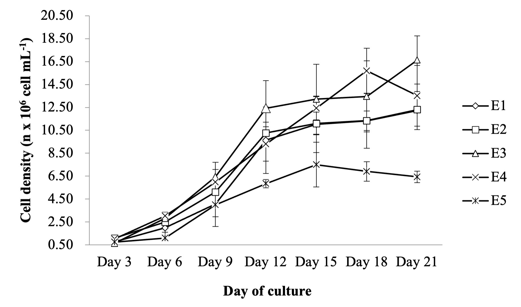
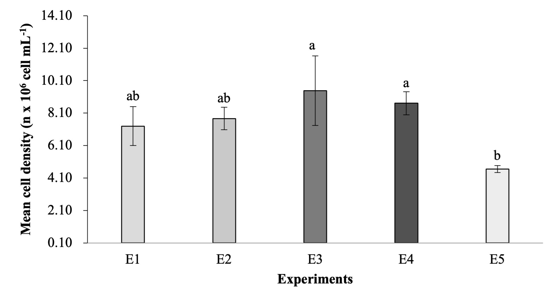
Figure 2 below demonstrates the cell density of Nannochloropsis sp. cultured at different concentrations of AMPEP in a nutrient medium. The initial density of Nannochloropsis sp. was started at 2.94 x 10⁵ cells mL-1, and the culture was done in triplicate. Based on the result of this study the cell density of experiment 1, 2, 3, 4 and 5 were 12.23±1.67 x 10⁶ cell mL-1, 12.31±1.44 x 10⁶ cell mL-1, 16.64±2.10 x 10⁶ cell mL-1, 13.52±2.65 x 10⁶ cell mL-1, 6.44±20.50 x 10⁶ cell mL-1, respectively after 21 days of culture period. Analysis of variance (ANOVA) revealed that experiments 3 and 4 were significantly higher (p<0.05) than the control experiment. Additionally, the mean cell density of experiment 1 (7.30±0.01 x 10⁶ cell mL-1), experiment 2 (7.77±0.01 x 10⁶ cell mL-1), experiment 3 (9.48±2.14 x 10⁶ cell mL-1), and group 4 (8.71±0.71x 10⁶ cell mL-1) were significantly different (p<0.05) than the control experiment (4.65±0.22 x 10⁶ cell mL-1) (Figure 3).
_grown_under_different_concentration.png)
_grown_under_different_concentr.png)
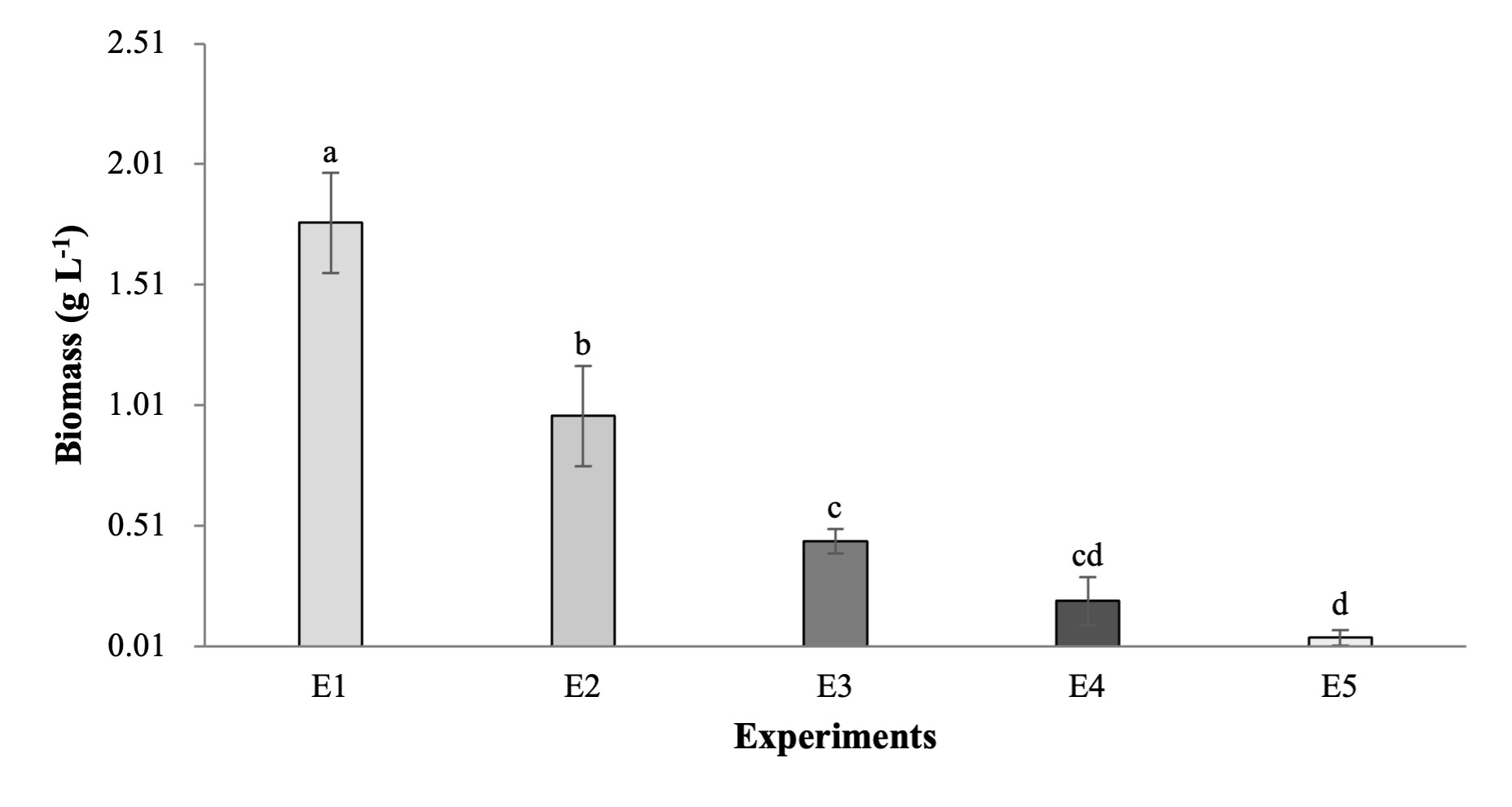
Specific Growth Rate and Biomass
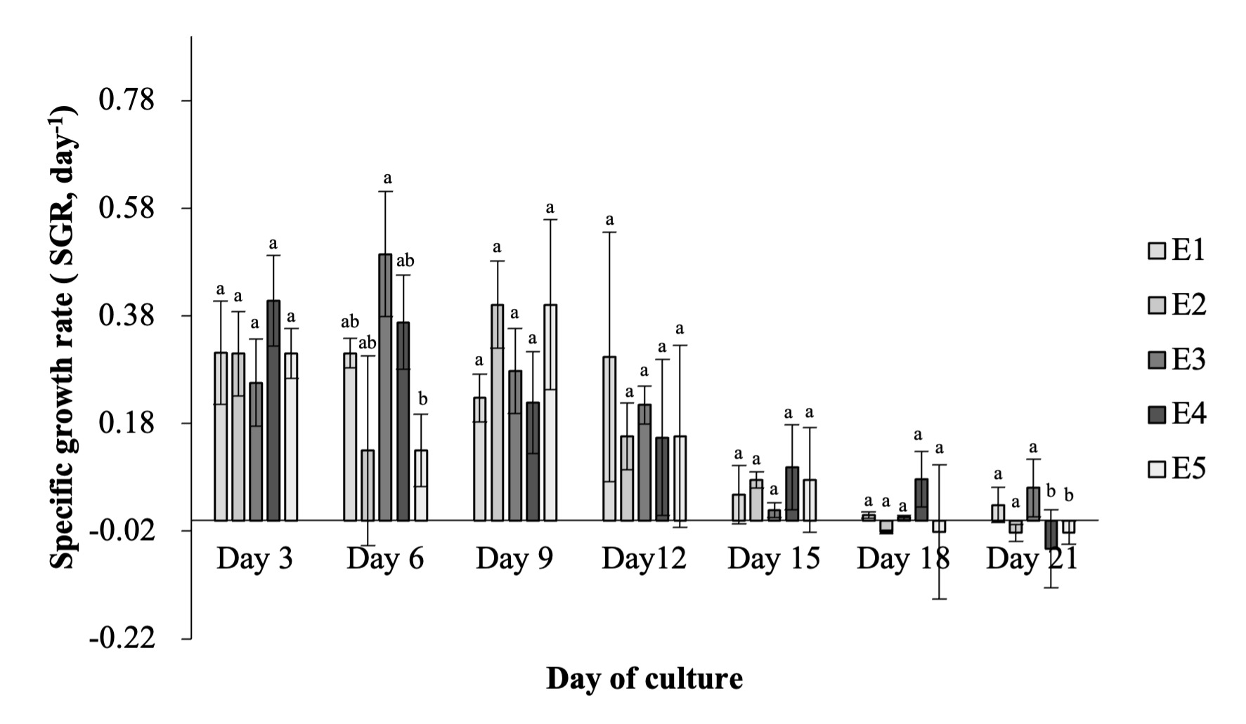
Figure 4 demonstrates the specific growth rate (SGR, day-1) of Nannochloropsis sp., cultured at different concentrations of AMPEP in nutrient medium. Based on the results of this study the maximum SGR were achieved in E3 (0.49±0.12 day-1) and E4 (0.37±0.09 day -1) significantly different (p<0.05) than the SGR in control experiment (0.13±0.07 day -1) as early as day 6 of culture period, however, decreases at the succeeding culture period. Moreover, Figure 5 showed that the mean SGR of E1, E2, E3, E4, and E5 were 0.29±0.01 day-1, 0.29±0.01 day -1, 0.30±0.003 day-1, 0.29±0.007 day-1, 0.26±0.01 day-1, respectively. ANOVA revealed that E1, E2, E3, and E4 significantly higher (p<0.05) than control experiment. Figure 6 shows the biomass (g L-1) of Nannochloropsis sp. cultured at different concentrations of AMPEP in a nutrient medium. Based on the results of the study, the biomass of E1, E2, E3, E4, and E5 was 8.00±0.46 g L-1, 8.00±0.10 g L-1, 6.00±1.00 g L-1, 4.67±0.15 g L-1, and 4.33±0.31 g L-1, respectively. ANOVA revealed that E1 and E2 were significantly (p<0.05) different than the biomass of the control experiment. This indicates that the addition of AMPEP concentration significantly influences the growth of Nannochloropsis sp., with E1 and E2 achieving higher biomass.


_of_*nannochloropsis*_sp.__grown_under_different_concentrations_of_acadia.png)
Pigments Accumulation
Figure 7 demonstrates the chlorophyll a pigment accumulation of Nannochloropsis sp., cultured at different concentrations of AMPEP in a nutrient medium. Based on the results of the study, the chlorophyll a pigment accumulation in E1, E2, E3, E4, and E5 was 4.64±0.37 µg mL-1, 4.16±0.25 µg mL-1, 7.40±0.95 µg mL-1, 7.25±0.57 µg mL-1, and 3.98±0.60 µg mL-1, respectively. ANOVA revealed that E3 and E4 were significantly different (p<0.05) from the control experiment, as indicated in the figure, which shows that increasing AMPEP concentrations enhanced chlorophyll a pigment accumulation in Nannochloropsis sp., cultures. Additionally, Figure 8 shows the total carotenoid pigment accumulation of Nannochloropsis sp., cultured at different concentrations of AMPEP in a nutrient medium. Based on the results of the study, the total carotenoid pigment accumulation in E1, E2, E3, E4, and E5 was 1.78±0.15 µg mL-1, 1.58±1.16 µg mL-1, 2.84±0.37 µg mL-1, 2.75±0.24 µg mL-1, 1.49±0.21 µg mL-1, respectively. ANOVA revealed that E3 and E4 were significantly different (p<0.05) from the control experiment; the figure indicated that increasing AMPEP concentrations enhanced the total carotenoid pigment accumulation in Nannochloropsis sp. cultures.
_of_*nannochloropsis*_sp.__grown_under_dif.png)
_of_*nannochloropsis*_sp.__grown_under_di.png)
Discussion
AMPEP, an extract derived from the brown macroalga Ascophyllum nodosum, comprises approximately 5–10% protein, 40–70% carbohydrates, and 2–7% lipids by dry weight, contributing to its distinctive nutritional profile and biochemical functionality.21,22,30–32 This study explored the effects of different AMPEP concentrations on the growth of Nannochloropsis sp. Results showed that adding AMPEP to the culture medium significantly boosted cell density. Notably, experiments E3 and E4, which received 150 mg L-1 and 200 mg L-1 of AMPEP, respectively, produced the highest cell densities. These findings suggest that these concentrations may be optimal for promoting cell growth and achieving higher cell density compared to the control group. This finding is consistent with previous studies showing that nutrient supplementation and optimized environmental conditions can significantly improve microalgal cell production. For instance, Erbil and Durmaz33 reported that adding 100 mg L-1 of myo-inositol to the culture medium increased the cell density of microalga Nannochloropsis oculata by 1.42-fold compared to the control. Interestingly, a higher concentration of 500 mg L-1 still resulted in a notable 1.28-fold increase, highlighting the potential of targeted nutrient enrichment in enhancing microalgal growth. Additionally, Aziz and Siti Mariam34 reported a high cell density of 32 × 10⁶ cells mL-1 in microalga Chlorella sp. cultures grown in f/2 medium enriched with a specific phosphate source. Similar studies have demonstrated the impact of different media compositions on microalgal growth. For example, Chia et al.35 observed a cell density of 2.38 × 10⁶ cells mL-1 in microalga Chlorella vulgaris cultures grown with varying phosphate concentrations. These findings support the idea that nutrient formulation plays a crucial role in optimizing algal biomass. In line with this, research has shown that moderate AMPEP concentrations, such as 150 mg L-1 can promote optimal growth in Nannochloropsis sp., while higher concentrations may cause stress, potentially leading to nutrient imbalances and decreased photosynthetic efficiency, ultimately limiting growth.
Nutrient availability plays a crucial role in microalgal growth.36 One way to assess the effects of nutrient conditions is by calculating the specific growth rate (SGR), which helps identify potential growth-limiting factors in cultures. In this study, the addition of AMPEP at concentrations of 150 mg L-1 and 200 mg L-1 significantly boosted the SGR of Nannochloropsis sp., reaching mean values of 0.49 day-1 and 0.37 day-1, respectively. These values were notably higher than those observed in the control group, indicating that AMPEP effectively enhances the growth performance of Nannochloropsis sp. cultures. Similar studies have also shown that nutrient availability can have a big impact on the specific growth rate (SGR) of microalgae. For example, Erbil et al.25 reported an SGR of just 0.078 day-1 for Chlorella sp. grown in standard BG-11 medium. In comparison, the results of this study suggest that adding AMPEP to the culture medium significantly boosts the growth rate, showing its potential as a more effective alternative to conventional nutrient media. Furthermore, the decrease in specific growth rate (SGR) after the initial peak observed in this study follows a common trend seen in microalgal cultures. This pattern has been well-documented in previous research. For instance, Dragone et al.37 noted that once microalgae reach their maximum growth rate, it’s typical for growth to slow down as nutrients in the medium become limited and light becomes less available due to increasing cell density. This natural slowdown highlights the importance of monitoring culture conditions closely to maintain optimal growth over time. As microalgae grow and multiply, they gradually use up the nutrients in the culture medium, and this drop in nutrient availability can slow down or even halt further cell division.38 In the same way, Sarri et al.4 observed that as cell density increases, microalgae can begin to shade themselves, known as self-shading, which reduces the amount of light reaching individual cells. This limits photosynthesis and contributes to the slowdown in growth rate over time. These observations are in line with previous research on microalgal culture dynamics, which shows that nutrient depletion and limited light availability are among the main reasons for the decline in growth rate after the initial phase of rapid, exponential growth.26 Hence, this study highlights the importance of nutrient availability, particularly the addition of AMPEP, in supporting the growth of Nannochloropsis sp. The observed increase in SGR with AMPEP supplementation demonstrates its potential as an effective growth enhancer when added as an extra nutrient to the BG-11 medium. At the same time, the decline in SGR following the initial growth phase reinforces the idea that nutrient depletion and light limitation are key factors that contribute to reduced growth rates in microalgal cultures.
The results showed that different concentrations of AMPEP had a noticeable impact on the biomass production of Nannochloropsis sp. culture. In particular, E1 and E2, both with 50 mg L⁻1 and 100 mg L⁻1 of AMPEP, produced the highest biomass at 1.77 g L⁻1 and 0.97 g L⁻1, respectively, outperforming E3 (150 mg L⁻1), E4 (200 mg L⁻1), and the control group. This suggests that the concentration of AMPEP plays a key role in regulating growth, and that lower concentrations may be more effective in promoting biomass accumulation in Nannochloropsis sp. culture. Similarly, Durmaz & Erbil39 reported that culturing Nannochloropsis oculata in a fiberglass-reinforced plastic panel photobioreactor using f/2 medium resulted in a dry biomass yield of 0.81 g L⁻1. Supporting this, Sarri et al.4 found that lower concentrations of AMPEP, specifically 125 mg L⁻¹, led to higher biomass production (2.57 g L⁻1) compared to both higher concentrations (625 mg L⁻1) and control groups. In another study, Briassoulis et al.40 demonstrated that continuous cultures of microalgae grown in helical tubular photobioreactors enriched with f/2 medium achieved even greater biomass productivities, reaching 2.02 and 3.03 g L⁻1. These findings highlight the importance of optimizing both nutrient levels and culture systems to maximize biomass yield. The results of this study align with previous findings, where lower AMPEP concentrations in the nutrient medium led to increased biomass in Nannochloropsis sp. cultures. In fact, the biomass produced in this study exceeded that reported by Feng et al.,41 who achieved a biomass of 0.90 g L⁻1 when cultivating Chlorella zofingiensis in BG-11 medium enriched with nitrogen and phosphate. These results highlight the effectiveness of using optimal AMPEP concentrations, particularly 50 mg L⁻1 and 100 mg L⁻1, in significantly enhancing both biomass and dry weight production in Nannochloropsis sp. cultures.
Interestingly, even though the lower AMPEP concentrations (50–100 mg L-1) led to the highest biomass levels, the higher concentrations (150–200 mg L-1) boosted pigment production instead. At first, this might seem contradictory, but it likely comes down to how microalgae respond to different nutrient conditions. When nutrients are available in moderate amounts, cells tend to focus on growing and dividing, which increases biomass. However, at higher AMPEP concentrations, the cells may experience mild stress or shifts in nutrient balance. In response, they often redirect energy toward producing protective compounds like chlorophyll a and total carotenoids. This kind of stress-induced pigment production is a common strategy in microalgae, helping them cope with changes in their environment.7,42 It’s also possible that high AMPEP levels affect the medium’s osmotic balance or nutrient ratios, which can slow growth but trigger higher pigment synthesis. These findings suggest that if the goal is to maximize either biomass or pigment content, AMPEP concentration needs to be carefully adjusted to match the desired outcome.
Chlorophyll a is one of the most important pigments in microalgae, playing a central role in photosynthesis. It helps absorb light energy and convert it into chemical energy and fueling the growth and metabolism of both algae and plants.42 Beyond its biological role, chlorophyll a has also caught attention for its potential health benefits. It acts as a powerful antioxidant, helping to neutralize harmful free radicals and protect cells from oxidative stress, which may help lower the risk of chronic illnesses such as heart disease and cancer.43 Other studies have also highlighted that chlorophyll a serves as a natural pigment with several benefits, and it can improve the look of products, boost their antioxidant properties, and even help extend their shelf life.7,44,45 In this study, chlorophyll a pigment accumulation increases with higher AMPEP concentrations, with E3 (150 mg L-1 AMPEP) and E4 (200 mg L-1 AMPEP) showing the highest value of 7.40 µg mL-1 and 7.25 µg mL-1, respectively. These findings align with those of other studies. For example, Ak46 tested different organic fertilizers to see how they affected chlorophyll a levels in microalga Spirulina platensis cultures. After five days, the treated groups showed small but noticeable increases in chlorophyll a, with concentrations rising to 2.45, 1.56, 1.90, 0.67, and 0.63 mg L-1 up from an initial level of 0.50 mg L-1 when using agricultural organic fertilizer. In contrast, Sarri and Elp1 found that nutrient media containing higher iron but lower phosphate levels promoted greater pigment accumulation in cells, reaching values as high as 1.378 pg cell⁻¹. Hence, the results of this study highlight the positive impact of AMPEP on chlorophyll a accumulation in Nannochloropsis sp. cultures, showing that higher concentrations like 150 mg L⁻¹ and 200 mg L⁻¹ can boost pigment production.
Carotenoids are a key group of pigments found in microalgae, playing important roles in photosynthesis and protecting cells through their strong antioxidant properties.47 They are also valued for their nutritional benefits, acting as precursors to vitamin A and helping shield the body from damage caused by free radicals. Because of this, carotenoids may help lower the risk of chronic illnesses like cancer and heart disease.48 Beyond their role in supporting algal health, carotenoids also add significant economic value to microalgae, making them a promising and sustainable source of bioactive compounds.42 This study showed that different AMPEP concentrations had a clear impact on total carotenoid accumulation in Nannochloropsis sp. cultures. The highest pigment levels were observed in E3 (2.84 µg mL-1) and E4 (2.75 µg mL-1), which received 150 mg L-1 and 200 mg L-1 of AMPEP, respectively. Moreover, previous studies have also reported increased total carotenoid accumulation in microalga Isochrysis sp. when cultured in f/2 medium, which is an enriched seawater medium widely used for growing microalgae.49 A similar study by Ogbonna et al.50 found that total carotenoid accumulation in Ankistrodesmus falcatus and Chlorella sorokiniana grown in Bold’s Basal Medium (BBM) increased steadily over time as the cultivation progressed. Thus, the findings suggest that AMPEP can effectively boost total carotenoid pigment accumulation in Nannochloropsis sp. cultures, with the highest levels observed at 150 mg L-1 and 200 mg L-1 concentrations. These results highlight the potential of AMPEP as a promising additive for enhancing total carotenoid production in microalgae.
While the findings from this study are encouraging, several limitations should be acknowledged. First, the relatively short cultivation period may not reflect the long-term effects of AMPEP on microalgal growth and pigment accumulation. Extending the culture duration in future experiments could provide deeper insight into biomass stability and productivity trends. Second, this study focused on cell growth and pigment production but did not include biochemical profiling, such as lipid, protein, or carbohydrate content, which is essential for fully assessing the bioresource potential of Nannochloropsis sp. Including such data in future work would provide a more complete understanding of how AMPEP affects cellular composition. Third, all experiments were conducted under controlled laboratory conditions. Evaluating AMPEP’s effects across varying light intensities, temperatures, and salinity levels will be necessary to determine its robustness and suitability for outdoor or industrial-scale applications.
In addition to these scientific considerations, practical factors such as scalability, cost, and environmental impact must also be addressed. Although AMPEP, a seaweed-derived and biodegradable product, is already used in agriculture and aquaculture, its large-scale use in microalgal systems would require assessments of supply consistency, cost-efficiency, and long-term ecological safety. While it is relatively affordable compared to synthetic alternatives, sustained use at industrial volumes may impact overall production costs. Further studies should explore AMPEP’s economic and environmental feasibility in large-scale and open-pond systems to evaluate its potential as a viable growth enhancer beyond the laboratory.
Conclusion
The findings of this study highlight the effectiveness of Acadian Marine Plant Extract Powder (AMPEP) in enhancing the growth and pigment production of the microalga Nannochloropsis sp. Optimal concentrations of 150 and 200 mg L⁻¹ AMPEP significantly improved key growth parameters, including cell density and specific growth rate, while lower concentrations (50–100 mg L⁻¹) were more effective in increasing dry biomass. Additionally, pigment analysis revealed a marked increase in chlorophyll a and total carotenoids, which are vital indicators of photosynthetic capacity and biomass quality. These measurable improvements collectively define what is referred to as “overall culture quality” in this study, encompassing not only higher productivity but also enhanced nutritional and physiological status of the microalgal biomass. Therefore, AMPEP demonstrates strong potential as a natural and effective growth enhancer for optimizing Nannochloropsis sp. cultivation in aquaculture and other biotechnological applications.
Authors’ Contribution
Conceptualization: Jurmin H. Sarri (Lead). Methodology: Jurmin H. Sarri (Equal), Khadiza H. Imlan (Equal), Al-Nasrif H. Kissae (Equal). Formal Analysis: Jurmin H. Sarri (Lead). Writing – original draft: Jurmin H. Sarri (Lead). Writing – review & editing: Jurmin H. Sarri (Equal), Khadiza H. Imlan (Equal), Nurmeta A. Ahajan (Equal), Melodina D. Hairol (Equal), Al-Nasrif H. Kissae (Equal), Nour Aley T. Yangson (Equal), Rizal Jhunn F. Robles (Equal), Emely M. Talaid (Equal). Data curation: Khadiza H. Imlan (Equal), Nurmeta A. Ahajan (Equal). Investigation: Khadiza H. Imlan (Equal), Al-Nasrif H. Kissae (Equal).
Competing Interest – COPE
The authors declare that they have no known competing financial interests or personal relationships that could have appeared to influence the work reported in this paper.
Ethical Conduct Approval – IACUC
For this type of research, the ethical approval is not required
Informed Consent Statement
All authors and institutions have confirmed this manuscript for publication.
Data Availability Statement
All are available upon reasonable request.