Introduction
China has a long history of aquaculture, dating from 2000 years. To date, China is now the world’s largest producer, contributing nearly 70% of global aquaculture production.1 In recent years, the growing demand for aquatic products has driven an increasing scale and density of aquaculture. With the rapid development of intensive aquaculture, subsequent outbreaks of various bacterial diseases have seriously obstructed the development of the aquaculture industry, leading to giant economic losses.2
Bacterial disease is considered as one of the main challenges in the sustainable development of aquaculture, especially amongst high-density. S iniae is a Gram-positive beta-haemolytic facultative anaerobic bacterium primely isolated from dolphins.3 In recent years, S iniae infection has been observed in a wide range of aquatic animals, such as American bullfrogs (Rana catesbeiana),4 Nile tilapia (Oreochromis niloticus),5 yellow catfish (Tachysurus fulvidraco)6 and Adriatic sturgeon (Acipenser naccarii).7 In infected aquatic animals, the most common clinical symptoms of S. iniae infection are panophthalmitis and meningitis.8 Furthermore, studies have shown that S iniae is also a potential zoonotic pathogen that can cause soft tissue infections and septicemia in humans.9 E. tarda, a gram-negative facultative anaerobic bacterium of the Enterobacteriaceae family,10 is the causative agent of edwardsiellosis in fish.11 This bacterium is widely distributed in natural ecosystems (lakes, streams, seawater, mud), and has a broad range of hosts.12 Edwardsiellosis, a typical disease caused by E. tarda, characterized by symptoms of ascites, hernia, and exophthalmia, is often found in aquatic animals, some as turbot (Scophthalmus maximus),13 barramundi (Lates calcarifer),14 giant mottled eel (Anguilla marmorata),15 gold fish (Carassius auratus),16 and hybrid fish (Salvelinus fontinalis).17 K. pneumonia is a Gram-negative, non-motile bacterium from the family Enterobacteriaceae. It is widely distributed in different environments18 and is the most common cause of pneumonia infections in hospitals. Hosts infected with K. pneumonia is associated with various symptoms, including septicemia, urinary tract infections, soft tissue infections, and meningitis. In addition to infecting humans, there have been increasing reports of mortality of aquatic animals caused by K. pneumonia in recent years, such as black-spotted frogs (Pelophylax nigromaculatus),19 Nile tilapia,20 and Chinese mitten crab (Eriocheir sinensis).21
At present, antibiotic treatment is the main strategy for aquatic animal diseases. Although the application of antibiotics can effectively control aquaculture diseases, the abuse of antibiotics has boosted the emergence of drug-resistant bacteria. Moreover, traditional antibiotics could pollute the environment and leave residues in aquatic products, thereby threatening human health.22 In this context, it is urgent to find a highly efficient, low-toxic, safe, and green drug that can effectively control diseases.
Recently, Chinese herbal medicine has emerged as an alternative therapy against aquatic disease, showing great potential for application. It showed that herbal bacterial inhibition research has become a prominent research field in aquaculture. Compared to conventional antibiotics, Chinese herbal medicine has many advantages, being safe without side effects, efficient, low residue, inexpensive, and environmentally friendly. Chinese herbals have anti-viral, anti-bacterial, anti-inflammatory activities in aquaculture due to the presence of different bioactive ingredients such as organic acids, terpenoids, alkaloids, phenolics, volatile oils and flavonoids.23 In addition, Chinese herbal medicine is often used as a feed additive to improve growth performance and immune function in aquatic animals.24
In this study, we investigated the in vitro antimicrobial effects of forty herbal aqueous extracts (HAEs) and their combinations against three pathogenic bacteria strains, such as S. iniae, E. tarda and K. pneumonia, which are etiologies of different diseases in aquaculture. These results provide a valuable reference for the development of herbal medicines and the prevention and control of aquatic bacterial diseases.
Materials and Methods
1.1. Preparation of pathogenic bacteria
Pathogenic bacteria used in the experiment were two types of Gram-negative rod (E. tarda and K. pneumoniae) and one type of Gram-positive coccus (S. iniae). They were isolated from the diseased aquatic animals and stored in our laboratory.6,19,25 All the cultures were preserved in brain heart infusion (BHI, HopeBio, China) containing 30% sterile glycerol at -80 °C. Bacteria were cultured at 28°C in BHI medium for 24 h, and then harvested by centrifugation at 10000 g for 10 min. Thereafter, the bacteria were wash with sterile phosphate-buffered saline (PBS) and the bacteria suspensions were adjusted to 3.0×10⁸ CFU/mL using a McFarland turbidity meter (LOOBO Qingdao, China).
1.2. Preparation of HAEs
Forty kinds of candidate herbals were purchased from Gutian Health Care Wholesale Market, Wuhan, China. The obtained herbals were taxonomically recognized and verified, and detailed information was listed in Table 1. Each herbal plant was grinded into powder and then used for preparation of candidate HAEs as described previously with minor modification.26 Briefly, 30 g of each herbal powder sample was soaked in 30 mL sterile distilled water for 10 min, extracted at 100 °C for 30 min, and centrifuged at 10000 g for 30 min at 28 °C. While, two herbals were mixed at 1:1, and treated as afore mentioned to obtain combination of HAEs. The HAEs were filtered and sterilized through a 0.22 μm microfilter and then stored at 4 °C for later use.
1.3. Antibacterial activity assay
The antibacterial activity of 40 HAEs against three pathogenic bacteria were determined using the plate perforation method with minor modification.27 In brief, 100 μL of bacteria suspension was swabbed onto BHI agar plates, and then wells of 6 mm diameter were punched with sterile cork borer into the agar medium and filled with 100 μL of each previously prepared HAEs (1000 mg/mL), followed diffusion at 28 °C for 1 h. The plates were then incubated at 28 °C for 24 h. Finally, the diameter of the inhibition zones around each well was measured in millimeter (mm). Three replicates were carried out for each extract against each of the test bacterium and the average values were recorded. Five classifications were made based on the zone of inhibition: no sensitive (0-1 mm), slightly sensitive (1-10 mm), moderately sensitive (10-15 mm), highly sensitive (15-20 mm) and extremely highly sensitive (> 20 mm).28
1.4. Bacteriostatic and bactericidal activity
The bacteriostatic or minimum inhibitory concentration (MIC) is generally regarded as the lowest concentration of a given HAEs that prevents growth of a bacteria after a specified incubation period. Based on the preliminary screening, HAEs showing highly and extremely highly sensitive inhibition against bacteria were examined and further tested to determine the MIC for each bacterial sample using twofold broth dilution method.29 Briefly, the HAEs (1000 mg/mL) were resuspended in sterile BHI to produce serial twofold dilutions in the range of 1.95-1000 mg/mL. The final concentration of each HAEs was 500, 250, 125, 62.5, 31.25, 15.62, 7.81, 3.91, 1.95, and 0.98 mg/mL. Then 1 mL of diluted HAEs were transferred to sterile culture tubes and mixed with 10 µL of suspended bacterial suspension (3.0 × 10⁸ CFU/mL). After incubation at 28 °C for 24 h, a spectrophotometer set to 600 nm was used to measure turbidity as the lowest concentration of HAEs that prevented the bacterial isolates in the test tubes from growing. The MIC value was defined as the lowest drug concentration corresponding to the tube concentration at which no turbidity was detected. Two test tubes including the HAEs with no bacteria and bacteria with no HAEs were considered as negative control and positive control, respectively. The above experiment was set up with three parallel groups. Each test was performed in triplicates.
Minimum bactericidal concentration (MBC) was determined by sampling 100 μL solution from all test wells that did not show any apparent growth and then inoculating them on BHI agar at 28 °C for 24 h. The lowest concentration at which there was no apparent bacterial colony was considered as the MBC values of the HAEs. All samples were done in triplicate.
1.5. Antibacterial, bacteriostatic and bactericidal activity of HAEs combinations
Based on the results of antibacterial activity assay, HAEs with inhibition zone diameters > 10 mm were selected and used for detection of combined effect of HAEs. Following the methodology described above, these compound formulations’ inhibition zones, MIC and MBC against three pathogenic bacteria were systematically analyzed.
1.6. Statistical analyses
All data were expressed as mean ± standard deviation (SD), and data were processed and analyzed using SPSS software version 23.0 for Windows.
Results
2.1. In vitro bacteriostatic effects of 40 HAEs against three pathogenic bacteria
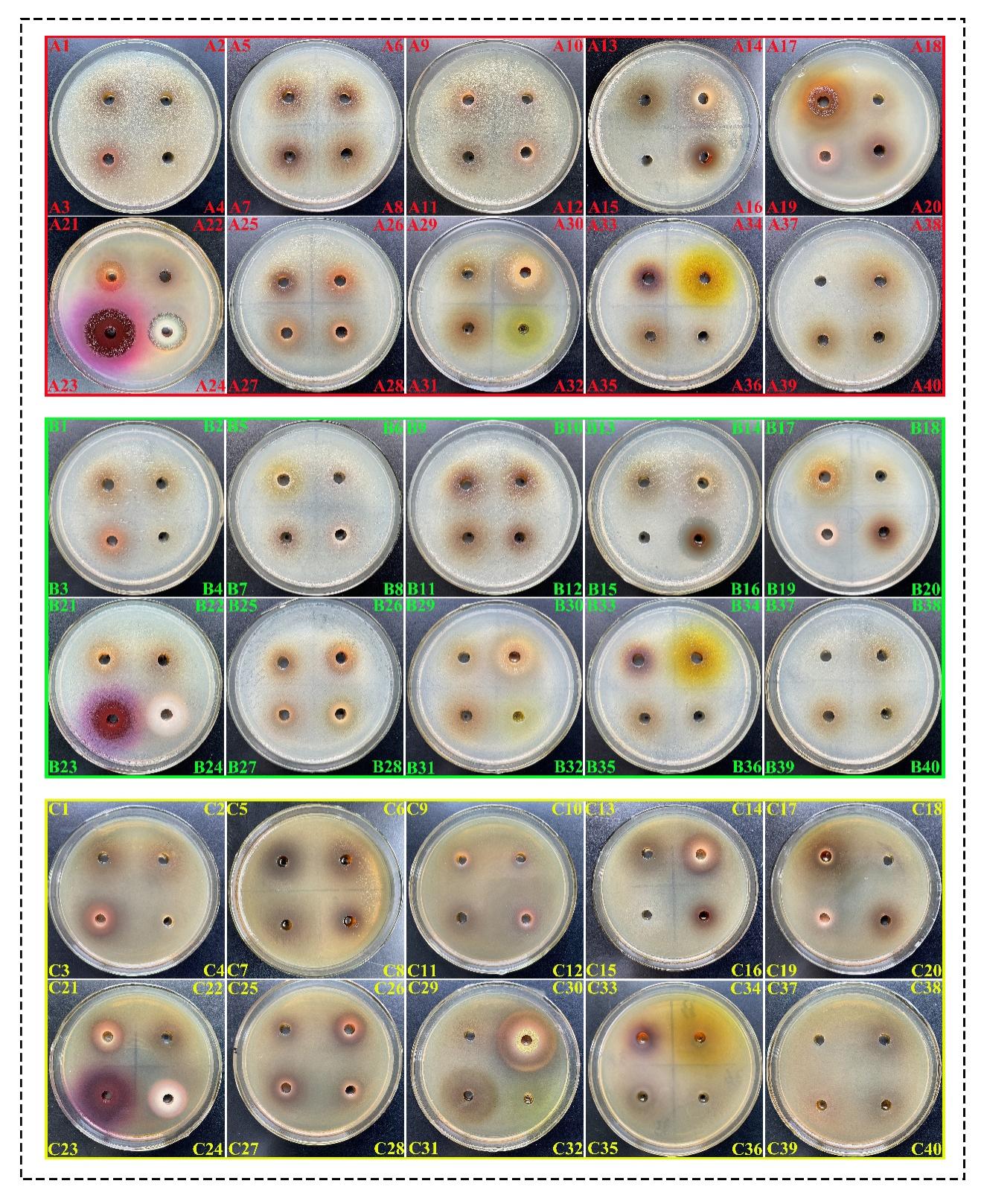
The HAEs of 40 herbals, as shown in Fig. 1, have different antibacterial activity utilizing the plate perforation method against three fish pathogenic bacteria. The results indicated that 10, 14 and 4 HAEs exhibited obvious inhibitory zones against E. tarda, S. iniae and K. pneumoniae, respectively. The diameters of inhibition circles of 40 HAEs against three pathogenic bacteria were shown in Table 2. For E. tarda, Caesalpinia sappan had the largest inhibition diameter (19.00 ± 4.00 mm), followed by Fructus mume, Rhus chinensis and Punica granatum, which had inhibition diameters of 17.00 ± 1.00 mm, 16.00 ± 3.00 mm, and 15.00 ± 1.00 mm, respectively. E. tarda was moderately susceptible to Terminalia chebula, Rubus idaeus and Radix paeoniae rubra, with inhibition diameters of 13.33 ± 4.33 mm, 11.67 ± 0.33 mm and 10.67 ± 4.33 mm, respectively. While E. tarda showed low sensitivity to Moutan cortex, Tuber Fleeceflower Stem and Semen Juglandis, and no sensitivity to the remaining HAEs, such as Scutellaria baicalensis, Scutellaria barbata and Syringa oblata Lindl. For S. iniae, F. mume and R. chinensis exhibited extremely antibacterial effects, with the inhibitory zones were 20.00 ± 0.33 mm and 20.00 ± 0.33 mm, respectively. Moreover, S. iniae was moderately susceptible to R. rhizoma rhei, S. baicalensis and P. granatum, with inhibition diameters of 13.50 ± 0.50 mm, 13.00 ± 0 mm and 12.67 ± 0.33 mm, respectively. The remaining HAEs showed low or no inhibition against S. iniae. Like S. iniae, F. mume also showed the strongest inhibitory effect against K. pneumonia, with an inhibition diameter of 17.00 ± 1.00 mm. Additionally, C. sappan, R. chinensis and P. granatum exhibited general antibacterial effects against K. pneumoniae, with inhibition diameters of 12.67 ± 1.33 mm, 11.00 ± 1.00 mm and 11.00 ± 1.00 mm, respectively. The remaining HAEs showed no inhibition effect.
_of_extracts_of_*eupatorium_japonicum*_thunb_(1)__*artemisia_carui.jpeg)
2.2. The MIC and MBC of 40 HAEs against three pathogenic bacteria
The HAEs with high and extremely high inhibitory effects were selected for further test of MIC and MBC against three pathogenic bacteria. The results obtained from the broth dilution assay demonstrated that F. mume, R. chinensis and P. granatum had bacteriostatic effects against all three pathogenic bacteria. The MIC of F. mume against the three strains ranged from 31.25 to 62.25 mg/mL, with the best antibacterial effect against S. iniae, having an MIC of 31.25 mg/mL. The MIC of R. chinensis against the three strains ranged from 62.5 to 250 mg/mL, with the best antibacterial effect against S. iniae and E. tarda, both with an MIC of 62.50 mg/mL. The MIC of P. granatum against the three strains ranged from 62.5 to 250 mg/mL, with the best antibacterial effect against S. iniae, having an MIC of 62.5 mg/mL. Moreover, C. sappan demonstrated the best antibacterial effect, with MIC for the E. tarda and K. pneumonia ranging from 31.25 to 125 mg/mL, and the best antibacterial effects against K. pneumonia, with an MIC of 31.25 mg/mL (Table 3).
The MBC results revealed that F. mume, R. chinensis and P. granatum had bactericidal effects at ≥ 500 mg/mL concentrations. F. mume had the best bactericidal activity compared with the other two herbs, which have the lowest MBC ranging from 62.5 to 125 mg/mL. The MBC of R. chinensis and P. granatum against the three strains ranged from 62.50–500 and 125–500 mg/mL, respectively. Additionally, the MBCs of C. sappan against the E. tarda and K. pneumonia were 62.50 and 125 mg/mL, respectively (Table 3).
2.3. In vitro bacteriostatic effects of HAEs compound against three pathogenic bacteria
The combination of C. sappan and F. mume exhibited the strongest inhibition against K. pneumoniae (24.50 ± 0.41 mm). Whereas, F. mume paired with R. chinensis demonstrated broad-spectrum efficacy, showing highest inhibitory circle against both S. iniae (24.50 ± 0.41 mm) and E. tarda (19.5 ± 0.41 mm). P. granatum combined with R. chinensis showed synergistic effects against all three pathogens, with inhibition zones of 21.17 ± 0.23 mm for S. iniae and K. pneumoniae. S. iniae displayed heightened sensitivity to formulations containing R. rhizoma rhei, such as F. mume + R. rhizoma rhei (23.17 ± 0.23 mm). K. pneumoniae was notably inhibited by tannin-rich combinations, including C. sappan + R. chinensis (21.83 ± 0.82 mm). Combinations involving R. idaeus or M. cortex exhibited minimal efficacy. For instance, R. idaeus + R. paeoniae rubra yielded weak inhibition against E. tarda (9.50 ± 0 mm). Several combinations showed no detectable inhibition, including C. sappan + Terminalia chebula against S. iniae and K. pneumoniae. (Table 4).
2.4. MIC and MBC of HAEs compound against three pathogenic bacteria
All results from MIC and MBC agree with the previous diameters of inhibition zones. As shown in Table 5, the combination of F. mume + R. chinensis exhibits the best antibacterial effect against E. tarda, with the MIC and MBC values being 15.625 mg/mL and 32.5 mg/mL, respectively. For S. iniae, the MIC and MBC of F. mume + R. chinensis were 7.8125 mg/mL and 15.625 mg/mL. In addition, the MIC and MBC of F. mume + R. chinensis against K. pneumoniae are both 31.25 mg/mL. Different from the above two pathogenic bacteria, the combination of C. sappan + F. mume shows the best antibacterial effect against K. pneumoniae, with the MIC and MBC values being 7.8125 mg/mL and 15.625 mg/mL, respectively.
Based on the results of the inhibitory zone, MIC and MBC tests involving the combined application of traditional herbal medicines, we found that the F. mume + R. chinensis group exhibited the best antibacterial effect against E. tarda and S. iniae. Additionally, C. sappan + F. mume showed the strongest antibacterial effect against K. pneumoniae. For both S. iniae and K. pneumoniae, the compound treatment was significantly more effective than a single application.
Discussion
Contemporary methods for controlling outbreaks of bacterial diseases in intensive aquaculture systems primarily rely on antibiotics, despite the evidence of their negative effects on the environment and human health.30 The increasing national awareness of antibiotics’ potential negative impact on health has spurred the search for alternative, more natural antibacterial medicines that can improve aquatic product safety and quality. Herbs are rich sources of antimicrobial substances for drug development, since its outstanding features of being natural, inexpensive, easy preparation, biocompatible and few side effects for aquatic animals.31 Herbs have the ability to damage cell walls, inhibit nucleic acid and protein synthesis and increase intracellular osmotic pressure.32 Some herbals, such as the Rhizoma coptidis,33 Ramulus Cinnamomi,34 Allium sativum,35 Portulaca oleracea L,36 Eryngium campestre,37 Eichhornia crassipes,38 Zingiber officinale,39,40 exhibit strong antibacterial effects against Aeromonas spp, Staphylococcus aureus, Vibrio spp and other common pathogens in medicine and aquaculture. In the present research, we determined the in vitro antimicrobial activity of 40 HAEs against three bacteria. Based on a systematic evaluation of the collected data, four HAEs exhibited optimal inhibitory effects against three bacterial pathogens, especially C. sappan against E. tarda as well as F. mume against K. pneumoniae and S. iniae.
F. mume, a traditional Chinese herbal medicine, has been used for chronic cough therapy in China for over a millennium.41 F. mume and its components, including organic acids, polysaccharides, flavonoids, amygdalin, terpenes and sterols, have been demonstrated to have potential antibacterial activity in vitro and in vivo.42 Previous studies have found that F. mume exhibits bactericidal activity against many pathogens, such as Streptococcus mutans, Salmonella spp, Staphylococcus aureus, Escherichia coli, and Helicobacter pylori.43,44 According to Chen,43 the composition of F. mume can significantly reduce bacterial biofilm activity, thereby inhibiting bacterial growth. Furthermore, the citric acid content in F. mume induces an acidic milieu (pH < 3.0), thus creating a bacteriostatic environment that suppresses microbial proliferation. In the present study, all three pathogens were successfully inhibited by F. mume with the circle of inhibition diameter ≥ 17.00 mm and MIC of 62.5 mg/mL.
C. sappan, belongs to the Leguminosae family, is widely distributed in China and Southeast Asia.45 Phytopharmacological analysis revealed that C. sappan consists of multiple bioactive components, including xanthones, coumarins, chalcones, flavonoids, isoflavonoids, and brasilin.46 Especially, brasilin, the main active component of C. sappan, has potent inhibition of many antibiotic-resistant bacteria due to the compound’s ability to block the synthesis of DNA and protein.47 In this research, C. sappan extract good inhibitory effect on K. pneumoniae and E. tarda, but showed no effect against S. iniae. The difference was probably related to components of bacterial wall.
P. granatum, belonging to the pomegranate family, is the dried peel of the pomegranate. P. granatum accounts for approximately 50% of the total fruit weight and is an important source of pharmacologically active constituents.48 Previous studies have demonstrated that P. granatum has antibacterial functions due to its multiple pharmacologically active ingredients, such as polyphenols, polysaccharides, terpenes and lignans.49 Zazharskyi et al.50 found that the ethanol extract of P. granatum have strong bactericidal activity on K. рneumonia, Salmonella typhimurium, Listeria monocytogenes, Escherichia coli, Corynebacterium xerosis, Proteus vulgaris.32 Moreover, the study carried out by Pandit and Vyas51 reported that P. granatum showed significant activity against Streptococcus spp (20.16 ± 0.76), similar to our findings.
R. chinensis, is a sac-shaped proliferative tissue structure induced by the parasitic activity of aphids on the leaves or petioles of Chinese sumac.52 According to phytochemical composition analysis, the main chemical components of R. chinensis include flavonoids, triterpenoids, geranylgeranyl derivatives, lignans, and phenolic acid glycosides (Wang, Sheng-Tian et al. 2024). Gallnut water extract was shown to have a good inhibitory effect on V. parahaemolyticus.32 In this study, we found that R. chinensis exhibited antibacterial activity against three aquatic pathogenic bacteria, confirming that R. chinensis has great promise for antibacterial applications.
Various studies have demonstrated that the combined effect of Chinese herbal prescriptions may be greater than the sum of the individual effects.32 Therefore, it’s of great significance to understand the compatibility and synergistic effects of herbal combinations, and then develop novel drug combinations.32 Based on our in vitro susceptibility test, C. sappan, F. mume, R. chinensis, P. granatum and their combinations with other herbs appear to be excellent candidates for the treatment of infections caused by K. pneumoniae and E. tarda. While, F. mume, R. chinensis, P. granatum and their combinations with other herbs appear to be excellent candidates for the treatment of S. iniae. Moreover, F. mume + R. chinensis exhibited the best inhibitory effect against E. tarda and S. iniae, while C. sappan + F. mume showed the strongest inhibitory effect against K. pneumoniae among all the HAEs combinations. However, assessing the efficacy and safety of HAEs are based on clinical practice rather than in a laboratory, it’s necessary to establish the clinical utility of HAEs for the treatment of K. pneumoniae, S. iniae and E. tarda infections in aquatic animals.
Conclusion
In this study, the in vitro bacteriostatic effects of forty HAEs and their compounds against three common aquatic pathogens were investigated. The results showed that four single HAEs (F. mume, C. sappan, P. granatum, R. chinensis) and their compounds (F. mume + R. chinensis and C. sappan + F. mume) have significant inhibitory effect and could be used for future treatment of aquatic diseases caused by S. iniae, E. tarda and K. pneumoniae. In our future research, the active components and antibacterial mechanisms of HAEs which have significant bacteriostatic and bactericidal activity will be investigated.
Acknowledgements
This study was supported by the Natural Science Foundation of Hubei Province (grant no. 2021CFB265) and Key Lab of Freshwater Biodiversity Conservation, Ministry of Agriculture and Rural Affairs of China (LFBC III0).
Authors’ Contribution
Conceptualization: Nengbin Zhu (Equal), Xuelei Qu (Equal). Methodology: Nengbin Zhu (Equal), Xuelei Qu (Equal), Eakapol Wangkahart (Equal). Writing – review & editing: Feiyang Gao (Equal), Rui Wang (Equal), Hongsen Xu (Equal), Huiping Ding (Equal). Supervision: Eakapol Wangkahart (Equal), Hongsen Xu (Equal), Huiping Ding (Equal). Formal Analysis: Qianrong Liang (Equal), Lin Zhang (Equal). Investigation: Qianrong Liang (Equal), Lin Zhang (Equal). Resources: Qianrong Liang (Equal), Hongsen Xu (Equal), Huiping Ding (Equal). Writing – original draft: Lihe Liu (Equal), Rui Wang (Equal), Hongsen Xu (Equal). Funding acquisition: Hongsen Xu (Equal), Huiping Ding (Equal).