



Introduction
The discovery of the Janus kinase (JAK)-signal transducer and activator of transcription (STAT) pathway arose from investigations of how cells respond to interferons (IFNs), and has been widely characterized from human to Drosophila.1–3 As a crucial component of the interferon immune response, the JAK/STAT signaling pathway comprises three key elements: tyrosine kinase-related receptors, JAKs, and STATs.4 In vertebrates, the canonical JAK/STAT signaling pathway is triggered upon ligand binding to corresponding transmembrane receptors.5 This binding event induces receptor dimerization, allowing the receptors’ intracellular domains to undergo transphosphorylation by their associated intracellular JAK kinases; in turn, cytoplasmic STAT proteins are phosphorylated and activated. Phosphorylated either homodimers or heterodimers via the Src-homology 2 (SH2) domain and phosphotyrosine interaction and translocate to the nucleus to function as transcription factors regulating immune responsiveness and other essential biological processes.6
Mammals possess seven members of the STAT family, including STAT1, STAT2, STAT3, STAT4, STAT5A, STAT5B, and STAT6. Similar to the mammalian antiviral response, STATs mediate the immune response in osteichthyes.7–9 Notably, crustaceans typically harbor only a single STAT gene.10,11 A STAT homologue, designated SpSTAT, was identified and cloned from the mud crab Scylla paramamosain. The full-length cDNA is 2952 bp, containing an open reading frame (ORF) of 2388 bp that encodes a 796-amino-acid protein with a predicted molecular weight of 91.29 kDa. SpSTAT shares high sequence identity with STAT proteins from other crustaceans, and its mRNA is predominantly expressed in the intestine and eyestalk, while showing markedly lower expression levels in the heart and muscle.10 The expression level of STAT in crustaceans can be induced by various stimuli, including lipopolysaccharide (LPS), S. aureus and poly (I:C), which further confirms the vital role of STAT in crustacean immune response. Activation of STAT in Litopenaeus vannamei results in responsiveness to white spot syndrome virus (WSSV) infection.12,13 Knockdown of STAT could promote the proliferation of mud crab reovirus (MCRV) in Scylla paramamosain.10 Infection with WSSV or injection of rMjVago-L protein could significantly enhance the phosphorylation and nuclear translocation of STAT, inducing Stat-regulated Ficolin to exert antiviral effects in Marsupenaeus japonicus.14 Despite these promising findings, the molecular mechanism of STAT-mediated antiviral responses in crustaceans remains poorly understood, particularly at the protein level, where studies are scarce. Proteins are the direct mediators of intracellular signaling and the functional executors of antiviral activity. Mechanistic conclusions without protein-level experimental support are largely descriptive at the transcriptional level and fail to provide convincing evidence for physiological functions and regulatory pathways.
Further investigation to determine the function of STAT requires its specific antibodies. However, few standardized anti-STAT antibodies are currently available. In this study, we expressed STAT in Escherichia coli and generated a polyclonal antibody against STAT by immunizing KM mice. The antibody demonstrated high sensitivity and specificity in immunoblotting assays. This provides a useful reagent for investigating STAT’s functional mechanisms.
Materials and methods
Animals and Bacterial Strains
Eight-week-old female mice were purchased from Haikou Yingyue Biotechnology Co., Ltd. and housed in a clean, temperature-controlled animal facility for acclimatization.
Mud crabs (30-50 g) were from an aquaculture farm in Hai Nan, China. Mud crabs were acclimated at 28 °C in a recirculating water tank system with air-pumped seawater (5‰ salinity) before experiments. E. coli strain DH5α was used for gene cloning, and E. coli BL21 (DE3) was used to express the fusion protein.
Bioinformatics analysis
The STAT protein sequences analyzed in this study were obtained from the National Center for Biotechnology Information (NCBI) databases (GenBank: MH924352.1). The physicochemical properties were predicted by ExPASy (https://www.expasy.org/). Transmembrane domains were predicted using TMHMM (https://dtu.biolib.com/DeepTMHMM). Phosphorylation sites were predicted with NetPhos 3.1 (https://services.healthtech.dtu.dk/services/NetNGlyc-1.0/). N-glycosylation sites were analyzed using NetNGlyc 1.0 (https://services.healthtech.dtu.dk/services/NetPhos-3.1). The secondary structure of the recombinant protein was predicted by SOPMA servers (https://npsa-prabi.ibcp.fr/cgi-bin/npsa_automat.pl?page=npsa_sopma.html).SWISS-MODEL (https://swissmodel.expasy.org/interactive) was also employed for 3D structure prediction.
Plasmid construction for pET-32a-STAT expression
Total RNA was extracted from mud crab gills by using Eastep® Super Total RNA Extraction Kit (Promega, USA) and reverse transcribed into cDNA by using a Reverse Transcription Kit Leaflet (AG, China). The full-length of STAT coding sequence was cloned into pET-32a vector using specific primers pET-STAT-F/R, and recombinant plasmid was transformed into E. coli strain BL21. The sequences of positive colonies were verified by sequencing at a commercial sequencing company (Beijing Qingke, China).
Expression of recombinant STAT protein
The culture from a single colony of E. coli strain BL21 containing the pET-32a-STAT was inoculated into 5 mL of LB medium containing 50 μg/mL ampicillin with 1:100 vol at 37 °C. When the cell density reached 0.6–0.8 (OD600), isopropyl-β-D-thiogalactoside (IPTG, Titan) was added to a final concentration of 1 mM to induce the expression of STAT protein. Productivity of recombinant STAT was evaluated at a variety of temperatures (16 °C, 20 °C, 25 °C, and 30 °C), and durations (24 h, 12 h, 8 h, and 6 h). The yield of recombinant proteins were evaluated using 10% SDS-PAGE gel.
Immunization of animals
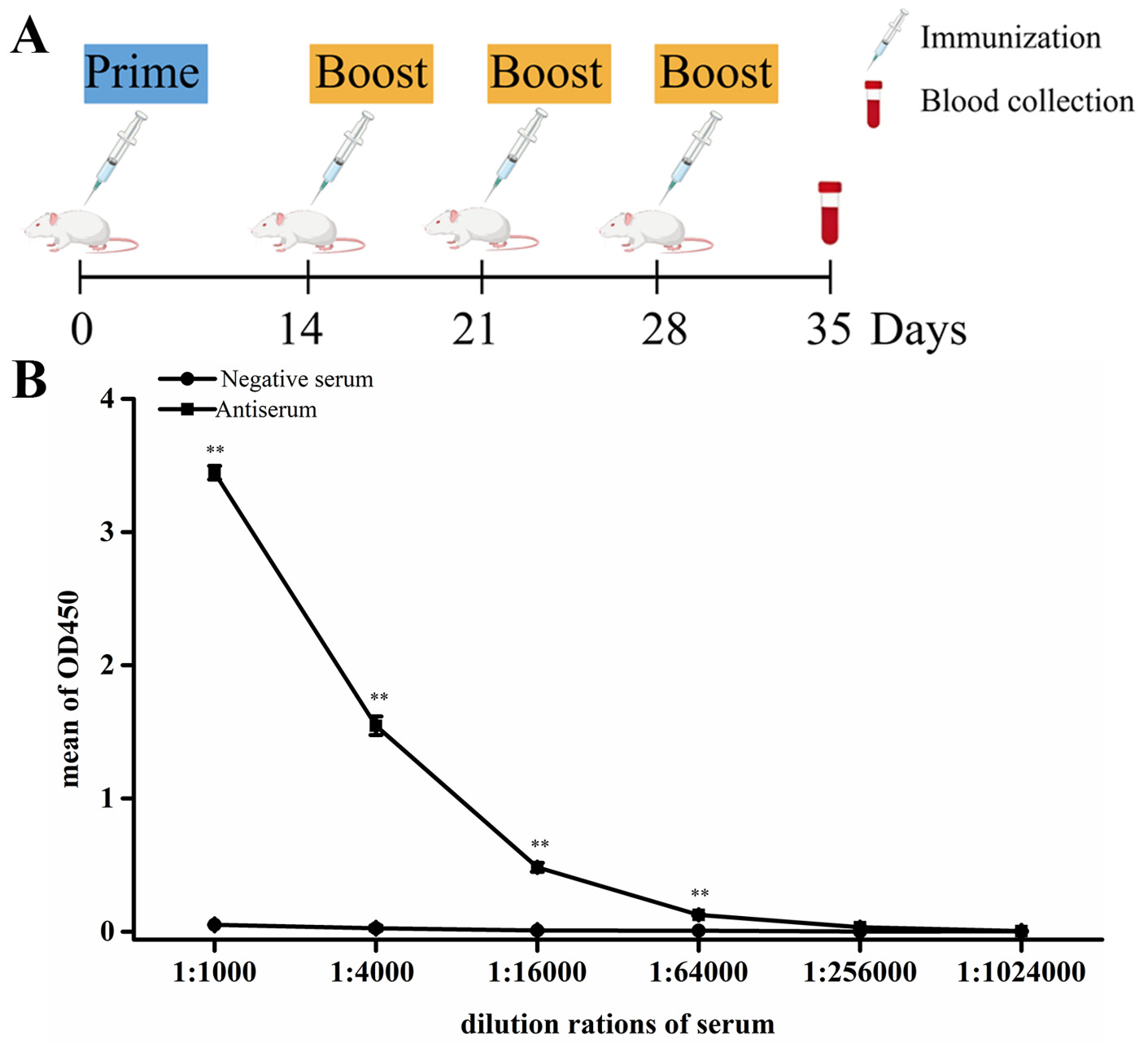
Eight-week-old mice from Haikou Yingyue Biotechnology Co., Ltd. were subcutaneously administered with 50 μg of target protein per mouse. The recombinant SpSTAT antigen was derived from bacterial inclusion bodies. The target protein was excised as gel slices following SDS-PAGE, fully denatured in SDS sample buffer, and used directly for immunization without additional solubilization or renaturation steps. The primary immunization was performed using complete Freund’s adjuvant (Sigma), and three booster immunizations were given on days 14, 21, and 28 with incomplete Freund’s adjuvant (Sigma). Mouse blood was collected by tail bleeding on day 35 and the sera were stored at -80°C until use.
ELISA Antibody Titer Assays
ELISA plates were coated at 4 °C overnight with recombinant protein (200 ng/well) resuspended in carbonate buffer. After three washes, the wells were incubated with blocking solution (10% nonfat milk in PBS containing 0.1% Tween 20) for 1 h at 37 °C. After five washes with PBST (PBS containing 0.1% Tween 20), 50 μL/well of diluted sera (serial dilutions, 200- to 409600-fold) was added and incubated for 2 h at 37 °C. After seven washes with PBST, the plates were incubated with HRP-labeled goat anti-mouse IgG antibody for 1 h at 37 °C. TMB Chromogen Solution (100 μL/well) was added after eight washes with PBST, and the plate was incubated for 5-10 min at room temperature. 50 μL/well of 2 M H2SO4 solution was added to each well to stop the reaction, and the optical density (OD) was immediately read at 450 nm. The antiserum was defined as positive when the ratio of the OD450 value of antiserum to that of negative serum exceeded 2.1, and the maximum corresponding dilution ratio was recorded as the antibody titer.
Western blot assays
To further verify the specificity of the prepared STAT antibody. The full-length coding sequence of SpSTAT was cloned into the pAc5.1-V5 vector using specific primers STAT-F/R, and the recombinant plasmid was transfected into Drosophila S2 cells. At 48 h post-transfection, the cells were harvested and lysed in ice-cold cell lysis buffer (50 mM Tris-HCl, pH 7.8, 150 mM NaCl, 1% Nonidet P-40) supplemented with a protease inhibitor cocktail (Sigma-Aldrich, USA). The cell lysates and prokaryotically expressed recombinant STAT protein were subjected to Western blot analysis using the prepared anti-STAT antibody at gradient dilutions of 1:2000, 1:5000, and 1:10000.
We also conducted tissue distribution analysis to further validate the reactivity of the prepared anti-STAT polyclonal antibody in vivo. Seven tissues, including stomach, muscle, heart, hepatopancreas, eyestalk, gill, and intestine, were dissected from three healthy mud crabs. Total protein was extracted from each tissue using RIPA lysis buffer (Beyotime, China) with protease inhibitors. Equal amounts of protein samples were separated by SDS-PAGE, transferred to PVDF membranes, and probed with the prepared anti-STAT antibody (1:2000). β-actin was used as the internal reference to normalize the protein loading.
Immunofluorescence assay
Hemocytes from three healthy mud crabs were collected via hemocoel puncture and seeded onto cell culture slides for cellular localization assays. For the challenge experiments, healthy mud crabs were injected with poly(I:C) (0.4 μg/g). Hemocytes from three crabs in each group were collected at 6 h, 12 h, 24 h, 48 h, 72 h and 96 h post-injection (hpi), and seeded onto cell culture slides. Indirect immunofluorescence was used to analyze the subcellular localization of SpSTAT. Immunostaining was performed using an anti-STAT antibody (1:1000). Afterwards, the cells were incubated with Alexa Fluor 488-labeled goat anti-mouse antibody (1:1000), then stained with Hoechst 33342 (Beyotime, China; 1:1000) and visualized under a confocal laser scanning microscope (Leica TCS SP8 STED 3X, Germany).
Statistical analysis
The mean and standard deviation (SD) from three detections was calculated. Student’s t-test was used to compare the two means using Microsoft Excel. The statistical procedures were performed using GraphPad Prism version 9.5.
Results
Bioinformatics analysis of SpSTAT
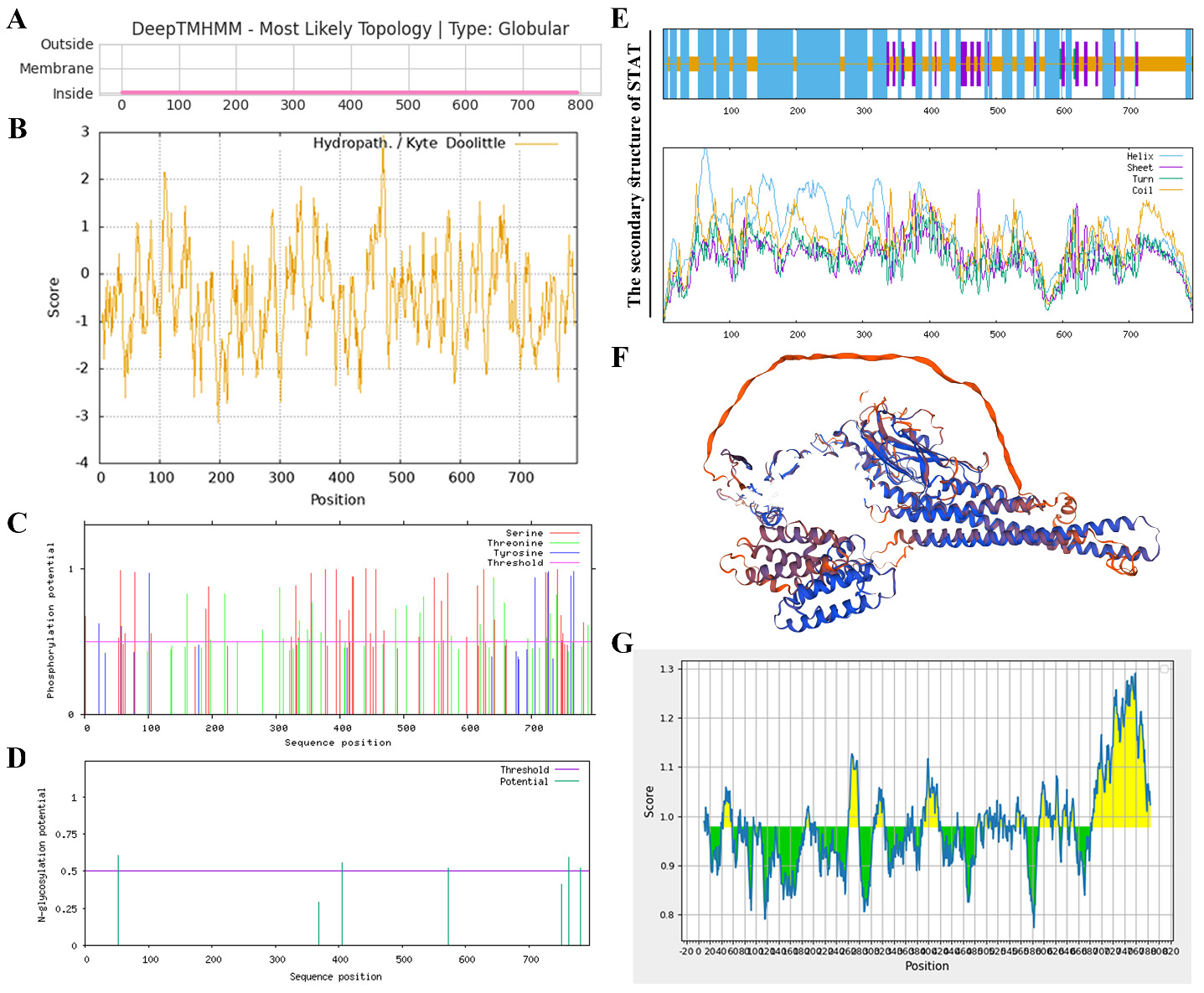
The characterization of the intrinsic biochemical properties of the STAT protein is essential for efficient antibody generation. TMHMM analysis revealed that all amino acids of the encoded protein are located outside the membrane (Figure 1A), and no transmembrane domains were detected. The physicochemical parameters of the protein were predicted using ProtParam, including a theoretical isoelectric point of 6.15, an instability index of 48.35, an aliphatic index of 80.43, and a solubility score of 0.710. The grand average of hydropathicity (GRAVY) of SpSTAT was -0.513 (Figure 1B), indicating that SpSTAT was predicted to be a hydrophilic protein. The NetPhos 3.1 server predicted 39 serine, 25 threonine, and 7 tyrosine phosphorylation sites in the SpSTAT protein (Figure 1C), whereas NetNGlyc 1.0 predicted five putative N-glycosylation sites (Figure 1D). According to the SOPMA server, the predicted structure comprises 53.46 % alpha helix, 8.05 % extended strand, and 37.86 % random coil (Figure 1E). 3D structure of SpSTAT predicted results were shown in (Figure 1 F) using SWISS-MODEL software. IEDB servers predicted continuous B-cell epitopes based on physicochemical properties. The results showed the multiple of STAT with antigenicity scores higher than 1.0 (Figure 1G).
Construction of plasmid and expression optimization of pET-32a-STAT recombinant protein
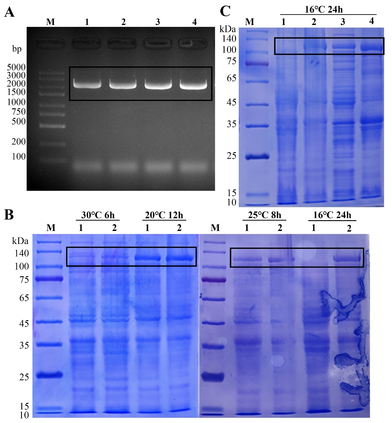
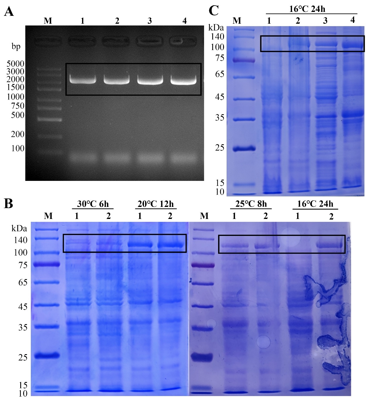
The successful construction of the recombinant plasmid was confirmed through double enzyme digestion of the pET-32a-STAT plasmid, which was further verified by sequencing (Figure 2A). The pET-32a-STAT plasmid was transformed into E. coli protein production strain BL21. To optimize the expression conditions, small-scale induction studies were performed at 16 °C, 20 °C, 25 °C, and 30 °C with 1 mM IPTG. The high level of recombinant STAT proteins was obtained when IPTG was added at a final concentration of 1.0 mM and induced for 24 h at 16 °C (Figure 2B). Ultrasonic treatment was employed to disrupt cells and extract proteins, followed by 10% SDS-PAGE analysis of supernatant and inclusion body proteins. The results clearly showed that the recombinant protein existed mainly in inclusion bodies (Figure 2C).
Preparation of polyclonal antibody against STAT
Five mice were immunized with the recombinant protein serving as the immunogen (Figure 3A). The titers of the resulting anti-STAT polyclonal antiserum were then determined by indirect ELISA (Figure 3B). Notably, the initial immunization with the recombinant protein elicited a robust IgG response specific to the immunogen. After four times immunization, a strong reaction occurred, and the antibody titer reached 1:64,000.
Specific detection of STAT polyclonal antibody
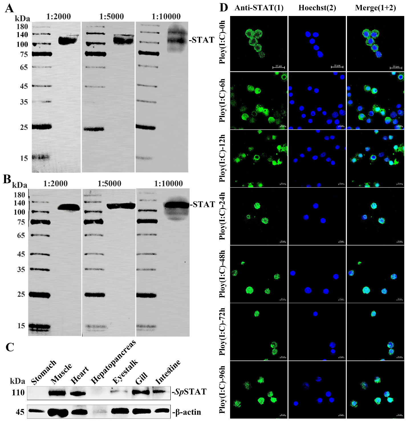
To verify the specificity of the prepared polyclonal antiserum against STAT, Western blot analysis was performed using prokaryotically expressed recombinant STAT protein (Figure 4A) and pAc5.1/V5-His-STAT expressed in Drosophila S2 cells (Figure 4B) as antigens. The antiserum was serially diluted to 1:2000, 1:5000, and 1:10000 for the assay. At a dilution ratio of 1:2000, the antibody exhibited favorable detection sensitivity with minimal non-specific binding, indicating that this dilution is suitable for subsequent experiments.
Furthermore, the reactivity and specificity of the prepared anti-STAT polyclonal antiserum were further evaluated by indirect immunofluorescence and Western blotting using total protein extracted from crab tissues. Western blot analysis revealed that the prepared antiserum could specifically recognize endogenous STAT in various mud crab tissues, including muscle, heart, eyestalk, gill, and intestine (Figure 4C). Indirect immunofluorescence was used to analyze the subcellular localization of SpSTAT in hemocyte at 6 h, 12 h, 24 h, 48 h, 72 h and 96 h post poly(I:C) stimulation. The results showed that the prepared antiserum effectively detected SpSTAT, which localized to the nucleus at all detected time points from 6 h to 96 h post-stimulation (Figure 4D). These results suggested that this polyclonal antibody had good reactivity and specificity against the SpSTAT protein and could be used for future studies.
Discussion
The production potency of polyclonal antibodies is comprehensively affected by multiple factors. Characterizing the protein’s intrinsic biochemical properties is essential for efficient antibody generation. Surface-exposed hydrophilic regions are the primary domains triggering immune responses, whereas buried hydrophobic segments rarely participate in antigen recognition.15–17 Random coils and extended strands tend to reside on the protein surface and form linear B-cell epitopes, while α-helices and β-sheets are usually buried in the protein core and less accessible to antibody recognition.18,19 Excessive N-glycosylation can shield linear and conformational B-cell epitopes via steric hindrance, thereby weakening humoral immune responses and lowering antibody titer and epitope specificity.20,21 Meanwhile, phosphorylation modification can alter protein spatial conformation and epitope characteristics, easily causing non-specific immune response and further limiting the improvement of effective antibody titer.22 Consistent with these principles, our pre-experimental bioinformatic analyses of SpSTAT identified multiple surface-exposed hydrophilic stretches that could serve as potential linear B-cell epitopes, alongside scattered predicted glycosylation and phosphorylation sites. The distribution of these features indicates that full-length SpSTAT possesses the structural basis to elicit antibody responses, while post-translational modifications may modulate epitope exposure under native conditions. Protein insolubility and aggregation tend to disrupt native epitope conformations during expression and purification, impair immune stimulation, and ultimately reduce antibody production potency.23,24The full-length protein exhibited an instability index of 48.35, a value above the 40 threshold that denotes intrinsically unstable polypeptides. Consistent with this feature, the recombinant protein was predominantly recovered as inclusion bodies under our prokaryotic expression conditions. Accordingly, we used unpurified inclusion bodies directly for immunization. Most conventional studies employ chromatographic purification of prokaryotically expressed proteins or inclusion bodies before mouse immunization.25,26 Encouringly, the antiserum titer of 1:64,000 achieved in this study is consistent with those reported in similar polyclonal antibody production efforts using prokaryotically expressed antigens, which typically range from 1:10,000 to 1:200,000.27–29 In addition to validation in a prokaryotic expression system, we verified the anti-STAT antibody in a eukaryotic expression system using native protein extracted from fresh Scylla paramamosain tissues, confirming that the prepared anti-STAT antibody recognizes the target protein in its native conformation with high specificity.
Polyclonal antibodies remain indispensable tools for studying protein expression, localization, and interactions in non-model organisms, where commercial antibodies are rarely available.30 Polyclonal antibodies have great potential for application in aquatic animal disease detection and immunological research. A VP28-specific polyclonal antibody has been utilized to develop lateral flow immunoassays (LFIA) for the on-site detection of white spot syndrome virus (WSSV), enabling timely disease screening and field-level surveillance in shrimp aquaculture.31 Similarly, antibodies against PirA and PirB toxins from Vibrio parahaemolyticus have supported the development of sensitive detection strips for acute hepatopancreatic necrosis disease (AHPND), which is critical for reducing economic losses in shrimp farming.32,33 Polyclonal antibodies have also advanced the understanding of adaptive and mucosal immunity in teleosts. Antibodies against IgM have been widely used in ELISA to evaluate serum and mucosal antibody responses after vaccination, providing a reliable indicator for vaccine efficacy.34 In crustaceans, such reagents remain particularly scarce for intracellular signaling molecules such as STAT (signal transducer and activator of transcription).
To date, few studies have systematically examined the tissue distribution of STAT proteins at the protein level; most relevant investigations have only detected STAT mRNA expression in crustaceans and fish by qRT-PCR.35–37 In the present study, a self-prepared SpSTAT polyclonal antibody was used to profile the tissue distribution of SpSTAT protein in healthy mud crabs. SpSTAT protein was detected in muscle, heart, eyestalk, and gill, while no specific protein band was observed in the stomach and hepatopancreas. However, previous research showed that SpSTAT mRNA was detectable in the hepatopancreas and stomach.10 This discrepancy occurred because the previous study examined mRNA levels, whereas the present study focused on protein abundance. While our antibody was validated for specificity, we cannot formally exclude the possibility that SpSTAT protein in these tissues falls below the detection threshold of Western blotting due to low expression levels. Similar to Litopenaeus vannamei,38 SpSTAT was found to translocate from the cytoplasm to the nucleus upon poly(I:C) stimulation in S2 cells.10 In the present study, the typical nuclear translocation of SpSTAT was also detected in crab hemocytes using our polyclonal antibody. By including unstimulated (0 h) controls and PBS-injected vehicle controls at each time point, we established that SpSTAT is predominantly cytoplasmic at baseline and that nuclear accumulation is specifically induced by the viral mimic, rather than reflecting constitutive nuclear localization or injection stress. These results further confirm the antibody’s high specificity and applicability to endogenous protein localization.
In conclusion, we performed bioinformatic prediction, prokaryotic antigen expression, and successfully generated specific anti-SpSTAT polyclonal antibodies, which provide a fundamental tool for studying SpSTAT immune function in mud crabs. Although the antibody was well validated for specific recognition of endogenous SpSTAT, further detailed investigations are still required to clarify the immune regulatory mechanism of SpSTAT. The antibody obtained in this study will support subsequent functional studies of crustacean STAT signaling.
Funding
This work was funded by the National Natural Science Foundation of China (32260924 and 32002440), the natural Science Foundation of Guangdong Province in China (2022A1515010958), Secondary Sub-projects of Scientific Research and Technology Demonstration Category in the National Modern Agricultural Industrial Park, Wanning City, Hainan Province, Innovational Fund for Scientific and Technological Personnel of Hainan Province (KJRC2023B22), Open Research Fund Program of Fujian Provincial Key Laboratory of Marine Fishery Resources and Eco-environment, Research Start-up Project of Hainan University (KYQD(ZR)-22119). The funders had no role in study design, data collection and analysis, decision to publish, or preparation of the manuscript.
Author Contributions
Data curation: Jiyu Hu (Equal), Qianying An (Equal). Formal Analysis: Jiyu Hu (Equal), Shaoni Fu (Equal), Peicheng Wang (Equal). Investigation: Jiyu Hu (Equal), Shuyan Zhang (Equal), Hu Lu (Equal). Validation: Jiyu Hu (Equal), Qianying An (Equal), Shaoni Fu (Equal). Visualization: Jiyu Hu (Equal), Hu Lu (Equal). Writing – original draft: Jiyu Hu (Lead). Software: Hu Lu (Equal), Shanshan Sang (Equal), Yongcan Zhou (Equal). Methodology: Shanshan Sang (Lead). Funding acquisition: Yongcan Zhou (Equal), Xing Zheng (Equal), Hengwei Deng (Equal). Conceptualization: Xing Zheng (Equal), Hengwei Deng (Equal). Project administration: Xing Zheng (Equal), Hengwei Deng (Equal). Resources: Xing Zheng (Equal), Hengwei Deng (Equal). Supervision: Xing Zheng (Equal), Hengwei Deng (Equal). Writing – review & editing: Hengwei Deng (Lead).
Conflicts of Interest
No competing interests were disclosed.
Ethical Conduct Approval
The animal experiment was approved by the Animal Ethics Committee of Hainan University.
Informed Consent Statement
All authors and institutions have confirmed this manuscript for publication.
Data Availability
All are available upon reasonable request.