


Introduction
Acute hepatopancreatic necrosis disease (AHPND) is currently considered one of farmed shrimp’s most dangerous bacterial diseases. This disease, caused by Vibrio parahaemolyticus,1 is reported to annually cost over 1 billion USD.2 AHPND has recently been more complicated and caused severe damage to the farmers. Therefore, it is essential to find solutions to prevent farmed shrimp from microbial diseases.3 Drugs, chemicals, and supplements containing antibiotics, disinfectants, and probiotics are often employed to prevent and treat AHPND.4–6 However, these substances have low treatment efficacy and may cause undesirable impacts such as adverse effects on the animal and human health, environment, and quality of aquatic products.7,8
Using herbal extracts brings certain benefits such as cheapness, ease of preparation and use, high effectiveness thanks to its ease of absorption, fewer/no side effects, and no impacts on the environment, and no serious harm to farmed animals and humans.9 Several previous studies showed the positive effects of using herbal extracts on increasing aquatic animals strengthen the immune system and inhibit pathogenic bacteria.10–13 In addition, many herbs are reported to inhibit bacteria, viruses, fungi, and parasites; stimulate growth and maturation; reduce stress and enhance the immune system.10
In Vietnam, many wild and planted herbs have been used to treat some common illness thanks to their medicinal characteristics (e.g., antioxidant activity). Recently, fewer herbal plants (e.g., myrtle leaf extract and seeds) have been determined to inhibit bacteria causing AHPND.14 The methanol extract of Eclipta alba was also tested for antibacterial activity against 12 strains of Vibrio spp. Isolated from the intestinal tract of black tiger shrimp.15 The studies indicate that herbal extracts can inhibit pathogenic bacteria on shrimp (Vibrio parahaemolyticus and V. harveyi), and reduce the incidence of AHPND.16 Moreover, the herbal extracts of Phyllanthus urinaria and Terminalia catappa were able to stimulate growth in whiteleg shrimp, enhance feed absorption, stimulate digestion, and increase survival.17,18 Therefore, herbal extracts are a suitable and effective in enhancing animal immunity.19 Herbal extracts are also believed to replace drugs and chemicals in aquaculture thanks to possessing bioactive substances.20 Although using herbs in aquaculture is potential and promising, their medicinal characteristics, bioactive components, and impacts have yet to be studied comprehensively. Furthermore, scientific information and evidence about the inhibition effect of herbal extracts on shrimp against pathogenic bacteria still need to be provided for shrimp farmers to employ them in their practical farms willingly.
Therefore, this present study was carried out to thoroughly investigate the potential of herbs, which are popular in South Vietnam, in inhibiting V. parahaemolyticus, causing acute hepatopancreatic necrosis disease, and possibly used in shrimp farming. To achieve this aim, the main experiments included screening bacterial inhibition activity of herbal extracts and examining the effect of herbal extracts on shrimp challenged with pathogenic bacteria were conducted. The research results will significantly contribute to scientific knowledge about the efficacy of herbal extract in the prevention and treatment of shrimp severe diseases.
Materials and Methods
Herbal extraction and determination of inhibition ability against bacteria causing AHPND
a. Herbal extraction
This study contains leaf samples of ten herbal plants, including 1. Hibiscus sabdariffa, 2. Mimosa pirga, 3. Eucalyptus globulus, 4. Melaleuca leucadendron, 5. Pithecellobium dulce, 6. Annona squamosa, 7. Sesbania sesban, 8. Limonia acidissima, 9. Averrhoa carambola, and 10. Piper sarmentosum were collected and used for extraction. The sampling area was the Tra Vinh provinces in the Mekong Delta, Vietnam.
Leaves of those herbs were washed and dried at 50oC in a laboratory drying oven, then ground into a powder. The powder samples were soaked with methanol solvent at a ratio of 1:10 for 4 days. Each sample was filtrated through filter paper (Whatman No.1). Methanol was removed via rotation evaporation at 50ºC, with a rotation period of 150 rpm.21 To complete the extraction process, extracted samples were placed in the oven at 50ºC to remove the solvent and dry to constant mass. The dried extracts were stored in the refrigerator at 4ºC for further experiments.
b. Determination of the antibacterial activity of herbs
A working solution of herbal extract, paper discs impregnated with herbal extract, and the culture of V. parahaemolyticus were prepared before determining the antibacterial activity. To make a working solution of herbal extract, 800 mg of dried herbal extract (the solvent was removed entirely) was dissolved in 2 mL of Dimethyl sulfoxide (DMSO) 20% (v/v). To prepare the herbal extract-impregnated paper discs, 50 µL of the working solution was dropped slowly into the 8 mm paper discs (Advantec, Tokyo, Japan), which were already sterilized by exposure to UV light for 30 minutes. The disc impregnated with herbal extract was let to dry slowly. Bacteria V. parahaemolyticus 22 stored at the pathology laboratory of Tra Vinh University was proliferatively cultured in TSB medium supplemented with 1.5% NaCl for 12 -18 hours at 28oC. Subsequently, the bacterial culture was streaked on the TSA supplemented with 1.5% NaCl to check the bacteria purity by observing colonies and gram stain.
The antibacterial activities of different herb species were tested through the agar diffusion method. V. parahaemolyticus at a density of 108 CFU/mL was evenly spread on the surface of a Petri plate of TSA medium containing 1.5% NaCl. Herbal extract-impregnated paper discs (prepared above) were put on the Petri plates. The paper disc impregnated with DMSO was used as the negative control. The paper discs impregnated with 30 μg Doxycycline (DOX) antibiotic were used as the positive control. The Petri plates were incubated at 28°C for 24 hours. Each treatment (herbal extracts and the controls) was triplicated. The antibacterial activity was determined by measuring the diameter of the bacterial inhibition zone.23 Based on the diameter, the antibacterial activity of the herbal extract is classified into three levels: resistance, intermediate, and sensitivity.24 If it is a “resistant” sample, the inhibition zone diameter is equal to or smaller than 9 mm. If it is an “intermediate” sample, the inhibition zone diameter ranges from 10 to 14 mm. If it is a “sensitive” one, its inhibition zone diameter must be over 14 mm.
Minimum inhibitory concentration (MIC) and minimum bactericidal concentration (MBC) assays for herbal extracts
a. Minimum inhibitor concentration (MIC) determination
The herbal extract samples with inhibition zone diameter in the sensitive range to V. paraheamolyticus (as described above) were chosen to perform MIC assay. In the broth dilution method, 1 mL of herbal extract in the TSB + 1.5% NaCl solutions was prepared by two-fold dilutions from 25 to 25/1024 (0.02) mg/mL (10 times) in the test tubes. One milliliter of a bacterial suspension at 2 x 10⁶ CFU/ml was transferred into the series of broth dilutions and inoculated at 28oC for 24 hours. MIC was determined as the herbal extract that completely inhibits the growth of bacteria at the lowest concentration.25
b. Minimum bactericidal concentration (MBC) determination
The tubes containing the culture medium showed no growth of the bacteria, and the last tubes showing turbidity in the MIC test were used for the MBC testing. The 100 µL incubated cultures of the MIC assay at the different dilutions, which could inhibit bacterial growth, were spread on TCBS agar plates to conduct an MBC assay. After incubating at 28ºC for 24 hours, bacterial colonies on the TCBS agar plates were counted. The MBC assay was triplicated for each treatment. The minimum bactericidal concentration of the herbal extract was determined to be the lowest concentration, in which no colonies could grow on the TCBS plate.23
Effect of herbal extracts on shrimp growth and AHPND resistance
a. Experiment on the effect of herbal extracts on growth and hematological parameters in whiteleg shrimp
Some preparation was conducted to test the effect of herbal extracts on growth and hematological parameters in whiteleg shrimp under the in-vivo scale. Regarding shrimp preparation, 15-day postlarvae P. vannamei were checked for negative results of white spot disease, microspore disease, and AHPND 26,27. These postlarvae were cultured in tanks at the experimental farm of the Department of Agriculture and Fisheries of Tra Vinh University until they reached 8 g in weight. The shrimps were tested by PCR to select ones free of white spot disease and AHPND again. Before starting the experiment, shrimps were cultured for 3 days to adapt to the new environmental condition of the experimental tank.
The water used in the experiment was bittern of seawater with a salinity of about 85 ppt purchased from Vinh Chau District of Soc Trang Province. The bittern was filtered through a filter bag to remove residues. Then it was disinfected with chlorine at a concentration of 20-30 mg/L and aerated vigorously and continuously for 24 hours. The chlorine residue was neutralized with Na2S2O3 at the ratio of 7:1. Then, the bittern water was diluted with fresh water to get a salinity of 15 ppt.
Preparation of shrimp feed coated with herbal extract: Three samples of herbal extracts, whose antibacterial activity was determined as “sensitive,” was employed at the concentration of 1% for this experiment. Feed pellets (42% protein, CP, Vietnam) were coated with herbal extracts at a concentration of 1% in weight.17 The feed pellets also were coated with a second layer of 2% squid oil in weight (bought from Vemedim company, Vietnam). For the control treatment, feed pellets were coated with only squid oil. The coated feed pellets were then stored at 4ºC.
In the in-vivo experiment, 60 individuals of whiteleg shrimps at 8 g in weight were cultured in a 500 L composite tank. The water in the experiment tanks had a salinity of 15 ppt and was aerated. Daily, shrimps were fed with coated herbal extract feed pellets (three treatments of the three different extracts, H. sabdariffa, E. globulus, and M. pirga) with 4 times a day: at 7 am, 11 am, 3 pm, and 9 pm. The dose of feed was based on shrimp’s demand at 7-10% of animal weight). The experiment was triplicated and lasted for 30 days.
During the experiment, the water quality parameters, including pH, temperature, NH3, KH, and NO2, were monitored once a day. The temperature was measured with a thermometer, while other ones were conducted via a Sera test kit.
Every 5 days during the experiment duration, the growth performance of shrimps such as the growth rate in weight and length, was recorded. Five shrimps were randomly caught to weigh and measure their length. Shrimp length was measured from the tip of the rostrum to the tip of the tall. Based on the weight and length of the shrimp, the growth performance of shrimps was calculated as the following formula (i), (ii), (iii), and (iv). At the end of the experiment (on day 30th), the survival rate of shrimp was calculated as the formula (v) mentioned below.
Daily weight gain (g/day): DWG = (Wfinal – Winitial)/t (i)
Special growth rate (%/day): SGR = (LnWfinal – LnWinitial)*100/t (ii)
Daily length gain (mm/day): DLG = (Lfinal – Linitial) /t (iii)
Special growth rate in length (%/day): SGRL = (LnLfinal – LnLinitial)*100/t (iv)
Survival rate of shrimp (%) = (Nfinal + Nsample)*100/Ninitial (v)
Where, Wfinal and Lfinal: Mean final weight (g) and mean final length (mm) of shrimp at the end of the experiment; Winitial and Linitial: Mean final length (g) and mean initial length (mm) of shrimp at the beginning of the experiment. t: Experimental period (day). Nfinal and Ninitial: Number of shrimps at the end and at the beginning of the experiment. Nsample: Number of shrimps sampled during the experiment.
b. Immune Response (Hematological Parameters) of Experimental Shrimp
During the in-vivo experiment, the total hemocyte count (THC) and the differential hemocyte count (DHC) were examined at three periods: on day 1st (initial), day 15th (middle), and day 30th (end). The counting was triplicated; therefore, each treatment had a total of nine samples.
The total hemocyte count (THC) was conducted according to Le Moullac et al.28 Shrimp blood (100 µL) was collected through a sterile 1 ml syringe containing 900 µL of anticoagulant solution (including AS- trisodium citrate 30 mM, NaCl 338 mM, glucose 115 mM, EDTA 10 mM). Blood cell density was determined via manually counting under a compound microscope (40X).
The differential hemocyte count (DHC) was performed according to the method of Cornick and Stewart (1978), which has been modified. A 200 µL formalin-AS pH 4.6 syringe was used to withdraw 200 µL shrimp blood. The mixture was put into a 1.5 mL Eppendorf tube, mixed well, and centrifuged at 5000 rpm for 5 minutes. The supernatant was removed, and the centrifuged pallet was mixed with 200 µL of formalin-AS. It was centrifuged, and the supernatant was discarded again. Finally, the blood cell fraction was mixed with 50 µl of formalin-AS solution to make hemocyte samples. For the DHC, one drop of the hemocyte sample was placed and spread on the surface of a slide, then let dry. The slide was soaked in ethanol for 5 min to fix the sample. After that, the slide was washed with distilled water and soaked in Giemsa dye for 30 min. The slide was washed with acetone and xylene to remove the redundant dye. Counting differential hemocytes was manually conducted under a microscope (100X).
c. Effect of herbal extracts on shrimp’s resistance to AHPND (VpAHPND)
After 30 days of being fed with herbal extracts, shrimps were infected with V. parahaemolyticus by following the method of Loc Tran et al.29 Shrimp individuals were soaked in V. parahaemolyticus suspension at a density of 2x10⁷ CFU/mL for 15 minutes. Shrimps were soaked in the sterilized TSB medium containing 1.5% NaCl for the negative control. Afterward, shrimp individuals were cultured in 150 L plastic buckets containing 120 L of seawater with a salinity of 15 ppt. The seawater in the tank was also added with V. parahaemolyticus to get a density of 106 CFU/mL. No bacteria V. parahaemolyticus were added to the seawater of the negative control bucket. The buckets were aerated, and the shrimps were cultured with feed pellets (CP, 40% protein) coasted with herbal extracts 3 times daily: at 7 am, 11 am, and 5 pm. Shrimps were challenged with pathogenic bacteria in the positive control bucket and fed only commercial feed pellets (without coasting with any herbal extracts). The amount of feed was based on shrimp’s demand, about 5 - 7% of body weight. The experiment included 5 treatments, with 20 shrimp individuals per treatment. Each treatment was triplicated.
In this experiment, the density of V. parahaemolyticus in the hepatopancreas of tested shrimps was measured. The hepatopancreas was sampled for the first time on the first day of the experiment (before V. parahaemolyticus infection). Afterward, samples were collected 3 times more: on the 5th, 10th, and 14th. In each bucket, three shrimps were randomly sampled. The number of bacteria in the hepatopancreas was determined based on the counting method on agar plates.30 Hepatopancreas sampling was conducted under aseptic conditions. A certain amount of the hepatopancreas sample was ground in saline solution for a homogenous mixture. The mixture was diluted into a range of different dilutions. After being diluted, samples were dropped onto agar plates of CHROM agar™ Vibrio medium (Himedia, Indian). These plates were incubated at 28ºC for 24 hours. Then, all colonies growing on the agar plate were manually counted. The density of V. parahaemolyticus in hepatopancreas (CFU/g) was calculated as the following formula: Where N (CFU): number of colonies; D: dilution level; V (mL): volume of sample dropped on the plate; M (g): weight of hepatopancreas sample.
On day 14 (the last day of the experiment), pathological signs and shrimp mortality were examined. All shrimps were harvested. Each treatment’s mortality (%) was calculated using the following formula: [S1-S2] x 100/S1. Where S1 was the total of experimental shrimps at the beginning, S2 was the total number of experimental shrimp alive at the end.
Histopathological analysis of the challenged shrimps was also conducted. Shrimp hepatopancreas was randomly collected on the day 3rd and 14th. Tissue samples of hepatopancreas were fixed in Davidson’s AFA solution (1:10) for 48h and then transferred to alcohol 70º.31 Before being cast into the block with paraffin, the sample was dehydrated with ethyl alcohol of 70%, 80%, 95%, and 100% and xylene, respectively. Then the samples were cut into long strips. The samples were put in water at 45-50ºC to make the paraffin stretch and easy to be stuck on the flames. The samples on the flames were then stained with Haematoxylin and Eosin (H&E) dyes. The shrimp hepatopancreas samples were observed and photographed under the microscope at the magnifications of 10X, 40X and 100X.
Statistical Analysis
Statistical analysis was conducted with SPSS vs. 22 (IBM Inc.). One-way analysis of variance (ANOVA) was used to analyze the data. Duncan’s multiple-comparison test was used to determine the significant difference among treatments. Statistically significant differences were considered at p<0.05.
Results
Antibacterial ability of herbal extraction against V. parahaemolyticus
a. Determination of inhibition activity of herbal extracts via agar diffusion assay
The inhibition activity of herbal extracts against V. parahaemolyticus was evaluated via agar diffusion assay. The inhibition zone diameter of treatments is presented in Table 1. In the positive control, the DOX antibiotic inhibited the bacterial growth and created an inhibition zone of 16mm in diameter, which was classified as “sensitive”. In contrast, DMSO (the negative control) did not resist the bacteria. All ten herbs exhibited the ability to prohibit the growth of V. parahaemolyticus, the leading cause of AHPND on whiteleg shrimps. The inhibition zone diameters ranged from 12.33 to 25.67 mm. Four of the ten herbs were classified into intermediate resistance to V. parahaemolyticus with inhibition zone diameters of 12.33 - 13.67 mm. The remaining ones (Hibiscus sabdariffa, Mimosa pirga, Eucalyptus globulus, Melaleuca leucadendron, Pithecellobium dulce, and Annona squamosa) were sensitive to the bacteria with diameters of 14.67 – 25.67 mm. H. sabdariffa (25.67± 0.58 mm), M. pirga (25± 0.0 mm), and E. globulus (24.67± 0.58 mm) were the most effective herbs in growth inhibition of V. parahaemolyticus (Figure 1). Three herb species, including H. sabdariffa, M. pirga, and E. globulus showed the best potential in preventing and treating acute hepatopancreatic necrosis disease; therefore, these samples were used to conduct further experiments.
__*m._pirga*_(b)__and_*e._globulus*_(c)_against_bac.png)
b. The MIC and MBC assay of the three different herbal extracts
H. sabdariffa, M. pirga and E. globulus, which had the highest level to inhibit V. parahaemolyticus, were tested in the MIC and MBC assay. The results are shown in Table 2. The three herb extracts shared the same minimum inhibition concentration (0.02 mg/mL). However, MBC results were different among the three herbs. MBC of E. globulus (0.08 mg/mL) was higher than those of H. sabdariffa and M. pirga (0.04 mg/mL). The MBC/MIC ratio of E. globulus was 4, while this ratio of H. sabdariffa and M. pirga was 2. If the ratio of MBC/MIC is less than or equal to 4, the extract can kill bacteria.32 The result indicated that the extracts of H. sabdariffa, M. pirga, and E. globulus owned inhibitory and bactericidal abilities.
Beneficial effects of the herbal extracts on shrimp growth and hematological parameters
a. The water quality parameters
During the culture experiment, NH3 and NO2 concentrations, temperature, alkalinity, and pH of the tank water ranged from 0 to 0.13 mg/L; 0 - 4 mg/L, 26.5 – 28.0ºC, 110 - 120 mg CaCO3/L and pH 7.5 – 8.0, respectively (Table 3). In general, the water quality parameters were well managed and did not negatively affect shrimp’s average growth and development during the experimental period. No significant differences in water quality parameters among the three treatments were shown.
b. Growth of experimental shrimps
Growth in length
The shrimp length increased gradually over the sampling periods. Although no significant differences between the herbal treatments and the control were seen on the first 15 days of the experiment, significant differences were on the day 20th onward (Table 4).
On days of the 20th and 25th of the experimental period, the shrimp in the treatment of H. sabdariffa was longer than the ones in the treatments of M. pirga, E. globulus, and the control (p<0.05). On day 25th, the shrimp length in the treatment of H. sabdariffa (13.47 mm) was significantly higher than in other treatments (p<0.05). On the last day of the experiment (day 30th), the shrimp length of the treatments supplemented with H. sabdariffa, M. pirga, and E. globulus was significantly higher than that of the control (p<0.05). Still, there was no significant difference among the treatments of different herbal extracts. In general, the results showed that adding herbal extracts increased the length of the experimental shrimp.
The specific growth rate in length (SGRL mm/day) and the daily growth gain (DLG %/day) shared the same trend. Significant differences existed between the treatments of herbal extracts and the control on day 15th and day 20th but not on the other sampling days. On day 15th, the SGRL and DLG of shrimps in the treatment of H. sabdariffa were significantly higher than those of the control but not significantly different in comparison with the other herbal extracts. On day 20th, the SGRL and the DLG in all three herbal extracts were higher than the control (p<0.05), but no differences among the treatments of herbal extracts. In short, the addition of H. sabdariffa and M. pirga significantly improved the SGRL and the DLG of the experimental shrimp.
The growth in shrimp weight
The shrimp weight and the growth rate in weight increased gradually over the sampling periods (Table 5). The treatments were not statistically different in the shrimp weight from each other in the first 25 days of experimental operation. Only on the last sampling day (the 30th day), significant differences in the shrimp weight between the treatments of herbal extracts and the control were observed (p<0.05). The highest average weight (21.67 g/shrimp) was found in the treatment with H. sabdariffa, followed by the treatment with M. pirga (21.52 g/shrimp) and E. globulus (20.85 g/day) (p>0.05). Those shrimp weights were significantly higher than the control’s (p<0.05). In summary, adding H. sabdariffa and M. pirga significantly improved the experimental shrimp’s average weight significantly. However, the differences among the treatments of herbal extracts were not significant.
The specific growth rate (SGR) and the daily weight gain (DWG) of shrimp were significantly different between the treatments of herbal extract and the control on the day 10th and 30th (Table 5).
On the day 10th day, the treatment supplemented with H. sabdariffa exhibited the highest level in the SGR and the DWG (2.25 g/day and 0.77%/day, respectively), significantly higher than the treatments of E. globulus and the control (p<0.05). The SGR and the DWG in the treatment of M. pirga reached 1.68 g/day and 0.19 %/day, but they were not significantly higher than those in the treatments of E. globulus and the control. On day 30th, M. pirga exhibited the highest level in the SGR and the DWG, significantly different from those of the control, but not significant as compared with the ones in the treatment of H. sabdariffa and E. globulus. In summary, the result indicated that the addition of the extract of H. sabdariffa and M. pirga was able to significantly improve the SGR and DWG of the experimental shrimp.
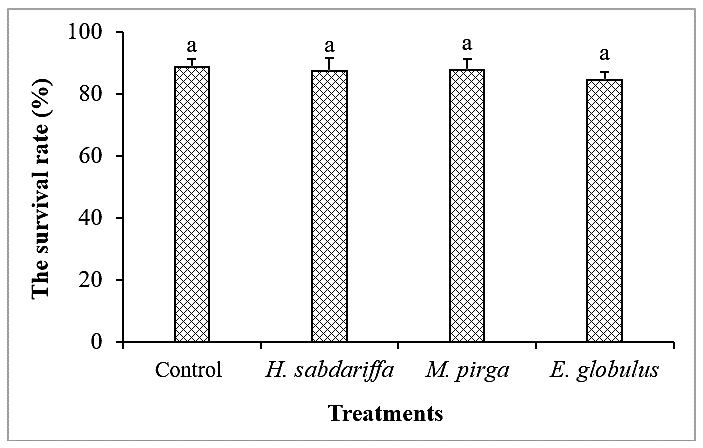
c. Survival rate of the experimental shrimp
The results in Figure 2 showed that the survival rate of shrimp in all treatments after 30 days ranged from 84.50 to 88.90%. Shrimps in all treatments had a very high survival ratio, but no significant differences in these ratios between the treatments were shown. The control had the highest rate (88.90%), followed by the treatment with the addition of M. pirga (87.78%). The lowest rate was found in the treatment with E. globulus (84.50%). In summary, adding herbal extracts into the feed did not negatively affect the normal survival of the whiteleg shrimps.

d. Immune parameters
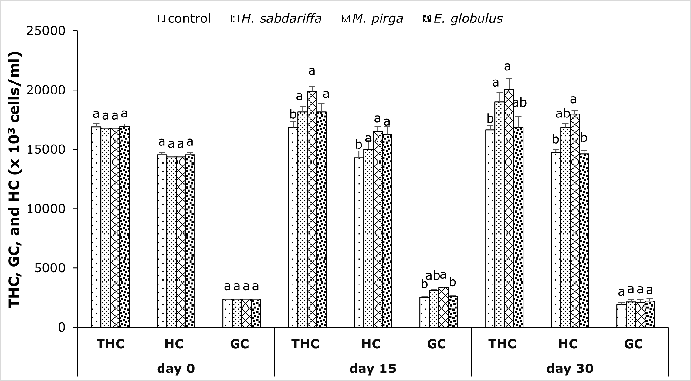
Total hemocyte count (THC) significantly increased when the shrimps were fed with 1% herbal extracts after 15 and 30 days of treatment. THC in M. pirga and H. sabdariffa were significantly higher than that in the control group (p<0.05). Specifically, after 30 days of treatment, the highest THC was found in the treatment of M. pirga, followed by H. sabdariffa and E. globulus, with 20090x103 cells/ml, 19000x103 cells/ml, 16855x103 cells/ml, respectively (Figure 3). The results indicated that the herbal extract slightly increased the total hemocyte count of shrimp in comparison to the control, but the hyaline cell and granular cell were not enhanced.
.png)
The differential hemocyte count (DHC) of shrimps in the three treatments and the control was also determined. The hyaline hemocytes count (HC) in M. pirga treatment was significantly higher than that in the control after 30 days of treatment. The granulocytes count (GC) in the treatment of M. pirga on the day 15th was enhanced and significantly different from those of the treatments with E. globulus and the control (p<0.05). However, on day 30th, there was no enhancement in the GC and no significant difference between the treatments and the control (Figure 3). In summary, the addition of M. pirga helped whiteleg shrimp increase the HC and the GC on the day 15th and 30th.
Effect of herbal extracts on shrimp’s resistance to AHPND (VpAHPND)
a. The density of Vibrio parahaemolyticus in the hepatopancreas of whiteleg shrimp
The density of V. parahaemolyticus in the liver of the challenged shrimp (infected shrimp) is shown in Table 6. Significant differences existed between the positive control and the other treatments on the 5th, 10th, and 14th days. The highest number of V. parahaemolyticus was found in the positive control at 2.8x10⁵, 1.1x10⁵, and 5.7x10³ CFU/g on the day 5th, 10th, and 14th, respectively. It was followed by adding H. sabdariffa (2.7x10⁴, 1.7x10³, and 9.3 x10² CFU/g). The lowest density was in the treatment with E. globulus (4.4x10², 2.3x10², and 2.4x10² CFU/g). It was clear that the bacteria density in the positive control was significantly higher than that in the treatments of herbal extracts. However, no significant difference was seen among the treatments of different herbal extracts. The results of this present study indicated that all three experimental herb extracts effectively decreased the infection of V. parahaemolyticus into the hepatopancreas of whiteleg shrimp.
b. Shrimp mortality
All the herbal extracts in the present study affected the mortality of the shrimp infected with pathogenic bacteria (Figure 4). Feeding herbal extracts to the whiteleg shrimps infected with V. parahaemolyticus significantly reduced the mortality rate of shrimps. The mortality rates in the treatments of different herbal extracts were significantly lower than that in the positive control (46.67%) (p<0.05). In the treatment of H. sabdariffa, the mortality was only 13.33%, the lowest among the three extracts, which shared a similar ratio to the negative control (about 10%). Similarly, the mortality in the treatment of M. pirga (18.33%) was as low as that of the control (p>0.05) but significantly lower than the positive control (p<0.05). The treatment with E. globulus also improved the survival rate, which was indicated by lower mortality than the positive control (Figure 4). In summary, the addition of H. sabdariffa and M. pirga extracts improved the survival rate of shrimp when infected with V. parahaemolyticus. H. sabdariffa, M. pirga extract was not affected by acute hepatopancreatic necrosis disease.
.png)
c. Histopathology of hepatopancreas
On days 3rd and 14th, the hepatopancreas of the shrimps challenged with V. parahaemolyticus, causing acute hepatopancreatic necrosis disease under the treatment with herbal extracts, and the positive control were sampled and stained with H&E dye. The optical microscopy images of the hepatopancreas showed that if hepatopancreatic tissue was normal (no damage, Figure 5a and 6a), the hepatopancreatic ducts (also called hepatopancreatic ampulla) had a stellate structure and were full of B, R, and F cells. On the day 3rd after V. parahaemolyticus, tissue of the shrimps in the positive control displayed typical characteristics of V. parahaemolyticus infection, such as the epithelial cells in hepatopancreatic tubule being atrophic and sloughing, especially lacking B, R, and F cells (Figure 5b). These characteristics were also found in the H. sabdariffa, E. globulus, and M. pirga treatments. However, the impact of bacteria on the histopathological structure under the herbal treatments was milder. Specifically, the atrophy and the sloughing of epithelial cells were only found at a low rate. Lacking B, R, and F cells per unit of hepatopancreas mass was less and not severe. The sloughing of epithelial cells in the hepatopancreatic tubule is also less. Moreover, there was no infection of pathogenic bacteria into hepatopancreatic tissue (Figures 5c, 5d, and 5e).


On day 14th, the survival shrimps in the treatments supplemented with herbal extracts showed a recovery in cellular structure characteristics. In particular, the pathogen was not detected in all treatments supplemented with H. sabdariffa, E. globulus, and M. pirga; therefore, the hepatopancreatic tubules were at normal state with some minus changes (Figures 6c, 6d, and 6e). These results were consistent to lower mortality and bacterial density in all the treatments of herbal extracts. Typical AHPND caused damage in the hepatopancreatic epithelium, necrosis of epithelial cells, and lacking B cells found in the positive control (Figure 6b). Those findings revealed the extracts of H. sabdariffa, E. globulus, and M. pirga were able to enhance the resistance of shrimps against V. parahaemolyticus infection.
Discussion
The mechanisms of phytochemicals in improving overall animal health have yet to be firmly understood since they certainly relate to the variety of complex interactions between the bioactive compounds and the hosts.33 These bioactive substances work as bactericidal and bacteriostatic agents. In other words, they are directly involved in killing pathogenic bacteria and preventing their growth. Specifically, phytochemicals affect the immune system to eliminate bacteria, preventing bacteria from entering the animal’s body.34 In addition, they are also growth promoters through effects on the villi structure in shrimp gut and inhibition of intestinal pathogenic bacteria in shrimp.35,36
In Vietnam, the effectiveness of herbal species in aquaculture has been evaluated.37–40 H. sabdariffa, M. pirga, and E. globulus are well-known to possess bioactive compounds to release human sickness; however, there are only a few studies in aquaculture. These herbs grow wildly or are planted. They can grow well in areas of freshwater and brackish-salty water. The habitat may affect the composition and content of chemical substances in these herbs. This study found these herbs to resist V. parahaemolyticus, causing acute hepatopancreatic necrosis disease in shrimp. Via agar plate diffusion assay, their antibacterial ring diameter ranged from 24.67 to 25.67 mm. Consequently, the methanol extracts of H. sabdariffa, M. pirga, and E. globulus can be considered “bactericidal agents.” According to Salleh et al.41 and Loi,42 H. sabdariffa has antiseptic, diuretic, and antioxidant abilities. Its composition contains gogoetine, hibiscin (anthocyanin), glucoside hibiscritin (flavanol); riboflavin, ascorbic acid, niacin, carotene, calcium, and iron (Duke, 198343; Lade et al.44). Previous studies show that H. sabdariffa extracts inhibit both gram-negative bacteria (Vibrio vulnificus, Aeromonas hydrophila, A. caviae, Escherichia coli, Salmonella enteric, Klebsiella pneumoniae, Proteus vulgaris and Pseudomonas aeruginosa) and Gram-positive bacteria (Staphylococcus aureus, S. cholermidis, S. mutans, and Bacillus cereus).44–46 The antibacterial activity of E. globulus has been demonstrated in previous studies.47,48 According to Nezhad et al.,49 the antibacterial activity of the extract of E. globulus is contributed to its bioactive components such as 1,8-cineole, citronellal, citronellol, citronellyl acetate, p-cymene, eucamalol, limonene, linalool, β-pinene, γ-terpinene, α-terpinol, oocimene and aromadendrene. Consequently, H. sabdariffa, M. pirga and E. globulus collected from other countries and Vietnam exhibited high levels of antibacterial activity thanks to possessing bioactive components. This feature makes them potential for pathogenic bacteria inhibition ability. Noticeably, the biological activity of the herbal extracts was greatly influenced by many factors such as extraction method, solvent, temperature, herbal age, and seasonal sampling.50
Feeding whiteleg shrimp with H. sabdariffa, M. pirga, and E. globulus brought the positive effects on the growth in weight and survival rate of the shrimp. Ghosh et al.51 reported that many herbal extracts (herbs) could stimulate growth, to enhance nutrient absorption and digestion, which results in improving the feed conversion index (FCR) in shrimp. Radhakrishnan et al.36 indicated that herbs can stimulate growth as they help to enhance the activity of digestive enzymes (protease, amylase, and lipase). Hence, in this experiment, adding the herb extracts with a dose of 1% into the feed caused a significant increase of the growth parameters in comparison with the control. In particular, the diet supplemented with these extracts did not negatively affect the survival rate of whiteleg shrimp after 30 days of treatment. According to Awaad,52 although natural compounds are usually milder than topical drugs, their activities may last longer. In this study, hematological parameters (THC, HC, and GC) were observed to determine the level of the cellular immunity of shrimp. The THC and HC of the shrimp in all three treatments were enhanced and significantly different from those in control (p<0.05). Blood cells play a vital role in the immune response of crustaceans; when a foreign object attacks the body, the blood cells perform the function of fighting against the foreign object, such as phagocytosis, encapsulation, ganglia, melanization, and activation of the Pro-PO system.53
The present study clearly shows that Penaeus vannamei shrimp fed with each extract of H. sabdariffa, M. pirga, and E. globulus (1% in the weight of feed) can resist acute hepatopancreatic necrosis disease, which is proved through the reduction in mortality and bacterial population, the improvement in histopathological parameters of shrimp’s hepatopancreas. It is reported that the dose and duration of the treatment with herbal extract are the main factors driving the enhancement in immune response and protection against pathogens. An immune response may be activated significantly at an optimal dose, but under a too-high dose, the immune response may not be enhanced or even suppressed.54 The duration of herbal supplementation is also one of the factors affecting the immune response of shrimp and fish. Therefore, the dose and duration of the herb and herbal products should be used to feed the fish and shrimp from 0.1% to 10% and from 14 days to 70 days, respectively.55 As a result, supplementing 1% H. sabdariffa, M. pirga, and E. globulus (in weight) in the present study helped shrimp improve the survival rate after being infected with V. parahaemolyticus. Especially, growth performance in all three herbs was significantly higher than the control (p<0.05). Moreover, the number of bacteria found in the shrimp hepatopancreas was also reduced, and the effect of the pathogen on hepatopancreas tissue lessened compared to the control. Generally, using herbs in aquatic animal husbandry at a proper dose and duration will positively influence the growth, non-specific immune response (including humoral and cell-mediated Immunity), survival ability, and resistance against pathogens.
Acknowledgments
The authors acknowledge financial support from Tra Vinh University.
Author contributions
Conceptualization: Thi T.L. Nguyen (Lead). Methodology: Thi T.L. Nguyen (Lead), Quoc P. Truong (Supporting), Mong H. Hong (Supporting). Formal Analysis: Thi T.L. Nguyen (Lead), Van D. Pham (Supporting), Mong H. Hong (Supporting). Investigation: Thi T.L. Nguyen (Lead), Van D. Pham (Supporting), Mong H. Hong (Supporting). Writing – original draft: Thi T.L. Nguyen (Lead), Thi T.H. Luu (Supporting), Thanh T. Nguyen (Supporting), Mong H. Hong (Supporting). Funding acquisition: Thi T.L. Nguyen (Lead). Resources: Thi T.L. Nguyen (Equal), Mong H. Hong (Supporting). Supervision: Thi T.L. Nguyen (Lead). Writing – review & editing: Thi T.H. Luu (Supporting), Trong N. Nguyen (Supporting), Mong H. Hong (Supporting).