
Introduction
Arachidonic acid (ARA), an n-6 highly unsaturated fatty acid (n-6 HUFA), serves as an essential fatty acid for most marine species, similar to docosahexaenoic acid (DHA) and eicosapentaenoic acid (EPA). However, studies on ARA have been partially overlooked, mainly because of its relatively minor content in membranes compared to DHA and EPA.1,2 Indeed, over the last few years, more attention has been paid to ARA as an essential fatty acid (EFA) in marine fish nutrition, predominantly due to its role as an eicosanoid precursor. It is well established that ARA serves as the primary precursor for the synthesis of eicosanoids, including prostaglandins, leukotrienes, and thromboxanes.
In recent years, a series of studies have been conducted to assess the nutritional role of ARA in aquaculture, accompanied by progressive clarification of the physiological actions mediated by eicosanoids. These signaling molecules are biologically active even at low physiological concentrations and play critical roles in regulating various biological processes, particularly growth and immune modulation.3,4 Shahkar et al. observed that the polynomial regression analysis of weight gain indicates that the optimum dietary ARA level could be 0.92% diet, and the optimum dietary ARA level could elevate the growth and immunity of Japanese eel.5 Medagoda and Lee demonstrated that 0.3%-0.9% supplemented ARA may improve the growth, immune, and hematological parameters of olive flounder (Paralichthys olivaceus).6 Meanwhile, a series of studies on fish have also reported that appropriate levels of dietary ARA contributed to enhancing tissue fatty acid profiles,7,8 lipid metabolism,9–11 regulation of reproduction,12–14 and disease resistance15,16 Besides, several studies in crustacean species also pointed to the effectiveness of an appropriate level of dietary ARA in the modulation of growth and reproductive performance.17–19 These studies all show that a certain amount of ARA is necessary to maintain growth and biological processes in aquaculture species.
Sea cucumber, Apostichopus japonicus, is one of the high-production marine-cultured animals in northeast China and has considerable market and nutritional value. Recent studies on the nutrient requirements of sea cucumber have focused on protein and amino acids,20,21 lipids and n-3 HUFA,22,23 carbohydrates,24 and vitamins.25,26 Up to now, there are few studies on the effects of ARA on the growth and non-specific immunity of the sea cucumber. In particular, there is a lack of assessment regarding the nutritional requirements of sea cucumbers for ARA. Interestingly, our previous work found that the ratio of ARA to total fatty acids on the body wall of sea cucumber (in the range of 4% to 8%) was significantly higher than that in marine fishes (in the range of 0.5% to 1%). This finding implies a potentially greater physiological significance of ARA in the regulatory biology of sea cucumbers. Consequently, this study was designed to systematically evaluate the effects of dietary ARA supplementation in sea cucumber, with specific emphasis on growth metrics and related gene expression changes, and parameters of non-specific immunity. The overarching aim is to define the optimal dietary ARA level in this echinoderm and delineate its specific roles in modulating growth and innate immune function.
Materials and Methods
Diet preparation
Six isonitrogenous and isolipidic diets were formulated at an average crude protein level of 19.88% and an average crude lipid level of 4.98%.23,26 Feed ingredients and main nutrients contents are presented in Table 1. The diets used fish meal and fermented soybean meal as protein sources and wheat meal as a sugar source. ARA-enriched oil (ARA content, 48.70% of total fatty acid; in the form of ARA-methylester; Cabio Biotech Co., Ltd., Wuhan, China) was supplemented separately to the diets at the expense of triglyceride stearate to provide six diets containing 0.02% (the control group), 0.17%, 0.36%, 0.51%, 0.59% and 0.98% ARA (dry weight), respectively. The fatty acid compositions of the diets are listed in Table 2.
All dry ingredients were ground into powder (0.08 mm) and mixed thoroughly. ARA-enriched oil was added to the powder and mixed thoroughly, then appropriate distilled water was added to form a dough. Feed pellets were prepared using a moist pelletizer and dried at 60 °C for 24 h in an oven and ground into desirable particle sizes (0.18~0.25 mm). All diets were packed and stored at -20 °C.24
Experimental design
Experimental sea cucumbers were obtained from Shandong Anyuan Aquaculture Co., LTD (Penglai, China) and transported into the laboratory at 4 °C. Prior to the beginning of the experiment, juvenile sea cucumbers were reared in a recirculating aquatic system for 2 weeks to acclimate to the experimental conditions and fed a basal diet during this period. At the beginning of the trial, sea cucumbers were starved for 24 h and weighed. Then, 720 sea cucumbers (initial weight of 10.78±0.41 g) were randomly allocated to 18 300 L blue cylindrical fiberglass tanks (75cm × 80 cm), with equal numbers (n = 40) per group, forming six groups in triplicate. Water depth was kept at 60 cm, and the feeding trial lasted for 56 days. There were two culture baskets with 20 corrugated boards in each tank, and the baskets were replaced every 2 weeks. Each tank was fed once daily (16:00) by hand, and the feeding amount was approximately 3% of total wet weight.
During this period, the temperature ranged from 18.0-19.5 °C, the water flow rate was kept at 2 L·min-1, the salinity was held at 28-30, the pH was 7.5-8.1, the dissolved oxygen was above 6 mg·L-1, and the total ammonia nitrogen was maintained at less than 0.05 mg·L-1. Every other day, a thorough cleaning of the tank bottom was performed through siphoning with a pipe. The trial was performed in low light (~150 lx).
Sampling collection and chemical analysis
At the end of the experiment, the sea cucumbers were starved for 24 h, and the total number and body weight of sea cucumbers in each tank were then measured. After weighing, ten sea cucumbers per tank were randomly selected and dissected on ice. The separated intestinal tracts were thoroughly put into 1.5 ml tubes (RNAase-Free; Axygen). The separated body walls were placed in a -20 °C freezer for biochemical analysis. All samples were frozen in liquid nitrogen and stored at -80 °C for further analysis. The dissection tools used for the sample processing were pretreated at 180 °C for 4 h to eliminate RNAase.
Proximate composition analyses of experimental diets and body wall samples were performed using standard methods.27 Crude protein (N × 6.25) was determined by the Kjeldahl method after acid digestion. Crude lipid was measured by the ether extraction using the Soxhlet method. Moisture was determined by oven-drying at 105 °C overnight. Ash was determined by combustion at 550 °C in a muffle furnace. The fatty acid composition of the diets and body wall of sea cucumbers was analyzed using the procedures described by Xu et al. and using a GC-2010 gas chromatograph (Shimadzu, Japan).28
The intestines of the sea cucumber were homogenized with a 9-fold cold physiological saline solution after cutting in an ice bath. The homogenized solution was collected by centrifugation at 4000×g for 10 min at 4 °C using a High-Speed Refrigerated Centrifuge (Hitachi CRG Series, Japan). The supernatant was aliquoted into a 2 ml centrifuge tube and stored frozen at -80 °C until the activity assay.
Total antioxidant capacity (T-AOC), superoxide dismutase (SOD), catalase (CAT) activities, and malondialdehyde (MDA) content were measured using commercial kits from Nanjing Jiancheng Bioengineering Institute, Nanjing, China, according to the manufacturer’s instructions. Alkaline phosphatase (AKP), acid phosphatase (ACP), aspartate aminotransferase (AST), alanine aminotransferase (ALT), lactate dehydrogenase (LDH) and albumin (ALB) activities were analyzed by the commercial kits provided by Shanghai Enzyme-linked Industrial Co., Ltd., Shanghai, China.
RNA extraction and real-time quantitative polymerase chain reaction (QRT-PCR)
Three intestines per tank were used to detect gene expression profiles. Total RNA was extracted from the intestine using RNAiso Plus (TaKaRa, Japan) according to the manufacturer’s specifications, and was electrophoresed on a 1.0% denaturing agarose gel to test the integrity. The quantity and quality of the RNA were assessed with the NanoDrop ND-1000 UV Spectrophotometer (NanoDrop Technologies, Wilmington, DE, USA). Total RNA from each sample was reverse-transcribed into cDNA using the PrimeScript™ RT reagent Kit (Takara, Japan) according to the manufacturer’s protocol.
qRT-PCR was carried out using the Roche LightCycler® 480Ⅱ instrument (Roche Inc., Switzerland). The reactions of three replicates were conducted with a total volume of 20 uL containing 10 μL SYBR®Premix Ex TaqII (Tli RNaseH Plus) (2×) (TaKaRa, Janap), 0.8 ul of 10 umol L-1 each forward and reverse primer, and 2 ul cDNA template and 6.4 uL Milli-Q water. The qPCR profiles contained an initial activation step at 95 °C for 30 s followed by 40 cycles of 95 °C for 5 s and 60 °C for 20 s. The melting curve was performed according to the default procedure. Melting curve analysis was performed on the PCR products at the end of each run to ensure that a single product was amplified. The primer sequences for β-actin, Cyclin B, Follistatin and Tensilin were listed in Table 3. The amplification efficiency of each primer pair was monitored using a dilution series of cDNA. All amplification efficiencies were between 99% and 101%. A relative quantification method was used to calculate the gene expression values using the comparative Cq method (2−ΔΔCq) described in the literature.29
Statistical analysis
All data were expressed as the means ± SD of three replicates. The date were subjected to one-way analysis of variance (ANOVA), using the SPSS program version for windows (Release 20.0), and the significant differences among treatments were examined with Tukey test for multiple comparisons at the significance level of 0.05 (P<0.05). The optimal dietary ARA requirement of sea cucumber was estimated with Second-order polynomial regression model on the basis of specific growth rate (SGR).30
Results
Growth
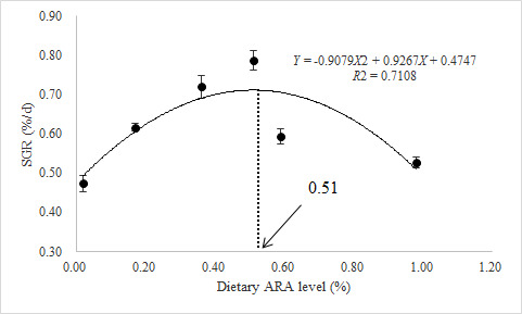
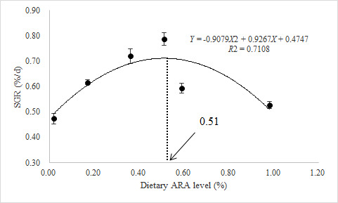
The WGR and SGR of sea cucumber were significantly higher in the 0.51% group than in the 0.02% group, and then gradually decreased thereafter. The WGR and SGR in the 0.36% and 0.51% groups were significantly higher than those in other groups (P<0.05). Feed efficiency (FE) showed significant differences among experimental diets, with a trend of initially increasing and then decreasing, similar to WGR and SGR (P<0.05). However, there were no differences in SR among all groups (P>0.05) (Table 4).
Based on SGR, the appropriate supplementation of dietary ARA levels for sea cucumber was estimated to be 0.51% of diet by using a second-order polynomial regression model (Y = -71.831X2 + 73.387X+30.194, R2 = 0.6968)(Figure 1).
Proximate composition of the body wall
There were no significant differences in crude protein, crude ash, and moisture contents of the body wall among all groups. The crude lipid contents exhibited obvious shifts, with the lowest values in 0.51% group. The crude lipid contents of 0.51% group were significantly lower than 0.02%, 0.17% and 0.98% groups, while there were no significant differences with 0.36% and 0.59% groups (Table 5).
Fatty acid profiles of the body wall
There were no significant differences in the sum of saturated fatty acid contents of the body wall among all groups. With increasing dietary ARA levels, ARA and n-6 PUFA contents increased, while EPA and DHA decreased. Meanwhile, the sum of polyunsaturated fatty acids increased initially and then decreased as dietary ARA levels increased, with the lowest values observed in the 0.02% group (Table 6).
Antioxidant and metabolic enzymes
SOD activity increased significantly at first and then decreased, with the highest values in the 0.51% group. Similar trends were also observed in T-AOC and CAT activities. MDA contents in 0.59% and 0.98% groups were significantly higher than those in other groups, and there were no significant effects in groups from 0.02% to 0.51% (Table 7).
The ALT and AST activities first increased and then decreased, reaching a maximum in the 0.51% group. With increasing dietary ARA levels, both ACP and AKP activities increased initially and then decreased, with the highest values in the 0.51% group, which was significantly higher than those in the 0.02%, 0.17%, and 0.98% groups. The LDH activities had a similar trend to the ACP and AKP. However, ALB activities in the intestinal significantly decreased in 0.36% and 0.51% groups compared to the other groups (Table 8).
Gene expression
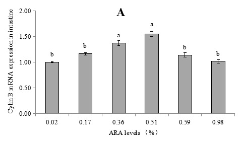
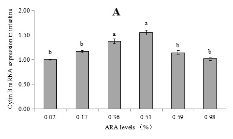
The expression levels of Cyclin B mRNA in the 0.36% and 0.51% groups were significantly higher than those of other groups, while there were no differences among other groups. The expression levels of Follistatin mRNA decreased initially and increased afterward with increasing dietary ARA levels, and the 0.36% and 0.51% groups were significantly lower than the 0.02% and 0.98% groups. There were no differences on expressions of Tensilin mRNA among all groups (Figure 2).
Discussion
Effects of dietary ARA levels on growth performances of the sea cucumber
Arachidonic acid is one of the essential fatty acids that play important roles in the survival and growth of most marine fish. Many studies have illustrated that dietary optimum ARA can stimulate the growth performances, survival, non-specific immune responses, and resistance to acute stress of gilthead seabream (Sparus aurata),31 Japanese seabass (Lateolabrax japonicas),28 European sea bass (Dicentrarchus labrax),32,33 Half-smooth tongue sole (Cynoglossus semilaevis),7 Japanese eel (Anguilla japonica),5 and juvenile yellow catfish (Pelteobagrus fulvidraco),34 olive flounder (Paralichthys olivaceus)6 and yellowfin seabream (Acanthopagrus latus).16 However, to our knowledge, the dietary ARA requirement of sea cucumber has not been reported so far. In our study, sea cucumbers fed diets containing 0.36%-0.51% ARA exhibited significantly higher SGR than the control group and the 0.98% group, indicating that sea cucumbers, like most marine fish, require moderate dietary ARA to maintain normal growth and physiological functions. Some researchers have presumed that dietary ARA affects growth performance by altering the ratio of the two prostaglandins, prostaglandin E2 and prostaglandin F2α,28 and have speculated that a similar mechanism may exist in sea cucumber.
The optimum ARA requirement level was determined by polynomial regression analysis based on the SGR and estimated at approximately 0.51% of the diet. This requirement level is lower than those reported for most fish, for instance, half-smooth tongue sole (1.07%-1.42%),7 Japanese eel (0.92%),5 and Chinese sturgeon (1.00%).8 The differences were probably due to experimental species and body size, study method, and experimental conditions.
In addition, this study found that excess dietary ARA (0.59%-0.98%) negatively affected sea cucumber growth performance. This result was consistent with many previous studies in several fish species.7,8 This inhibitory effect of excess dietary ARA might be due to potential inhibitory effects on EPA bioconversion.12 However, some studies have also suggested that ARA has no significant influence on the growth of aquaculture species. There were also no particular growth-promoting effects of ARA in the giant tiger prawn (Penaeus monodon)35 and red snapper (Lutjanus malabaricus).36 The differences might be due to specie’s long-term evolutionary adaptation to natural diets containing different ARA content and their different biosynthesis capacity.19
There is an intimate relationship between ARA and growth performance in aquatic animals. However, to date, no study has examined the regulation of ARA growth-related genes in the sea cucumber. Therefore, in an effort to elucidate the mechanisms by which dietary ARA modulates the growth performance of sea cucumber, the effects of dietary ARA concentrations on growth-related gene (Cyclin B, Follistatin, and Tensilin) mRNA expression were investigated. Cyclin B, Follistatin, and Tensilin are related genes involved in growth regulation and play an important role in the growth and development of the sea cucumber. Cyclin B, as a cyclin, can form a heterodimer with a cyclin-dependent kinase, primarily by controlling the G2/M transition to regulate cell division, a key step in the eukaryotic cell cycle, and plays a key role in the operation of regulatory factors.37 It was meant that the organism would have exuberant mitosis and better growth when the Cyclin B gene has a high expression level. In the present study, Cyclin B mRNA expression levels in the intestine of sea cucumbers fed a diet containing 0.36%-0.51% ARA were significantly higher than those of the other four groups. It seemed that the change patterns of the WG and SGR were consistent with the change patterns in Cyclin B expression in diets containing different levels of ARA, indicating that a suitable dietary ARA can up-regulate Cyclin B gene expression and consequently promote the growth of sea cucumber. The results were consistent with the aforementioned indicators, such as WGR and SGR, further confirming that ARA plays an important role in the growth of sea cucumbers.
Follistatin, a rich glycosylated single polypeptide chain containing cysteines, is one of the transforming growth factor superfamily β and inhibits the secretion of follicle-stimulating hormone (FSH).38 Previous studies had shown that the overexpressed Follistatin could promote the development of skeletal muscle in zebra fish and mice.39,40 However, in the present study, Follistatin mRNA expression decreased initially and then increased with increasing dietary ARA levels, in contrast to Cyclin B mRNA expression. The study speculates that it would be more beneficial for growth when Follistatin mRNA is expressed at low levels, due to the lack of a skeleton in sea cucumbers. In addition, the Follistatin mRNA expression values across all treatments in the present study were nowhere near overexpression. Besides, no significant difference was observed in the mRNA expression of Tensilin, indicating that Tensilin gene was not regulated by dietary ARA, at least on the transcriptional level. Tensilin, a collagenous fibronectin-like protein, is synthesized in the mutable dermis of sea cucumbers. Previous studies have reported tissue-specific expression of Tensilin, with lower levels in the intestine and coelomic fluid and higher levels in the body wall.41,42 The mechanisms linking Follistatin and Tensilin gene expression to growth performance in sea cucumber remain unclear, so more studies are needed to investigate this mechanism.
Effects of dietary ARA levels on the proximate composition of the body wall of the sea cucumber
In this study, the body wall crude lipid was significantly influenced by dietary ARA. Both the 0.36% and 0.51% groups were lower than the other groups. Consistent with our results, studies on grass carp and Synechogobius hasta juveniles have also indicated that dietary optimum ARA could decrease whole-body lipid content.43–45 The study results indicated that appropriately dosed dietary ARA could promote fat cell activation and fat metabolism, thereby reducing lipid content. Besides, Norambuena et al. had reported that dietary ARA may reduce lipid accumulation and inhibit the biosynthesis of fatty acids.46 These results also indicate that a consistent trend for lower lipid accumulation exists in relation to increased dietary ARA levels.43
However, one noteworthy consequence of this study may be that lipid content of the body wall increases significantly when dietary ARA levels are increased from 0.51% to 0.98%. Similar results were also observed in Xu et al.'s investigations.28 Those results may suggest that excess dietary ARA caused an imbalance of fatty acids or excessive organic acids in the diet, which leads to an inability to digest and absorb fatty acids completely, resulting in fat deposition in the body. And the excess HUFA caused lipid peroxidation and decreased antioxidant enzyme activities in sea cucumber (see discussion below on antioxidant enzyme activities), which can partially account for this phenomenon.22
Changes in the fatty acid profiles of aquatic animals in response to different dietary ARA levels have also been reported in previous studies. Generally, the fatty acid profiles of bodies and tissues reflect those of the diets, particularly in marine fish species with reduced LC-PUFA biosynthetic activity.15 It was also observed in the present study. In the present study, fatty acid concentrations, including C16:0, ARA, and n-6 PUFA, were positively associated with their respective dietary compositions. Despite almost constant EPA (about 1.0%) in the diet, EPA content was negatively correlated with dietary ARA levels, as reported in other aquatic animals45,47 Tocher and Sargent suggested that the EPA and ARA exist in competition during phospholipid esterification in fish, which may be one reason for the lower EPA contents in the ARA groups. A similar competition mechanism may exist in sea cucumbers, but further studies are needed to investigate this mechanism.48 It is suggested that the competition mechanism between EPA and ARA in sea cucumber resembles those observed in fish. Notably, sea cucumber fed the lowest ARA diets (control group) showed a higher content of ARA (12%) in body wall, significantly higher than the content of EPA (4.61%) and DHA (3.00), which implies its essential physiological role in sea cucumber.
It was also found that SFA contents were lower than those in the diets, whereas MUFA contents showed the opposite trend. Yuan et al. and Wu et al. also found similar results. On the one hand, it implied that the SFA may have been preferentially metabolized due to excessive SFA levels in the diets.7,8 On the other hand, part of the SFA might be desaturated and elongated to form MUFA.49,50
Effects of dietary ARA level on the enzyme activities of the sea cucumber
Sea cucumbers mainly rely on their innate immune systems against pathogens, as do other marine invertebrates that lack adaptive immunity.42 It has been demonstrated that the antioxidant enzymes (SOD, CAT, T-AOC) all play important roles in the immune defense of the sea cucumber. SOD and CAT can clear reactive oxygen species (ROS) generated internally in immune cells, reducing their toxic effects, and also play an important role in maintaining the oxidation-antioxidation equilibrium in organisms; hence, they are indicative parameters of immune function.7,51 Therefore, SOD and CAT activities can reflect the abilities of the body’s resistance to oxidative stress to a certain extent, and indirectly reflect the immune levels of aquatic animals.22,52 The role of ARA in improving the antioxidative capacity of animals has been confirmed by some studies.5,53 In the present study, significant improvements were observed on the SOD and CAT activities in the intestine of sea cucumber fed diets supplemented with 0.36%-0.51% of ARA compared to the 0.02% and 0.98% groups. The T-AOC activity was significantly higher in the 0.51% group than in the 0.02% and 0.98% groups. These results indicated that optimal dietary ARA enhanced the scavenging of oxygen-free radicals and the antioxidant capacity of sea cucumber. The mechanism by which ARA affects fish immune function was believed to involve alterations in eicosanoid production.54 ARA-derived eicosanoids, mainly PGE2 and leukotriene B4 (LTB4), play an important role in mediating ARA’s effects on immune cells.55 However, the activities of antioxidant enzymes were significantly decreased in 0.98% group than 0.36%-0.51% groups. It may be due to excess ARA being oxidized, producing a certain amount of oxyradicals that could consume some antioxidant enzymes.18,28 This suggests that appropriate dietary ARA supplementation improved the antioxidative capacity of sea cucumber, whereas excessive supplementation had negative effects.
MDA is produced from the lipid peroxidation of polyunsaturated fatty acids. The content of this aldehyde might indirectly reflect the degree of damage and the antioxidant capacity of aquatic animals, and MDA is used as a biomarker to measure oxidative stress in an organism.56 In the present study, MDA content in 0.59% and 0.98% groups was significantly higher than in the other groups, indicating that the lipid peroxidation was enhanced by excess dietary ARA levels. Similarly, Li et al.53 in large yellow croaker and Shahkar et al.5 in Japanese eel observed a significant increase in the MDA content when fed with high levels of dietary ARA. Moreover, this is because ARA contains four double bonds, which are prone to lipid peroxidation via free radical attack, thereby causing oxidative stress and impairing the functional integrity of cell membranes.53 However, the study of Macrobrachium nipponense revealed that the MDA content of the hepatopancreas exhibited a generally increasing trend in response to elevated dietary levels of ARA.57 This discrepancy could be attributed to the different abilities among species of counteracting the increased oxidative stress by mobilizing antioxidants.
Transamination is one of the main pathways for amino acid synthesis and deamination and is also a valuable diagnostic tool for stress responses in several aquatic animals.58 Phosphatase could accelerate the hydrolysis and synthesis of organic esters of phosphoric acid and the transfer of phosphate groups to other compounds. The increase in phosphatase activity reflected increased phosphorus metabolic activity in cells.59,60 Both transaminases and phosphatases are important physiological enzymes in the sea cucumber.21 It has also been demonstrated that the enzyme activities of transamination and phosphatase all play important roles in the immune defense of echinoderms.61 In the present study, dietary optimal ARA (0.36-0.51%) can promote the enzyme activities of AST, ALT, AKP, and ACP significantly higher than 0.02% and 0.98%. In addition, as with growth results, ARA exerted its effects on the immune response in a dose-dependent manner across several fish species.28,60,62 These implied that 0.36-0.51% of dietary ARA could improve the immune function of the sea cucumber.
Conclusions
In conclusion, the optimal dietary ARA content for juvenile sea cucumber (initial bodyweight 10.78 g) was 0.51% based on the second-order polynomial regression models of SGR. 0.36%- 0.51% dietary ARA could significantly enhance the growth performance and non-specific immunity of sea cucumbers. These results also indicate that dietary ARA may be an essential fatty acid in Apostichopus japonicus.
Acknowledgments
Funding: This work was supported by the Industry and Technology System of Sea Cucumber in Modern Agricultural Industry and Technology System of Shandong Province, China (SDAIT-22-06) and Fund of Yantai Key Laboratory of Quality and Safety Control and Deep Processing of Marine Food (Grant No. QSCDP202404).
Author contributions
Conceptualization: Chengqiang Wang (Equal), Jian Zhang (Equal). Data curation: Chengqiang Wang (Equal), Xiaoyan Wang (Equal), Zhiwei Xiang (Equal). Methodology: Chengqiang Wang (Equal), Xiaoyan Wang (Equal), Tiantian Hao (Equal), Lu Li (Equal), Jinjian Zang (Equal), Yongzhi Sun (Equal). Writing – original draft: Chengqiang Wang (Lead). Writing – review & editing: Min Shen (Equal), Zhidong Song (Equal), Jian Zhang (Equal). Funding acquisition: Jian Zhang (Lead). Supervision: Jian Zhang (Lead).
Competing of Interest – COPE
Declaration: We have no known competing financial interests or personal relationships that could have appeared to influence the work reported in this paper.
Ethical Conduct Approval – IACUC
The following information was supplied regarding ethical approvals (i.e., approving body and any reference numbers): The feeding trial management and sampling were approved by the Shandong Marine Resource and Environment Research Institute Aquatic Experimental Animal Ethics Committee (No. 202526). All measures were taken to reduce stress and pain in the experimental animals.
Informed Consent Statement
All authors and institutions have confirmed this manuscript for publication.
Data Availability Statement
All are available upon reasonable request.