
1. Introduction
Taurine (2-aminoethanesulfonic acid, C2H7NO3S) is a sulfur-containing β-amino acid that is widely distributed in animal tissues. As a functional amino acid, taurine has received considerable research attention and has been widely used in the diets of aquatic animals.1 In fish, taurine participates in osmoregulation, bile acid conjugation, antioxidant defense, appetite regulation, and several immune responses and maintenance of intestinal homeostasis.2 The capacity for endogenous taurine synthesis differs among species and is often limited in carnivorous fishes.1 The content of fish meal in carnivorous fish feed is usually high, and fish meal is the main natural source of taurine. However, taurine is a water-soluble amino acid and is prone to moisture loss during fish meal processing and feed preparation.2,3 Moreover, the background taurine levels in different fish meal sources vary widely, and relying solely on a high addition of fish meal cannot ensure that the taurine content in the feed meets the fish body’s requirements.
Eels (Anguilla spp.) are economically important species in freshwater aquaculture worldwide. Their flesh is highly valued for its palatability and nutritional quality and is often referred to as “water ginseng” in China, making eel an important aquaculture commodity.4 With the decline in natural recruitment of Japanese eel (Anguilla japonica) and European eel (Anguilla anguilla), American eel (Anguilla rostrata) has become increasingly important in eel farming in the southeastern coastal regions of China.4 The American eel is a typical carnivorous fish. Its feed contains a high amount of fish meal. However, there are currently no relevant reports on the dose-response or mechanistic data for taurine in high fish meal diets.
Previous studies have shown that the dietary taurine requirement of aquaculture animals is approximately 0.79%.5 In studies on carnivorous fish, the dietary taurine requirement generally ranges from 0.6% to 1.5%.6–8 Furthermore, research on Anguilla marmorata showed that adding 0.6% taurine (total content of 1.308%) resulted in higher weight gain and specific growth rates than in the other groups.6 The combined proportion of red fish meal and white fish meal in the basal diet of this study accounted for 43%. Based on a weighted calculation using general reference values for taurine in fishmeal, the taurine content in the basal diet was only approximately 0.138%. In this study, 6 g/kg (i.e., 0.6%) taurine was added to the basal diet; this supplementation level is at the lower end of the dietary taurine requirement range for carnivorous fish and represents a conservative yet effective approach. We hypothesized that dietary taurine supplementation would improve growth performance and overall physiological and intestinal health in juvenile American eel. Therefore, the present study evaluated the effects of supplementing a high fishmeal diet with 6 g/kg taurine on growth performance, serum biochemical and immune-related indices, intestinal morphology, and intestinal microbiota of juvenile American eel. The findings are expected to provide a reference for further studies on taurine supplementation and taurine requirements in this species.
2. Materials and Methods
2.1. Experimental fish and design
The animals used in the experiment were juvenile American eels, purchased from Karenda Fisheries Co., Ltd. in Mingxi County, Sanming City, Fujian Province. After three weeks of acclimation, 240 healthy fish with an average weight of (11.84±0.03) g were randomly assigned to the control group and the taurine group. Each group has four repetitions, and each repetition consists of 30 fish. Then a 10-week formal experiment was carried out.
2.2. Experimental diets and feeding trial
The basal diet contained 45.83% crude protein, 6.26% crude lipid, 9.53% ash, 6.85% moisture, 2.31% total calcium, and 1.40% total phosphorus. The treatment diet was prepared by adding 6 g/kg taurine (purity 99%; Xiamen Longli Industrial and Trade Co., Ltd., Xiamen, China) to the basal diet. Diets were mixed for 20 min before use. The ingredient composition and proximate composition of the basal diet are shown in Table 1. The 6 g/kg value refers to the formulated amount of exogenous taurine added to the basal diet.
During the feeding trial, fish were fed twice daily at 06:00 and 18:00 at a daily feeding rate of approximately 1-3% of body weight. The feed was mixed with water at 1.2 times its weight and kneaded into dough before feeding. The feeding amount was adjusted according to apparent feed consumption. Uneaten feed was collected 20 min after feeding, weighed, and recorded. Water temperature was maintained at 25-26 ℃, dissolved oxygen at 6.6-7.6 mg/L, pH at 7.8-8.2, and ammonia nitrogen below 0.25 mg/L.
2.3. Sample Collection
After the experiment, the juvenile American eels in each tank were counted and weighed. After fasting for 24 hours, 15 fish from each tank were selected at random and narcotized with 2 grams per liter of eugenol for about 5 minutes. Then, their body weight and body length were measured. Blood was drawn from the caudal vein of the fish using a disposable sterile syringe. The blood samples were placed in a 4℃ refrigerator for 12 hours, then taken out and centrifuged (3000 × g, 4℃) for 10 minutes. The supernatant was aspirated and divided into centrifuge tubes, and kept in a refrigerator at -80 degrees for serum biochemical and immune analysis. After collecting blood, the eels were dissected and eviscerated, then the intestine was isolated using surgical scissors. Two intestinal tracts measuring 3 mm were randomly collected from each tank, spread on filter paper, and placed in electron microscope fixative for scanning electron microscopy. The foregut of three fish was placed in a frozen tube containing Bouin’s solution for section observation and stored in a refrigerator at 4 ℃. For intestinal microbiota analysis, midgut samples were collected from four fish per tank, but ultimately only two samples per tank were used, with the remaining ones kept as backups. Samples were not pooled, resulting in eight individual samples per treatment and 16 samples in total. A summary of the sampling scheme, including intestinal region, within-tank sample number, total sample number, and statistical unit, is provided in Table 2.
2.4. Growth performance parameters calculation
After the feeding trial, all fish in each tank were weighed, and growth performance (WGR, SGR, FI, FE and SR) was calculated as follows:
Weight gain rate (WGR, %) = (final weight (g/fish) - initial weight (g/fish)) / (initial weight (g/fish)) × 100
Specific growth rate (SGR, %/d) = [ln (final weight (g/fish)) - ln (initial weight (g/fish))] / number of days × 100
Feed intake (FI, g/fish) = feed consumption / number of final fish
Feed efficiency (FE, %) = (final weight (g/fish)-initial weight (g/fish)) / feed consumption × 100
Survival rate (SR, %) = number of fish at the end / number of fish at the start × 100
2.5. Serum biochemical assay
Serum immunoglobulin M (IgM), complements C3 (C3), D-lactate acid (D-lac), diamine oxidase (DAO), glutamic-oxalacetic transaminase (GOT), glutamic pyruvic transaminase (GPT), acid phosphatase (ACP), alkaline phosphatase (AKP) and lysozyme (LZM) were measured with commercial assay kits (Nanjing Jiancheng Bioengineering Institute, Nanjing, China) according to the manufacturer’s instructions. For each tank, two within-tank serum measurements were obtained for each biochemical or immune-related variable. The two values were averaged to generate one tank-level value for statistical analysis.
2.6. Intestinal morphology and microscopy
Foregut samples from three fish per tank were dehydrated through graded ethanol, cleared in xylene, embedded in paraffin, sectioned at 5 µm using a KD-TS3A microtome (Beijing Century Kexin Instrument Co., Ltd., Beijing, China), and stained with hematoxylin and eosin. Sections were observed with an Olympus BX80-JPA light microscope and analyzed using Image-Pro Plus 6.0 (Media Cybernetics, Silver Spring, MD, USA).9 Villus length and muscular layer thickness were summarized as tank level values for statistical analysis.
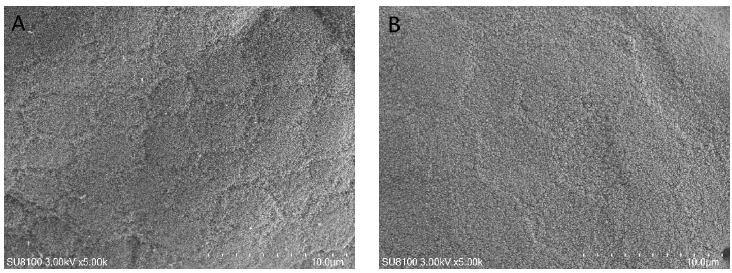
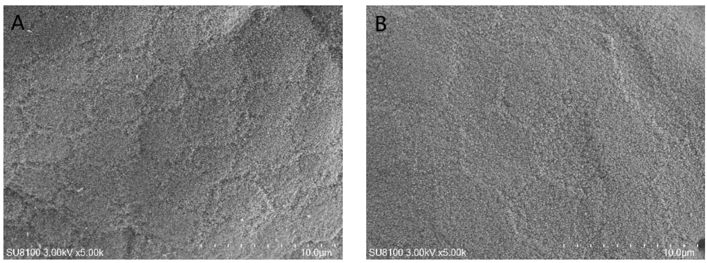
For scanning electron microscopy, intestinal samples were fixed in glutaraldehyde, washed three times in phosphate buffer, dehydrated through a graded ethanol series, critical-point dried (K850, Quorum Technologies, UK), sputter-coated with gold (MC1000, Hitachi, Tokyo, Japan), and examined with an SU8100 scanning electron microscope (SEM, Hitachi, Tokyo, Japan).10 The micrographs were used as representative qualitative images and were not subjected to quantitative microvillus density analysis.
2.7. Intestinal microbiota analysis
A total of 16 individual midgut samples were subjected to 16S rRNA sequencing, comprising two samples from each of the eight tanks. Intestinal bacterial DNA extraction, quality assessment, amplification of the bacterial 16S rRNA V3-V4 region, and the initial bioinformatic workflow followed Shi et al.11 Sequencing was conducted using an Illumina MiSeq PE300 platform by Beijing Allwegene Technology Co., Ltd. (Beijing, China).
2.8. Statistical analysis
The tank was considered the experimental unit for all dietary treatment comparisons. Growth performance variables were calculated at the tank level, providing four independent observations per treatment (n = 4 tanks). For serum biochemical and immune-related variables, the two within-tank measurements were averaged to obtain a single tank-level value, yielding n = 4 independent observations per treatment. For intestinal histological variables, measurements from fish within the same tank were similarly summarized to obtain a single tank-level value (n = 4 per treatment). The results were presented as mean ± SD and analyzed using one-way ANOVA to assess statistical significance (P<0.05) in SPSS 26.0 (IBM Corp., Armonk, N.Y., USA). The gut microbiota was analyzed using QIIME (version 1.8.0) and R (version 3.6.0; R-Statistical Corp., Vienna, Austria). The LDA score threshold was set to 3. Since only two treatments were compared, no multiple comparisons were necessary.
3. Results
3.1. Growth performance
As shown in Table 3, final body weight, feed intake, weight gain rate, specific growth rate, and feed efficiency were higher in the taurine group than in the control group (P<0.05). Initial body weight was not significantly (P>0.05), and survival was 100% in both groups.
3.2. Serum biochemical and immune-related parameters
The effects of taurine supplementation on serum biochemical and immune-related parameters of juvenile American eels are shown in Table 4. Serum GOT, GPT, DAO, and D-lactate were lower in the taurine group (P<0.05). IgM and the activities of LZM, AKP, and ACP were higher in the taurine group (P<0.05). The complement C3 was not significantly affected between treatments (P>0.05).
3.3. Intestinal morphology
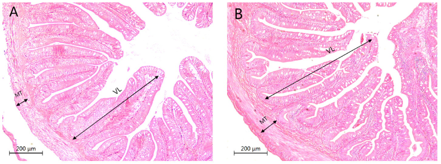
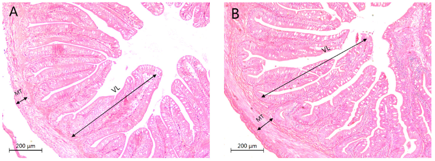
The results of intestinal morphology parameters are presented in Table 5 and Figures 1-2. Intestinal villus length (VL) and muscular layer thickness (MT) were greater in the taurine group than in the control group (P<0.05; Table 5 and Figure 1). As shown in Figure 2, the intestinal epithelial cells of the taurine group were closely arranged, and the thickness of microvilli was uniform. Representative SEM images showed qualitative differences in the appearance of the intestinal mucosal surface between the two groups. No quantitative microvillus density analysis was performed.
3.4. Alpha diversity of the intestinal microbiota
No significant differences were observed in ACE, Chao1, Shannon, Simpson, or coverage between the control and taurine groups (P > 0.05; Table 6). Coverage exceeded 99% in both groups, indicating adequate sequencing depth.
3.5. Intestinal microbiota composition at the phylum level
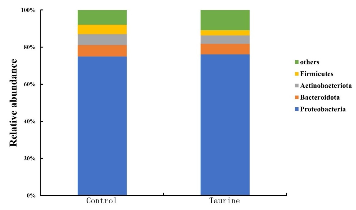
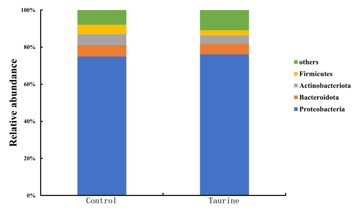
As shown in Figure 3, Proteobacteria, Bacteroidota, Actinobacteriota, and Firmicutes were the predominant phyla in both groups. In the taurine group, the relative abundances of Firmicutes and Actinobacteriota exhibited a downtrend, whereas the Proteobacteria and Bacteroidetes showed no significant alterations.
3.6. LEfSe analysis of intestinal microbiota at the genus level
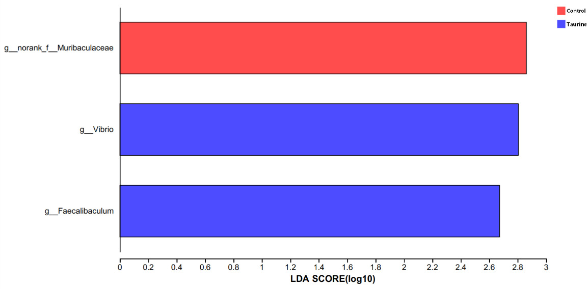
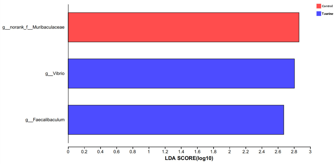
LEfSe analysis identified the family Muribaculaceae as more abundant in the control group and the genera Faecalibaculum and Vibrio as more abundant in the taurine group (LDA score > 3.0; P < 0.05; Figure 4).
4. Discussion
The results of this experiment show that adding 6 g/kg of taurine to the feed can observably increase the FBW, WGR, SGR, FI and FE of juvenile American eel, indicating that taurine can further the growth performance for juvenile American eel. Similar growth-promoting responses have been reported in several fish species, although the magnitude and direction of the response vary with species, physiological stage, basal diet composition, and taurine supplementation level.8,12–14 In the present study, FI increased by approximately 4.2%, FBW by 4.1%, and FE by 3.3%, suggesting that greater feed intake may have contributed to the growth response, while the concurrent increase in FE indicates that changes in feed utilization may also have been involved. In contrast, supplementation with 9.7 g/kg taurine reduced growth performance and increased production costs in Nile tilapia (Oreochromis niloticus),15 demonstrating that higher taurine inclusion does not necessarily confer greater benefits. Collectively, these findings indicate that responses to taurine are likely influenced by species specific metabolism, physiological stage, basal dietary composition, endogenous taurine supply, and supplementation level.
The activities of GOT and GPT are often used as indicators for judging the degree of liver injury, and they are mainly scattered among the mitochondria of liver cells. Cellular injury may increase membrane permeability and promote the release of intracellular transaminases into the circulation, resulting in elevated serum GOT and GPT activities.16,17 In the present study, dietary taurine supplementation significantly decreased serum GOT and GPT activities in juvenile American eels, suggesting reduced cellular enzyme leakage and a potentially improved physiological status. Similar decreases in serum transaminase activities following taurine supplementation have also been reported in rice field eel,18 Japanese flounder (Paralichthys olivaceus)19 and Cynoglossus semilaevis.20 This effect may be related to taurine’s ability to maintain cellular membrane integrity and alleviate tissue stress, thereby reducing the release of intracellular transaminases into the bloodstream.
Serum IgM, complement C3, LZM, ACP, and AKP are commonly used to characterize immune status in fish. IgM is the predominant immunoglobulin involved in the humoral immune response of teleost, whereas C3 is a central component of the complement system and participates in innate immune defense and inflammatory regulation.21 In the present study, dietary taurine supplementation significantly increased serum IgM concentration, while C3 was not significantly affected. Similar enhancement of serum IgM following taurine supplementation has been reported in yellow catfish (Pelteobagrus fulvidraco),22 channel catfish (Ictalurus punctatus),23 rice field eel,24 and golden pompano (Trachinotus ovatus).25 The higher IgM concentration suggests that taurine supplementation was associated with enhanced selected humoral immune related responses in juvenile American eel. However, the unchanged C3 concentration in the present study indicates that the effect of taurine was not uniform across all circulating immune components, and therefore does not support a generalized stimulation of the complement system. The differential responses of IgM and C3 may reflect differences in their regulation, physiological functions, and sensitivity to dietary taurine.
Dietary taurine supplementation also significantly elevated serum LZM, ACP, and AKP activities. LZM is an important component of the innate antibacterial defense system and contributes to the hydrolysis of bacterial cell walls,26 whereas ACP is a canonical lysosomal enzyme that eliminates and degrades microbial pathogens and AKP is a polymorphous enzyme within the fish immune defense system, with the capacity to promote phagocytosis via modification of pathogen surface molecules.27 Increased LZM activity following taurine supplementation has also been reported in longfin yellowtail (Seriola rivoliana),28 turbot (Scophthalmus maximus),29 and yellow catfish,30 while increased ACP and AKP activities have been observed in Culter alburnus31 and longfin yellowtail.32 Collectively, the simultaneous increases in IgM, LZM, ACP, and AKP indicate that 6 g/kg taurine enhanced selected humoral and innate immune-related responses in juvenile American eels. Nevertheless, the immunomodulatory effect of taurine appears to be dose- and species-dependent. For example, higher taurine supplementation levels have been associated with decreased LZM activity in black carp (Mylopharyngodon piceus),33 suggesting that greater taurine inclusion does not necessarily produce stronger immune responses. Therefore, the present findings support an immunomodulatory role of taurine at the tested supplementation level, although the underlying mechanisms and the optimal dose for immune regulation in American eel still require further investigation.
The intestine is a major site of nutrient digestion and absorption in fish, and maintenance of its structural integrity is essential for normal intestinal function.34 Serum D-lac concentration and DAO activity are commonly used as indirect indicators of intestinal mucosal integrity and permeability. Damage to the intestinal mucosa may increase intestinal permeability, facilitating the translocation of bacterially derived D-lactate and mucosal DAO into the peripheral circulation.35 In the present study, dietary taurine supplementation significantly decreased serum D-lac concentration and DAO activity, suggesting improved intestinal barrier integrity and reduced intestinal permeability in juvenile American eels. Consistently, taurine supplementation significantly increased intestinal villus length and muscular layer thickness, which are commonly used as morphological indicators of intestinal health and may provide favorable structural conditions for nutrient digestion and absorption.10,36 Similar improvements in intestinal morphology have been reported in rice field eel.18 In addition, taurine supplementation has been reported to increase the expression of tight-junction-related genes in coho salmon (Oncorhynchus kisutch),37 channel catfish,23 and turbot,38 with comparable barrier protective effects also observed in growing piglets.39 Taken together, the reductions in serum D-lac and DAO, together with the improved intestinal morphology, support a beneficial effect of taurine on intestinal barrier-associated characteristics. Because tight junction proteins, oxidative stress markers, and nutrient digestibility were not directly measured in the present study, these findings should be interpreted as barrier associated changes rather than evidence of a defined mechanistic pathway. These changes may partly contribute to the improved feed utilization and growth performance observed in the present study.
The intestinal microbiota is closely involved in nutrient metabolism, mucosal homeostasis, and immune regulation in fish.25 In the present study, dietary taurine supplementation did not significantly affect the alpha diversity indices of the intestinal microbiota, suggesting that the overall richness and diversity of the bacterial community remained relatively stable. Nevertheless, marked differences were observed in microbial composition between the two dietary groups, indicating that taurine primarily altered the taxonomic structure rather than the overall alpha diversity of the intestinal microbiota. At the phylum level, taurine supplementation decreased the relative abundances of Actinobacteriota and Firmicutes. Previous studies in fish have associated changes in these phyla with intestinal physiological status and growth performance,11,40 and reduced Firmicutes abundance has also been reported in American eels supplemented with oligomeric proanthocyanidins or Macleaya cordata extract.9,41 Nevertheless, both Actinobacteriota and Firmicutes comprise functionally diverse taxa; therefore, their decreased relative abundances cannot by themselves be interpreted as evidence of improved intestinal health. Rather, these changes further indicate that dietary taurine modified the intestinal microbial composition of juvenile American eels.
LEfSe analysis identified several differential bacterial taxa between the two dietary groups. Muribaculaceae was more abundant in the control group, whereas Faecalibaculum and Vibrio were more abundant in the taurine group. Muribaculaceae is a functionally diverse bacterial family that typically plays a positive role in maintaining gut health, but its effects may vary across host species and dietary environments.42 Although existing studies have demonstrated that when the gut microbiota is dominated by Muribaculaceae, the risk of pathogen infection in mice will increase, and even sepsis may be induced,42 no relevant reports have been documented in fish species to date. Therefore, its higher abundance in the control group cannot be interpreted as evidence of increased infection risk. Similarly, the genus Vibrio contains commensal, opportunistic, and pathogenic members, and genus level 16S rRNA sequencing does not allow the functional identity of the increased taxa to be determined. The role of Faecalibaculum in the intestine of American eel also remains poorly characterized. Therefore, these LEfSe results support a taurine associated shift in intestinal microbial composition but do not establish whether individual differential taxa were beneficial or detrimental to the host.
Collectively, dietary taurine supplementation was associated with lower serum D-lac concentration and DAO activity, improved intestinal morphology, and a distinct intestinal microbial composition in juvenile American eels. Taken together, these changes were associated with a more favorable intestinal physiological profile involving mucosal structure, barrier-associated characteristics, and microbial composition. These responses may partly contribute to the observed improvements in feed utilization and growth, but a direct causal relationship cannot be established from the present design. Moreover, from a practical perspective, supplementation with 6 g/kg taurine produced modest but significant improvements in growth under the present high-fishmeal feeding conditions, including approximately 4.1% higher FBW and 3.3% higher FE than the control. These findings indicate that additional taurine may have practical value even in a diet containing 43% fishmeal. However, because the lack of a feed cost-benefit analysis, the present study does not provide sufficient evidence to recommend routine commercial supplementation at 6 g/kg. Further dose-response trials combined with economic evaluation under commercial farming conditions are required to determine whether the observed biological benefits translate into a cost-effective feeding strategy.
5. Conclusion
In conclusion, supplementation with 6 g/kg taurine improved growth performance and was associated with favorable changes in selected serum biochemical and immune-related parameters, intestinal morphology, and intestinal microbial composition in juvenile American eels fed a high fishmeal diet. Because only one supplementation level was evaluated, the present study cannot determine an optimal taurine inclusion level or the dietary taurine requirement; the exact amount required still needs further study.
Acknowledgments
This study was financially supported by the earmarked fund for China Agriculture Research System (CARS-45).
CRediT Authorship Contribution Statement
Data curation: Yuting Song (Lead). Formal Analysis: Yuting Song (Lead), ShaoWei Zhai (Lead). Investigation: Yuting Song (Lead). Methodology: Yuting Song (Equal), Yichuang Xu (Equal), ShaoWei Zhai (Equal). Validation: Yuting Song (Lead). Visualization: Yuting Song (Lead). Writing – original draft: Yuting Song (Lead). Resources: Yichuang Xu (Equal), ShaoWei Zhai (Equal). Supervision: Yichuang Xu (Equal), ShaoWei Zhai (Equal). Writing – review & editing: Yichuang Xu (Equal), ShaoWei Zhai (Equal). Conceptualization: ShaoWei Zhai (Lead). Project administration: ShaoWei Zhai (Lead).
Conflict of Interest Statement
No competing interests were disclosed.
Ethical Conduct Approval – IACUC
The study was conducted according to the guidelines of the Declaration of Helsinki and was approved by the Animal Care Advisory Committee of Jimei University (Approval No. 2019-0906-003). This committee is the institutional ethics body responsible for reviewing and approving animal research protocols at our university.
Informed Consent Statement
All authors and institutions have confirmed this manuscript for publication.
Attestation Statements
Data related to any of the subjects in the study has not been published previously.
All study and manuscript data will be made available to the journal editors upon request before and/or after manuscript publication for review or query
Data Availability Statement
Data can be obtained from the corresponding author.