
Introduction
Tilapia is one of the most economically important freshwater fish species in China, owing to its rapid growth rate, strong environmental adaptability, and favorable processing characteristics (e.g., absence of intramuscular bones). It is widely cultivated in southern China.1 However, the rapid expansion of intensive aquaculture has been accompanied by an increasing prevalence of bacterial diseases. Among these, streptococcosis caused by Streptococcus agalactiae is considered one of the most serious threats to tilapia farming, often resulting in mass mortality and causing substantial economic losses.2
Currently, disease management for streptococcosis in tilapia mainly relies on improved aquaculture practices, environmental optimization, and immunoprophylaxis. Chinese herbal medicines have attracted considerable attention as promising candidates for aquatic disease control, owing to their abundant resources, relatively lower toxicity than synthetic antibiotics, and lower risk of inducing bacterial resistance. However, it should be noted that many medicinal plants contain bioactive compounds with substantial toxicity, and safety is compound-, dose-, species-, and exposure-dependent.3 Their antibacterial mechanisms are generally characterized as multi-target and multi-pathway in nature, including disruption of bacterial cell wall and membrane integrity, inhibition of nucleic acid and protein synthesis, interference with energy metabolism, suppression of biofilm formation, and disruption of quorum sensing systems.4–9 Despite these promising features, systematic screening of Chinese herbal medicines specifically against tilapia-derived S. agalactiae remains limited, and the underlying metabolic regulatory mechanisms are still poorly understood.
Cortex Moutan, the root bark of Paeonia × suffruticosa Andrews, is a widely used traditional Chinese medicine with a long history of use for treating inflammatory and infectious diseases.10 Its major bioactive constituents include paeonol and paeoniflorin, which exhibit antibacterial, anti-inflammatory, and antioxidant activities. Paeonol, in particular, has been reported to inhibit several Gram-positive and Gram-negative bacteria, including Staphylococcus aureus and Escherichia coli. However, studies on the antibacterial activity of Cortex Moutan against aquatic pathogens, especially S. agalactiae, remain limited.11
Traditional approaches to studying the antibacterial activity of herbal medicines have largely been confined to phenotypic screening (e.g., inhibition zone diameter and MIC determination), which provide limited insight into their mechanisms of action. As a key component of systems biology, metabolomics enables the comprehensive detection of global changes in small-molecule metabolites, thereby facilitating the elucidation of drug–pathogen interaction networks. This approach has been successfully applied to investigate the mechanisms of various herbal medicines against aquatic pathogens.10 However, to the best of our knowledge, based on a comprehensive literature search of PubMed, Web of Science, and CNKI, the metabolomic regulatory mechanism of Cortex Moutan against S. agalactiae has not been reported previously.
In the present study, 35 commonly used Chinese herbal medicines were screened for in vitro antibacterial activity against S. agalactiae isolated from tilapia. The most effective extract was further investigated using untargeted metabolomics to elucidate its metabolic regulatory mechanisms. We hypothesized that Cortex Moutan exerts its antibacterial activity against S. agalactiae through both direct antimicrobial action of its bioactive constituents and disruption of central carbon and amino acid metabolism. This study aims to provide preliminary in vitro evidence and candidate compounds for the future development of plant-derived alternatives to antibiotics in tilapia aquaculture.
Materials and Methods
Test strain and reagents
The S. agalactiae strain used in this study was isolated from naturally infected Nile tilapia, then identified by biochemical and molecular characterization, and stored in the laboratory. S. agalactiae was grown in brain-heart infusion agar (BHIA) (Qingdao Hope Bio-Technology Co., Ltd., China) at 30 °C for 48 h. Sterile drug-sensitive tablets have a diameter of 6mm, a thickness of 0.7mm, and a maximum liquid absorption of 20 µl (Changde Bimanke Biotechnology Co., Ltd., China). All herbal materials (Table 1) were purchased from Beijing Tongrentang, Chongzuo City, Guangxi, China. The botanical identity of each herb was authenticated according to the Chinese Pharmacopoeia. The 35 herbal medicines were selected based on three criteria: (i) traditional use in Chinese medicine for treating infectious or inflammatory diseases; (ii) reported antibacterial activity against aquatic or other bacterial pathogens; and (iii) availability in local markets in Guangxi, China. This selection strategy aimed to balance ethnopharmacological rationale with practical applicability in tilapia aquaculture.
Preparation of liquid medicine
Each herbal powder (20 g) was soaked in 500 mL of distilled water for 60 min, boiled for 60 min, and filtered through four-layer gauze. The residue was re-decocted, and the combined filtrates were concentrated to 20 mL (1 g crude drug/mL), then autoclaved at 121°C for 20 min and stored at 4°C.
Preparation of bacterial suspension
A single colony of S. agalactiae was inoculated into 1.5 mL BHI broth and incubated overnight at 28°C with shaking (180 r/min). A 5 μL aliquot was transferred into 5 mL fresh BHI broth and incubated for 6 h under the same conditions.
Antimicrobial susceptibility testing
The Kirby–Bauer disk diffusion method was used. Bacterial suspension (1 × 10⁸ CFU/mL, 100 μL) was spread onto BHI agar plates. Blank disks (6 mm) served as negative controls. Plates were incubated at 28°C for 24 h. Inhibition zones were measured with a vernier caliper in triplicate. Susceptibility criteria: >20 mm (extremely sensitive), 15–20 mm (highly sensitive), 10–15 mm (moderately sensitive), ≤10 mm (weakly sensitive), no zone (resistant).
Antimicrobial susceptibility testing of herbal extracts
The disk diffusion method was used for herbal extracts. BHI agar plates were prepared. S. agalactiae suspension (OD₆₀₀ = 1.0, ≈1 × 10⁹ CFU/mL) was swabbed onto plates. After drying (3–5 min), sterile paper disks impregnated with 20 μL herbal decoction (1 g/mL) were placed on the agar. Sterile water served as negative control. Plates were incubated at 28°C for 24 h. All antibacterial assays were performed in three independent biological replicates. Inhibition zones are presented as mean ± standard deviation (SD). Statistical analyses were performed using GraphPad Prism 9.0 (GraphPad Software, USA). Differences among groups were analyzed using the Kruskal–Wallis test followed by Dunn’s post hoc test. P < 0.05 was considered statistically significant.
Determination of minimum inhibitory concentration (MIC) and minimum bactericidal concentration (MBC) of selected herbal extracts
Selected herbal extracts were evaluated by broth dilution. Two-fold serial dilutions of stock solution (1 g/mL) were prepared in BHI broth, yielding concentrations from 0.5 to 0.001953 g/mL. Positive (no herbal) and negative (no bacteria) controls were included. Each tube received 3 μL bacterial suspension (~1 × 10⁶ CFU/mL) and was incubated at 28°C for 18–24 h. MIC was the lowest concentration with no visible turbidity. For MBC, 100 μL aliquots from clear tubes were spread on BHI agar and incubated at 28°C for 18 h. MBC was the lowest concentration yielding <5 colonies. MIC and MBC values were determined from three independent biological replicates.
Metabolomics sample preparation
S. agalactiae was cultured to log phase, then treated with Cortex Moutan extract at MIC or equal volume PBS (control), and incubated at 28°C, 180 r/min for 10 h. Three biological replicates (50 mL/tube) were collected. Bacterial pellets were obtained by centrifugation (4500 r/min, 10 min), quenched in liquid nitrogen, and stored at –80°C.
Metabolite extraction and UPLC-Q-TOF-MS analysis
Each pellet (50 mg) was extracted with 500 μL ice-cold 80% methanol, vortexed, sonicated, and incubated at –20°C for 30 min. After centrifugation (4°C, 20,000 × g, 10 min), the supernatant was collected and centrifuged again for 5 min. Equal aliquots were pooled as QC samples.
Chromatography used an ACQUITY UPLC HSS T3 column (100 × 2.1 mm, 1.8 µm; Waters). Mobile phase: (A) 5 mM ammonium acetate + 5 mM acetic acid in water, (B) acetonitrile. Gradient: 0–0.8 min (2% B), 0.8–2.8 min (2–70% B), 2.8–5.0 min (70–90% B), 5.0–5.5 min (90–100% B), 5.5–7.5 min (100% B), 7.5–7.6 min (100–2% B), 7.6–10.0 min (2% B). Flow rate: 0.35 mL/min, injection: 4 μL, column temperature: 40°C.
Mass spectrometry used ESI in positive and negative modes. Ion source: 350°C. Capillary voltages: +3.8 kV (positive), –3.4 kV (negative). Gas flows: purge (1 Arb), auxiliary (15 Arb), sheath (50 Arb). Data acquisition: full scan (70–1050 Da, resolution 60,000) with DDA. Top four ions (>5,000 intensity) were selected for MS/MS (resolution 15,000). Dynamic exclusion: 4 s.
Data processing
Raw data were converted to mzML format using ProteoWizard. Peak alignment, retention time correction, and peak area extraction were performed using XCMS (parameters: centWave m/z = 20 ppm, peakwidth = 5–25 s, snthresh = 6, prefilter = c(4, 5000), noise = 1000; grouping: binSize_R = 0.4, gapInit = 0.3, binSize_G = 0.015, bw = 5, minfrac = 0.5). Metabolites with missing values >50% in QC or >80% in experimental samples were removed. Remaining missing values were imputed by KNN, outliers removed, and PQN normalization applied. Data from positive and negative ESI modes were processed separately and then combined for multivariate analysis. Redundant annotations across modes were removed by matching retention time and m/z within 10 ppm and 0.1 min. Only unique features were retained for downstream analysis. Differential metabolites were selected using VIP ≥ 1.0 and T-test P < 0.05. KEGG pathway enrichment analysis was performed using a hypergeometric test with FDR correction (Q-value ≤ 0.05).
Results
In vitro antibacterial activity of 35 Chinese herbal extracts against S. agalactiae
The inhibition zones of all 35 herbal extracts are summarized in Table 2. Only three extracts produced detectable inhibition zones: Cortex Moutan (15.63 ± 0.52 mm), Folium Sennae (14.38 ± 0.45 mm), and Fructus Foeniculi (14.75 ± 0.12 mm). The remaining 32 extracts produced no detectable inhibition zone. The negative control (sterile water) showed no inhibition zone (0 ± 0 mm).
MIC and MBC of selected herbal extracts against S. agalactiae
Based on the inhibition zone diameters obtained from the disk diffusion assay, three herbal extracts—Cortex Moutan, Folium Sennae, and Fructus Foeniculi—that exhibited the most potent antibacterial activity against S. agalactiae were selected for the determination of MIC and MBC using the broth dilution method. The results are presented in Table 3. The MIC values of Cortex Moutan, Folium Sennae, and Fructus Foeniculi were 0.125, 0.125, and 0.25 g/mL, respectively. All three extracts exhibited an MBC of 0.5 g/mL.
Quality assessment of metabolomics data
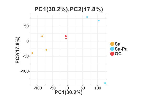
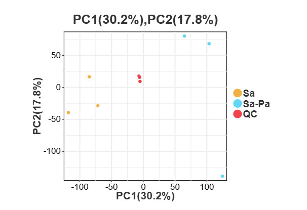
PCA was performed for an initial assessment of data quality and overall metabolic patterns (Figure 1). PC1 and PC2 explained 30.2% and 17.8% of the total variance, respectively. The treatment group (Cortex Moutan aqueous extract) was separated from the control group along the PC1 axis. However, PCA separation alone requires further validation. QC samples clustered tightly around the origin. Biological replicates within each group also showed good clustering.
Multivariate statistical analysis
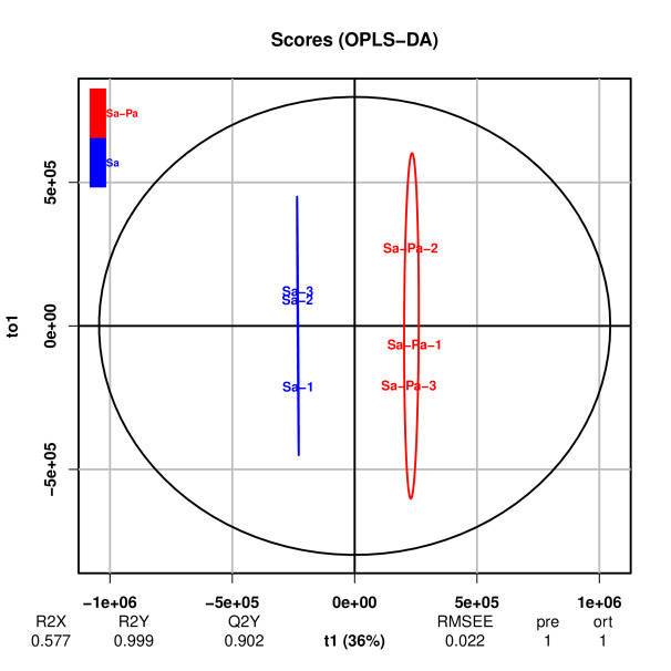
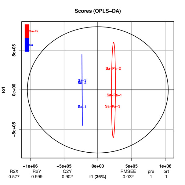
OPLS-DA was performed to further discriminate the metabolic profiles between the control and treatment groups (Figure 2). The OPLS-DA model showed a separation between the control and treatment groups. The three biological replicates within each group clustered tightly, demonstrating good reproducibility. However, given the small sample size (n = 3 per group), the OPLS-DA results should be interpreted with caution.
Screening and identification of differential metabolites
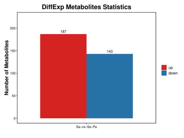
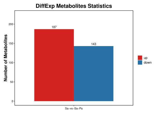
A total of 23,541 features were detected across both positive and negative ESI modes. Using the criteria of VIP ≥ 1.0 and T-test P < 0.05, 330 differential metabolites were identified between the control and treatment groups (Figure 3), including 187 up-regulated and 143 down-regulated metabolites. The slightly higher number of up-regulated metabolites may suggest that the treatment induced both metabolic inhibition and stress responses in S. agalactiae.
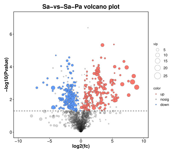
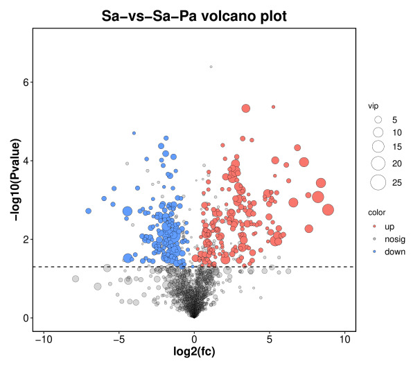
The volcano plot (Figure 4) displayed the distribution of these metabolites, with the x-axis representing log₂(fold change) and the y-axis representing –log₁₀(P-value). The metabolic profile of S. agalactiae was extensively altered, with fold changes ranging from –6 to +6 and the highest significance at –log₁₀(P) = 3.8 (P ≈ 1.6 × 10⁻⁴). Down-regulated metabolites (left) showed larger changes and greater significance, suggesting stronger inhibitory effects, whereas up-regulated metabolites (right) were more numerous but less significant, possibly reflecting bacterial stress responses.
VIP analysis
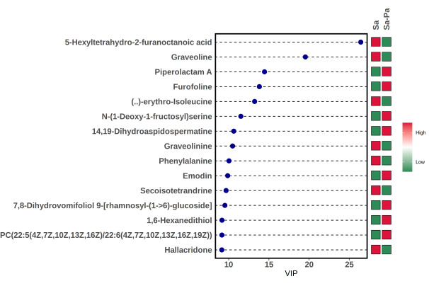
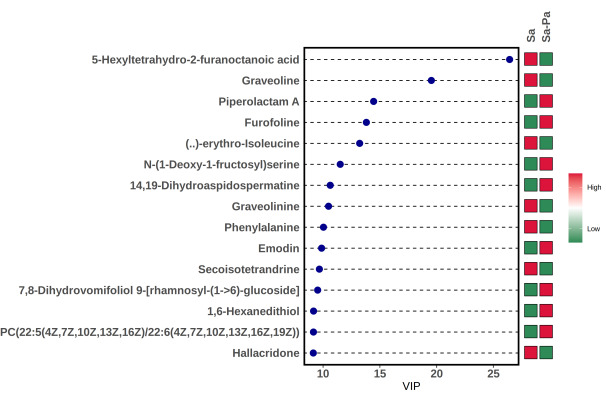
The top 15 metabolites contributing to the group separation are shown in the VIP bar plot (Figure 5). The highest VIP scores were observed for 5-hexyltetrahydro-2-furanoctanoic acid (VIP = 22), graveoline (VIP = 18), piperolactam A (VIP = 16), erythro-isoleucine (VIP = 14), phenylalanine (VIP = 10), and emodin (VIP = 9). Based on MS/MS spectral matching and database comparison, graveoline, piperolactam A, and emodin were tentatively identified and detected at higher levels in the treatment group, while isoleucine and phenylalanine were detected at lower levels.
Metabolite set enrichment analysis (MSEA) of differential metabolites
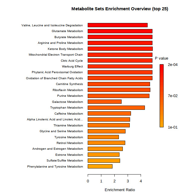
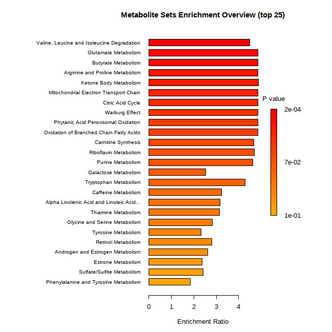
To further explore the biological functions of the identified differential metabolites, metabolite set enrichment analysis (MSEA) was performed, and the results are presented as a clustered bar chart (Figure 6). As shown in Figure 6, the differential metabolites were significantly enriched in multiple metabolic pathways, including valine, leucine and isoleucine degradation (P = 2.0 × 10⁻⁴), glutamate metabolism, butyrate metabolism, arginine and proline metabolism, ketone body metabolism, the electron transport chain (ETC), and the tricarboxylic acid (TCA) cycle. These pathways were primarily associated with branched-chain amino acid (BCAA) catabolism, core energy metabolism (TCA cycle and butyrate oxidation), and nitrogen metabolism. In addition, purine metabolism, riboflavin metabolism, and tryptophan metabolism exhibited moderate enrichment trends, although they did not reach statistical significance (P = 0.07). In contrast, pathways such as α-linolenic acid metabolism and glycine/serine metabolism showed no significant enrichment (P > 0.05).
Discussion
In this study, 35 Chinese herbal medicines were screened against S. agalactiae. Cortex Moutan, Folium Sennae, and Fructus Foeniculi showed inhibitory effects, with Cortex Moutan exhibiting the strongest activity.
Among the 35 herbal extracts tested, only three showed detectable activity against S. agalactiae, with Cortex Moutan being the most effective. This low hit rate contrasts with some previous screening studies and may reflect differences in extraction methods, bacterial strains, and testing conditions. In particular, the aqueous decoction used here may have extracted fewer lipophilic antibacterial compounds than organic solvents, which could explain why several herbs previously reported to be active against S. agalactiae showed no activity in our assay.
Cortex Moutan contains paeonol and paeoniflorin as its major active components, which exhibit antibacterial, anti-inflammatory, and antioxidant activities.11However, its activity against S. agalactiae has not been previously reported. To the best of our knowledge, based on a comprehensive literature search of PubMed, Web of Science, and CNKI, this is the first report of the antibacterial activity of Cortex Moutan specifically against S. agalactiae. Folium Sennae contains anthraquinones such as sennosides and emodin, while Fructus Foeniculi contains volatile oils such as anethole and anisaldehyde.12,13Studies on the application of these two herbs against aquatic pathogens remain limited, and the present study expands their potential use in aquaculture.
Notably, most of the tested herbal extracts (32 out of 35) showed no activity against S. agalactiae, which contrasts with previous findings. For example, Wang reported that Sappan wood and Phellodendron chinense exhibited good antibacterial activity against S. agalactiae.3 Several factors may explain this discrepancy. First, strain variation may play a role, as different S. agalactiae isolates may have distinct susceptibility profiles.14 Second, the discrepancy with Wang et al. may be partly explained by the extraction method. Organic solvents are generally more efficient than water at extracting lipophilic antibacterial compounds such as alkaloids and terpenoids, whereas the aqueous decoction used here mainly extracts polar compounds.15 Third, the concentration of the herbal extracts (1 g/mL) may be suboptimal for some herbs.16 Fourth, differences in culture conditions, such as medium, pH, and temperature, may also influence the results.17
Based on MS/MS spectral matching and database comparison, graveoline (VIP = 18), piperolactam A (VIP = 16), and emodin (VIP = 9) were tentatively identified and detected at higher levels in the treatment group. Among these, emodin has been reported to possess anti-Gram-positive bacterial activity by disrupting bacterial cell membranes and inhibiting nucleic acid synthesis.11,18Additionally, graveoline exhibits a broad spectrum of biological activities, including antibacterial and anti-inflammatory effects. These compounds may be derived from the Cortex Moutan aqueous extract; however, it remains unclear whether they were taken up by bacterial cells or simply remained associated with the extracellular extract.19 Furthermore, 5-hexyltetrahydro-2-furanoctanoic acid (VIP = 22) was identified as the metabolite with the highest VIP score among all differential metabolites. However, its biological function remains largely unclear. It is hypothesized that this metabolite may be associated with bacterial fatty acid metabolism or stress responses, warranting further investigation.
On the other hand, bacterial metabolism was significantly disrupted. Isoleucine (VIP = 14) and phenylalanine (VIP = 10) were down-regulated in the treatment group. BCAAs and aromatic amino acids are essential for protein synthesis and serve as TCA cycle substrates. MSEA showed significant enrichment of TCA cycle, ETC, and valine/leucine/isoleucine degradation (P = 2.0 × 10⁻⁴). Succinate, a TCA cycle intermediate, was also down-regulated. These results may suggest that Cortex Moutan inhibits bacterial growth by disrupting energy metabolism and amino acid synthesis. However, these interpretations are speculative and require further validation.
In conclusion, Cortex Moutan, Folium Sennae, and Fructus Foeniculi exhibited in vitro antibacterial activity against S. agalactiae, with Cortex Moutan being the most effective. Integrative metabolomics and VIP analyses may suggest that its antibacterial action arises from both the activity of its bioactive metabolites and the suppression of bacterial central carbon and amino acid metabolism. However, these interpretations require further validation. These findings provide preliminary mechanistic insights for exploring plant-derived alternatives to conventional antibiotics in aquaculture. Future studies should extend this work in several directions: testing multiple S. agalactiae isolates, increasing the metabolomics sample size, confirming compound identities with authentic standards, including an extract-only control and intracellular quantification, and evaluating the cytotoxicity and safety of Cortex Moutan extracts in tilapia host tissues.
Acknowledgments
This study was supported by the Basic Ability Improvement Project for Young and Middle-aged Teachers in Guangxi Universities (Grant No. 2025KY2146).
CRediT authorship contribution
Conceptualization: Mengqi Wang (Lead). Methodology: Mengqi Wang (Equal), Yuanyuan Lin (Equal). Writing – original draft: Mengqi Wang (Lead). Writing – review & editing: Shengyu Zhang (Lead). Investigation: Fangmei Liang (Equal), Xinwen Nong (Lead). Resources: Lianhui Pang (Lead). Funding acquisition: Chunlan Lu (Lead). Project administration: Chunlan Lu (Lead). Supervision: Chunlan Lu (Lead).
Declaration of competing interests
The authors declare no conflict of interest.
Ethical Conduct Approval
All procedures were carried out in accordance with the Institutional Animal Care and Use Committee Guide at Guangxi Vocational and Technical College of Agricultural Engineering.
Informed Consent Statement
All authors and institutions have confirmed this manuscript for publication.
Data Availability Statement
All data generated or used during the study appear in the submitted article.