
1. Introduction
Seawater pH can be altered by natural and anthropogenic drivers, and low-pH conditions can impose substantial physiological challenges on marine organisms.1 Seawater pH fluctuations have been recognized as important environmental stressors affecting marine ecosystems.2 The reduction in seawater pH not only alters the calcium carbonate saturation state of seawater,3 but also directly interferes with critical physiological processes in marine organisms, including acid-base regulation,4 ion transport,5 respiratory function,6 energy metabolism, and immune defense,7,8 thereby affecting their growth, development, reproduction, and population dynamics.9 Among marine vertebrates, fish are particularly dependent on precise regulation of internal homeostasis and may therefore exhibit complex physiological responses to low-pH stress.
Previous studies have shown that many fish possess considerable capacity to regulate acid–base balance; however, such tolerance does not necessarily indicate the absence of physiological costs.6,10 A number of evidence indicates that both chronic and acute low-pH stress can induce oxidative imbalance, inflammatory responses, and immune dysregulation in fish.11 Particularly during the juvenile stage, when physiological regulatory systems are still undergoing development, fish exhibit heightened sensitivity to environmental changes, and the impacts of low-pH stress may be more profound and far-reaching.12 Therefore, relying solely on adults may overlook sublethal physiological impacts of low-pH stress.
Yellowfin tuna (Thunnus albacares), as a pelagic migratory fish, holds significant ecological and economic importance in tropical and subtropical waters.13,14 However, due to their distinctive migratory habits and sensitivity to changes in the marine environment, juvenile yellowfin tuna may be particularly vulnerable to environmental fluctuations due to their active metabolism and developmental requirements.15 Previous studies have demonstrated that low-pH stress can induce oxidative stress and inflammatory responses in fish by lowering blood pH and interfering with ion transport and acid-base regulation, affecting physiological processes in these tissues.16–18 As the primary respiratory and ion-exchange organ in fish, the gill serves as the first point of contact with environmental changes.5 The liver is the primary metabolic and detoxification organ, and its functional status influences energy metabolism and immune homeostasis in fish.19 In the context of global climate change, both wild populations and aquaculture systems of yellowfin tuna may face potential challenges from future changes in seawater chemistry.20 However, molecular responses of tuna species to low-pH stress remain poorly understood.
The antioxidant defense system serves as a critical barrier for maintaining cellular homeostasis.21 Low-pH stress may disturb redox homeostasis and alter reactive oxygen species (ROS) production in fish. If the antioxidant system fails to scavenge excessive ROS in a timely manner, it will result in lipid peroxidation and protein damage.22 Among antioxidant-related genes, superoxide dismutase (sod), catalase (cat), and glutathione peroxidase (gpx1a) are widely used molecular markers reflecting transcriptional regulation of antioxidant defenses under environmental stress.23–25 In fish coping with low-pH stress, the expression levels of antioxidant genes provide molecular indicators of antioxidant-related transcriptional responses in tissues.26 Because the gill is directly exposed to external water conditions, whereas the liver plays a central role in metabolism and detoxification, these tissues may exhibit distinct antioxidant responses during low-pH stress. We hypothesized that the gill would show increased expression of antioxidant-related genes under low-pH exposure, whereas the liver might exhibit a different or comparatively reduced transcriptional response.
Inflammatory and immune responses are key physiological processes that reflect immune modulation and the maintenance of immune homeostasis in organisms.27 Toll-like receptor 3 (tlr3) is a pattern recognition receptor involved in innate immune signaling, while cytokines such as interleukin-1β (il-1β) and tumor necrosis factor α (tnf-α) are commonly used indicators of pro-inflammatory response.28,29 Interleukin-6 receptor (il6r) participates in cytokine-mediated signaling pathways, whereas interleukin-10 (il10) and transforming growth factor β (tgf-β) contribute to the regulation of inflammatory processes.30,31 We hypothesized that the gills would show increased expression of pro-inflammatory genes in response to direct environmental exposure, whereas the liver might exhibit a distinct or comparatively reduced immune-related transcriptional response. Changes in the expression of these immune-related genes can provide insights into tissue-specific immune modulation during low-pH stress.
Energy metabolism also represents a critical component of the stress response, as fish may adjust metabolic pathways to maintain physiological homeostasis under environmental challenges.32 Malate dehydrogenase (mdh2) participates in the tricarboxylic acid (TCA) cycle and is associated with aerobic metabolic processes, whereas phosphoenolpyruvate carboxykinase (pck2) regulates gluconeogenesis and may contribute to metabolic adaptation under stress conditions.33,34 Diacylglycerol acyltransferase (dgat1b) is involved in triglyceride synthesis and lipid metabolism, and is closely linked to energy balance and cellular maintenance.35 Given the central role of the gill in ion and acid–base regulation, we anticipated increased expression of metabolism-related genes in the gill under low-pH exposure. In contrast, the liver, as a major metabolic organ, was expected to exhibit a distinct and potentially selective transcriptional response in metabolism-related genes. Accordingly, the expression patterns of these metabolic genes in the liver and gills may inform tissue-specific metabolic regulation under low-pH stress.36
Currently, studies on the physiological responses of fish to low-pH stress have predominantly focused on model species and coastal fishes, whereas molecular response data for economically important pelagic species, such as juvenile yellowfin tuna (Thunnus albacares), remain limited. To address this knowledge gap, we conducted a 48-h exposure experiment using wild juvenile yellowfin tuna collected from the South China Sea, with four pH treatments (8.1, 7.6, 7.1, and 6.6). The expression patterns of the aforementioned genes involved in antioxidative defense, inflammatory responses, and energy metabolism were examined in liver and gill tissues to test our tissue-specific prior hypotheses. By comparing transcriptional responses between these two tissues under acute low-pH stress, this study aims to elucidate the distinct physiological strategies employed by high-metabolic-rate pelagic fish in response to abrupt environmental pH fluctuations, thereby providing a theoretical foundation for assessing molecular sensitivity and guiding future investigations into long-term exposure effects.
2. Materials and methods
2.1. Experimental Animals, Acclimation, and Experimental Design
Juvenile yellowfin tuna used in this study were collected in June 2023 from the waters near Xincun Harbor in the South China Sea (18°22′31.9″N, 109°58′28.9″E), with the assistance of Sanya Tropical Fisheries Research Institute. A total of 72 healthy juveniles with intact body surfaces and active swimming behavior were selected, with a mean body length of 18.21 ± 1.09 cm and a mean body weight of 307.49 ± 49.38 g. The juveniles were first acclimated for 7 days in circular tanks (5000 L) located within the workshop of the Tropical Aquaculture Research and Development Center. During the acclimation period, the fish were fed once daily at 8:00 a.m. to satiety with ice-preserved trash fish. Water quality parameters during both the acclimation and formal experimental periods were maintained as follows: water temperature at 28.5 ± 1 °C, dissolved oxygen > 7.5 mg/L, sodium nitrite < 0.05 mg/L, and ammonia nitrogen below the detection limit (< 0.01 mg/L). The experimental seawater used was sand-filtered natural seawater. After acclimation, the juveniles were randomly assigned to four experimental groups, each with three replicates and six fish per replicate, for a total of 72 individuals. Natural seawater at pH 8.1 was designated as the control group. Three low-pH treatment groups were established with pH values of 7.6, 7.1, and 6.6 to cover a range from moderate to severe low-pH conditions.37,38 The experiment lasted for 48 h, during which the fish were not fed, and the photoperiod was maintained at 12 h light:12 h dark cycle. Seawater pH was continuously monitored throughout the experimental period using a pH meter (Orion Star A211, Thermo Scientific, USA). The instrument was calibrated according to the NBS scale, and standardization was performed every 2 h using commercial buffer solutions (pH 4.01, 7.00, and 10.01). The pH of each treatment group was maintained within ± 0.1 by adding 1.0 mmol/L NaOH or 1.0 mmol/L HCl solutions.
It is worth noting that HCl/NaOH was used to adjust seawater pH instead of CO₂ bubbling, as the core objective of the experimental design was to evaluate the physiological and transcriptional effects of experimentally induced low-pH conditions on juvenile yellowfin tuna, rather than to investigate changes in the full carbonate chemistry system. Therefore, mineral acid/base adjustment provided a direct and stable means of achieving the target pH gradients, which was advantageous for isolating the biological responses associated with experimentally induced low-pH conditions.39
2.2. Sample Collection
At the end of the 48-h exposure, all six juvenile yellowfin tuna in each tank were sampled for gene expression analysis. The mean expression value of the six fish was used as the representative value for that tank in statistical analyses, with the tank serving as the statistical unit (n = 3). Prior to sampling, the juveniles were euthanized with an overdose of buffered MS-222 at 250 mg/L buffered with sodium bicarbonate. After a complete loss of motor activity and cessation of opercular movement, death was confirmed, and the fish were immediately dissected.40 All six fish in each tank were sampled at the end of the 48-h exposure, and both liver and gill tissues were collected from the same individuals for gene expression analysis. The tissues were then placed into 2 mL cryogenic vials and stored at −80°C until further use. The entire sampling procedure was performed on ice to minimize tissue degradation and exogenous contamination, thereby reducing potential interference with subsequent analytical results.
2.3. Total RNA Extraction and cDNA Synthesis
Total RNA was extracted from the liver and gill tissues using Trizol reagent (Lan Jieke Technology Co., Ltd., Anhui, China) according to the manufacturer’s instructions. RNA concentration and purity were determined using a Nano-300 spectrophotometer (Allsheng Instruments Co., Ltd., Hangzhou, China), and RNA integrity was evaluated by agarose gel electrophoresis (DYY-6C, Beijing Liuyi Instrument Factory, Beijing, China). Only RNA samples with A260/A280 ratios between 1.8 and 2.0 and no apparent degradation were used for subsequent analyses. A total of 1 μg of total RNA was used for first-strand cDNA synthesis following the protocol of the EasyScript® All-in-One First-Strand cDNA Synthesis SuperMix (TransGen Biotechnology Co., Ltd., Beijing). Each 20 μL reaction contained 1 μg total RNA, 4 μL 5× EasyScript® All-in-One SuperMix for qPCR, 1 μL gDNA Remover, and RNase-free water, and the reaction conditions were as follows: 37 °C for 15 min, followed by 85 °C for 5 s. The synthesized cDNA was stored at −20°C for subsequent use.
2.4. Gene Expression Analysis
Relative gene expression was quantified using real-time quantitative PCR (qTOWER,3 Analytik Jena GmbH, Germany) with SYBR Green fluorescent dye (Tiangen Biotech Co., Ltd., Beijing). Based on the gene sequence information of yellowfin tuna available in the NCBI database, specific primers for each target gene and the reference gene were designed using Primer Premier 5.0 software,41 and their specificity was validated by BLAST alignment against the yellowfin tuna genome. The primer sequences are listed in Table 1. Each 20 μL reaction contained 10 μL of 2 × RealUniversal PreMix, 0.6 μL of each primer, 2 μL of cDNA template, and 6.8 μL of ddH₂O. The amplification program was set as follows: initial denaturation at 95 °C for 15 min, followed by 40 cycles of denaturation at 95 °C for 10 s, annealing at the primer-specific temperatures listed in Table 1 for 20 s, and extension at 72 °C for 30 s. Following amplification, melting curve analysis was performed to evaluate product specificity, and the results showed that all primers yielded a single melting peak, with no non-specific amplification or primer-dimer signals detected. Primer amplification efficiency was validated by the standard curve method: serially diluted cDNA templates were used to generate linear regression between Ct values and logarithmic template concentrations, and the efficiency was calculated according to the formula E = [10(-1/slope)-1] × 100%. The results indicated that the amplification efficiencies of all primers ranged from 90% to 110%, with correlation coefficients (R2) all exceeding 0.98. The stability of β-actin expression was evaluated separately in the liver and gill across the four pH treatments. Because β-actin Ct values showed low variation and no significant differences among treatments, β-actin was used as the reference gene. Each cDNA sample was analyzed in triplicate, and the mean Ct value of the three technical replicates was used for subsequent analysis. The relative mRNA expression levels of target genes were calculated using the 2-ΔΔCt method, with the expression level of the pH 8.1 group used as the calibrator.
2.5. Statistical Analysis
Experimental data were organized using Excel 2010 software, and statistical analyses were performed with SPSS 25.0 software. For all detected genes (including antioxidant-, immune-, inflammatory-, and metabolism-related genes), the tank was used as the statistical unit (n = 3), and the mean value of the six fish sampled from each tank was used as the representative value for that tank. This procedure was applied consistently for both liver and gill tissues. Normality of the data was assessed using the Shapiro-Wilk test, and homogeneity of variances was evaluated using Levene’s test. One-way analysis of variance (one-way ANOVA) followed by Duncan’s multiple range test was employed to determine significant differences among treatments. Data are presented as mean ± standard deviation (Mean ± SD). A significance threshold of p < 0.05 was considered statistically significant. Bar graphs were generated using GraphPad Prism 9.5 software.
3. Results
3.1. Antioxidant-Related Gene Expression in the Liver and Gill
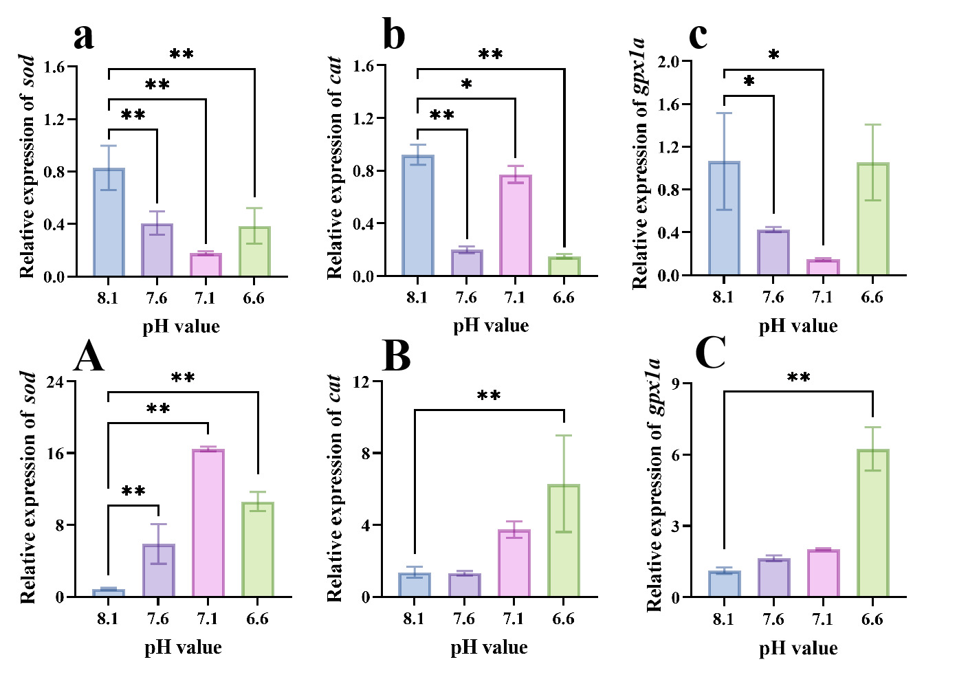
The mRNA expression levels of antioxidant-related genes exhibited distinct patterns between the liver and gill (Fig. 1). In the liver, sod expression was significantly lower in the pH 7.6, 7.1, and 6.6 groups than in the pH 8.1 control group (p < 0.01; Fig. 1a). Hepatic cat expression was significantly lower at pH 7.6 and 6.6 (p < 0.01) and at pH 7.1 (p < 0.05; Fig. 1b). The expression of gpx1a was significantly lower at pH 7.6 and 7.1 (p < 0.05), whereas no significant difference was detected at pH 6.6 (p > 0.05; Fig. 1c).
.png)
In the gill, sod expression was significantly higher in all three low-pH groups than in the pH 8.1 control group (p < 0.01; Fig. 1A). The expression levels of cat and gpx1a were significantly higher at pH 6.6 (p < 0.01), whereas no significant differences were detected at pH 7.6 or 7.1 (p > 0.05; Fig. 1B, C).
3.2. Immune- and Inflammation-Related Gene Expression in the Liver and Gill
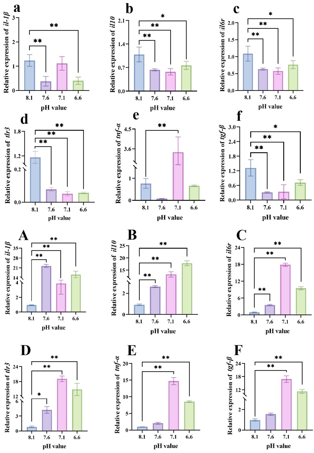
The mRNA expression levels of immune- and inflammatory-related genes differed between the liver and gill tissues (Fig. 2). In the liver, il-1β expression was significantly downregulated in the pH 7.6 and pH 6.6 groups compared with the control group (p < 0.01), whereas no significant difference was observed at pH 7.1 (p > 0.05; Fig. 2a). The expression levels of il10, il6r, and tgf-β were significantly decreased at pH 7.6 and pH 7.1 (p < 0.01), as well as at pH 6.6 (p < 0.05; Fig. 2b, c, f). Hepatic tnf-α expression was significantly upregulated at pH 7.1 (p < 0.01), but showed no significant changes at pH 7.6 or pH 6.6 relative to the control (p > 0.05; Fig. 2e). The expression of tlr3 was significantly reduced across all low-pH treatment groups (p < 0.01; Fig. 2d).
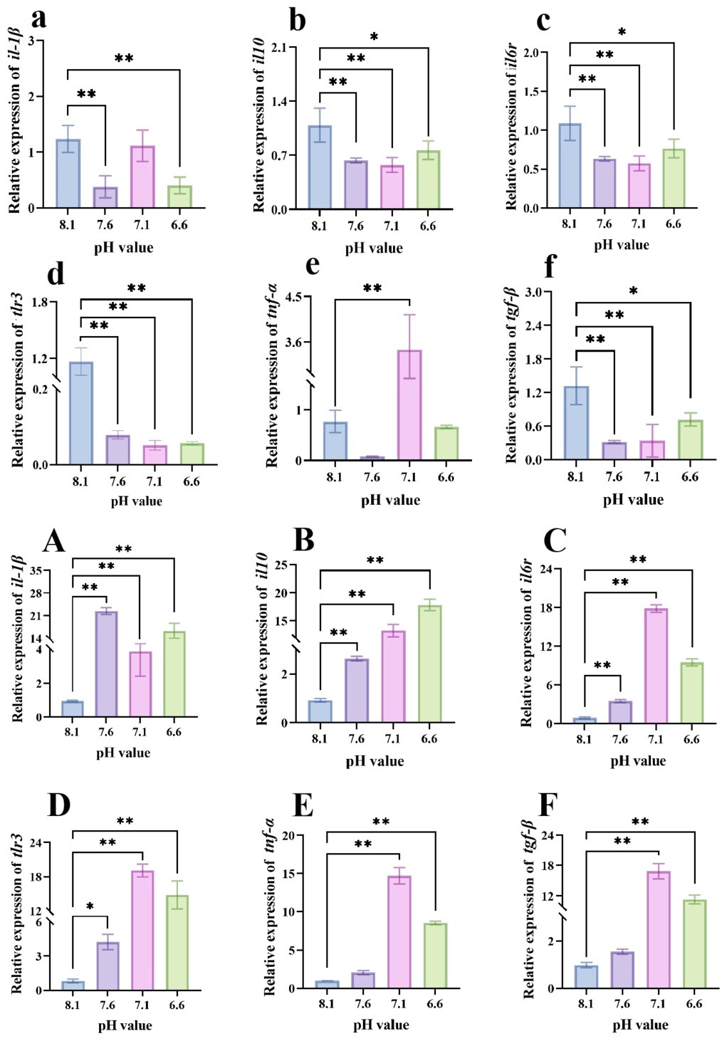
In the gill, the expression levels of il-1β, il10, and il6r were significantly elevated in all low-pH treatment groups compared with the control group (p < 0.01; Fig. 2A–C). tlr3 expression was significantly upregulated at pH 7.1 and pH 6.6 (p < 0.01), as well as at pH 7.6 (p < 0.05), relative to the control (Fig. 2D). The expression levels of tnf-α and tgf-β were significantly higher at pH 7.1 and pH 6.6 than in the control group (p < 0.01), whereas no significant differences were observed at pH 7.6 (p > 0.05; Fig. 2E, F).
3.3. Metabolic Gene Expression in the Liver and Gill
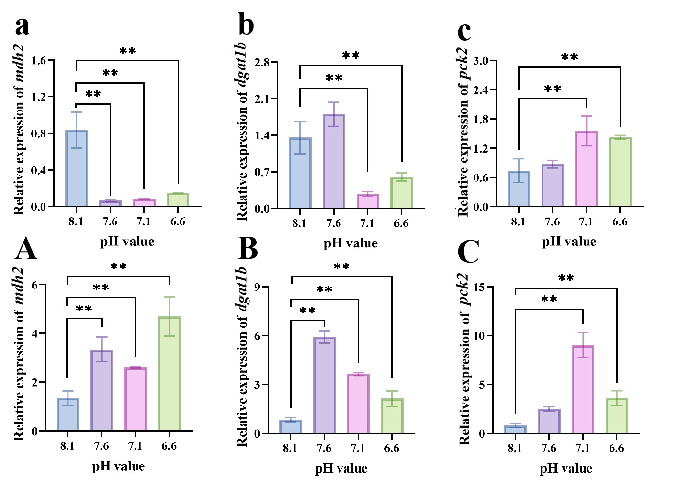
The transcriptional expression levels of metabolism-related genes differed between the liver and gill tissues under different pH treatments (Fig. 3). In the liver, mdh2 expression was significantly lower in all low-pH groups than in the control group (p < 0.01; Fig. 3a). The expression of dgat1b was significantly decreased at pH 7.1 and pH 6.6 (p < 0.01), whereas no significant difference was detected at pH 7.6 compared with the control (p > 0.05; Fig. 3b). pck2 expression was significantly increased at pH 7.1 and pH 6.6 (p < 0.01), but showed no significant change at pH 7.6 relative to the control (p > 0.05; Fig. 3c).
_l.png)
In the gill, the expression levels of mdh2 and dgat1b were significantly upregulated in all low-pH groups (p < 0.01; Fig. 3A, B). pck2 expression was significantly increased at pH 7.1 and pH 6.6 (p < 0.01), whereas no significant difference was detected at pH 7.6 (p > 0.05; Fig. 3C).
4. Discussion
In this study, we characterized the transcriptional responses of antioxidant-, immune-, inflammatory-, and metabolism-related genes in the liver and gill tissues of juvenile yellowfin tuna following short-term (48 h) exposure to different pH levels (8.1, 7.6, 7.1, and 6.6). The results revealed distinct tissue-specific response patterns under acute low-pH exposure. The gill generally showed increased expression of antioxidant-, immune-, and metabolism-related genes, whereas the liver exhibited predominantly reduced expression of antioxidant and immune-related genes with selective changes in metabolic genes. These contrasting expression patterns suggest tissue-specific regulation in juvenile yellowfin tuna when responding to short-term environmental pH fluctuations.
The antioxidant defense system is an important component that maintains cellular redox balance under environmental stress. In the present study, the expression levels of sod, cat, and gpx1a in the gill were generally increased under low-pH exposure, with the strongest responses observed at pH 6.6. As a mucosal tissue directly exposed to surrounding seawater, the gill is likely to be the first organ sensing environmental pH fluctuations and may require rapid transcriptional regulation of antioxidant-related genes to maintain cellular homeostasis.5,42 Similar tissue-specific responses have been reported in large yellow croaker (Larimichthys crocea), where low-pH exposure resulted in pronounced transcriptional changes in gill tissues and highlighted the importance of gill-mediated regulation during environmental stress.43 In contrast to the gill, hepatic expression levels of sod, cat, and gpx1a were significantly downregulated under low-pH treatments, with the exception of gpx1a at pH 6.6, which showed no significant difference compared with the control. This tissue-specific difference suggests that the liver and gill may employ distinct transcriptional responses during acute pH stress. Under environmental stress, organisms may adjust resource allocation among different physiological processes to maintain essential functions, although the specific metabolic costs and trade-offs in juvenile yellowfin tuna require further investigation.36 Similar reductions in hepatic antioxidant-related responses have been reported in juvenile zebra seabream (Diplodus cervinus), where hepatic CAT activity decreased under acidic conditions.44
Immune and inflammatory responses are important mechanisms for responding to environmental disturbances and maintaining physiological homeostasis. In the gill tissue, several immune- and inflammatory-related genes showed increased expression under low-pH exposure. The upregulation of the pro-inflammatory cytokines il-1β and tnf-α suggests enhanced inflammatory-related transcriptional responses, while the concurrent increase in il10 and tgf-β may indicate activation of regulatory mechanisms that modulate immune responses.45 Proteomic studies on Atlantic halibut (Hippoglossus hippoglossus) also reported changes in immune-related proteins, including complement component C3 and fibrinogen, suggesting that low-pH stress can influence systemic immune responses.46 In contrast, hepatic immune-related genes exhibited distinct expression patterns: with the exception of an increase in tnf-α at pH 7.1, the expression of il10, il6r, tlr3, and tgf-β was significantly downregulated across all low-pH treatments, and il-1β was also significantly reduced at pH 7.6 and 6.6. These results indicate that acute low-pH stress induces alterations in hepatic immune-related transcriptional regulation. These results are comparable with a transgenerational low-pH study in European sea bass (Dicentrarchus labrax), where low-pH exposure affected hepatic transcripts involved in immune response, carbohydrate metabolism, and cellular homeostasis.17 Differences between acute exposure in juvenile yellowfin tuna and transgenerational exposure in European sea bass indicate that exposure duration may influence hepatic transcriptional responses to low-pH stress.
The tricarboxylic acid (TCA) cycle, gluconeogenesis, and lipid metabolism are important pathways that maintain cellular energy homeostasis.47 In this study, changes in hepatic metabolic gene expression indicated tissue-specific transcriptional regulation of metabolic processes under acute low-pH exposure. The reduced expression of mdh2 together with increased pck2 expression may indicate altered transcriptional regulation of TCA cycle-related metabolism and gluconeogenesis-associated processes in the liver, rather than directly reflecting changes in metabolic flux. These transcriptional changes suggest that hepatic metabolism may undergo regulatory adjustments at the transcriptional level under acute low-pH stress.48 In contrast, the increased expression of mdh2, pck2, and dgat1b in the gill suggests enhanced transcriptional regulation of multiple metabolic pathways under low-pH exposure. As an organ directly involved in ion exchange and acid-base regulation, the gill may require coordinated transcriptional responses to support physiological regulation during environmental pH fluctuations.49 The increased expression of mdh2 may be associated with elevated transcriptional demands related to mitochondrial processes and ion regulation, whereas increased pck2 expression may help meet cellular metabolic demands at the transcriptional level under stress conditions 46,.50
Notably, several genes exhibited non-monotonic expression patterns across the tested pH levels, with expression increasing at pH 7.6 or 7.1 and subsequently decreasing at pH 6.6. This pattern suggests that transcriptional responses to acute low-pH exposure may not vary linearly with decreasing pH. One possible explanation is that moderate and severe low-pH conditions elicit different magnitudes or phases of the stress response, as stress-related energetic and regulatory processes in fishes can vary with stress intensity and physiological state.51 Similar variability in responses across pH levels has also been reported in other fishes exposed to low-pH conditions.52–54 However, because the present study assessed only a single 48-h endpoint and measured mRNA abundance alone, it remains unclear whether the decline observed at pH 6.6 represents reduced compensatory capacity, temporal differences in gene regulation, or other mechanisms. Studies in other marine fishes further indicate that sensitivity to severe low-pH exposure can vary substantially among species.54,55 Therefore, time-course experiments integrating physiological, biochemical, and protein-level measurements are needed to clarify the biological basis of these non-monotonic transcriptional responses.
It should be noted that seawater pH in this study was adjusted using HCl/NaOH rather than through CO₂ equilibration. Therefore, the observed responses primarily reflect the biological effects of low pH per se (i.e., elevated proton concentrations), rather than the full suite of carbonate chemistry changes associated with ocean acidification. Accordingly, the conclusions of this study should be interpreted strictly within the context of acute low-pH stress.56 In addition, as the present study was conducted at a single 48 h endpoint, it is not possible to distinguish transient from sustained responses. Moreover, mRNA abundance is not equivalent to protein levels or enzymatic activities; therefore, the observed changes in gene expression should be interpreted as transcriptional-level responses and as evidence for hypothesis generation.57 In addition, each treatment group consisted of only three replicate tanks (n = 3), with the average value of six fish per tank used as the representative measurement. Due to the difficulty and high cost of obtaining juvenile yellowfin tuna, the sample size was relatively small, which may have limited the statistical power to detect subtle differences. Furthermore, the Shapiro–Wilk normality test has limited ability to effectively detect deviations from normality with such a small sample size.58 The transition from feeding during acclimation to fasting during the exposure period may constitute a nutritional confound that could influence hepatic metabolic gene expression. Future studies should integrate CO₂-driven acidification experiments, time-course sampling, protein-level analyses (e.g., Western blotting or enzyme activity assays), larger sample sizes, and extended exposure durations, as well as employ more complex statistical models (such as two-way ANOVA or regression analysis), to further disentangle the specific effects of pH from other carbonate system variables, to validate the functional consequences of these transcriptional changes, and to gain deeper insights into the non-monotonic response patterns observed in this study.
Integrating the responses of antioxidant-, immune-, and metabolism-related genes, this study highlights contrasting tissue-specific transcriptional responses of juvenile yellowfin tuna under acute low-pH exposure. The gill generally showed increased expression of genes involved in antioxidant defense, immune regulation, and metabolism, whereas the liver exhibited reduced expression of many antioxidant- and immune-related genes accompanied by selective changes in metabolic gene expression. These differences suggest that distinct tissues may regulate their transcriptional responses in accordance with their physiological roles during acute environmental stress. Such tissue-specific regulation may reflect the differential allocation of cellular resources among physiological processes under stress conditions.56 This study provides molecular insights into the response characteristics of juvenile yellowfin tuna, an economically important pelagic fish species, under acute low-pH exposure.
5. Conclusions
By examining the expression responses of antioxidant-, immune-, inflammatory-, and metabolism-related genes in the liver and gill tissues of juvenile yellowfin tuna exposed to different pH levels (8.1, 7.6, 7.1, and 6.6), this study characterized tissue-specific molecular responses under acute low-pH stress. The gill generally exhibited increased expression of genes associated with antioxidant defense, immune regulation, and metabolism, whereas the liver showed reduced expression of several antioxidant- and immune-related genes accompanied by selective changes in metabolic gene expression. These contrasting transcriptional patterns highlight distinct regulatory responses between tissues during short-term pH stress. This study provides molecular insights into the response characteristics of juvenile yellowfin tuna, an economically important pelagic fish species, under acute low-pH exposure. Future studies integrating long-term exposure experiments with multi-omics, physiological measurements, and histopathological analyses are needed to further clarify the functional consequences of these transcriptional responses and their implications for aquaculture management.
Acknowledgement
This work was supported by the Ministry of Agriculture and Rural Affairs of the People’s Republic of China Science and Technology Project Funding, the Central Public interest Scientific Institution Basal Research Fund; CAFS (2023TD58, 2027TD604, 2025XT03, 2026TS01); the 2025 Research on breeding technology of candidate species for Guangdong modern marine ranching (2025-MRB-00-001); the Hainan Province Science and Technology Special Fund (ZDYF2024XDNY247); the National Natural Science Foundation of China (32460927) and the Guangdong Province Overseas Distinguished Scholars Program (MS202500183).
CRediT Authorship Contribution Statement
Investigation: Yitao Feng (Equal), Yuzhuo You (Equal), Xiaoyan Wang (Equal). Data curation: Yitao Feng (Equal), Yuzhuo You (Equal). Formal Analysis: Yitao Feng (Equal), Zhengyi Fu (Equal). Visualization: Yitao Feng (Equal), Zhengyi Fu (Equal). Writing – original draft: Yitao Feng (Equal), Zhengyi Fu (Equal). Methodology: Yuzhuo You (Equal), Xiaoyan Wang (Equal), Zhengyi Fu (Equal). Validation: Yuzhuo You (Equal), Xiaoyan Wang (Equal). Resources: Xiaoyan Wang (Equal), Zhenhua Ma (Equal). Conceptualization: Zhengyi Fu (Equal), Zhenhua Ma (Equal). Supervision: Zhengyi Fu (Equal), Zhenhua Ma (Equal). Project administration: Zhengyi Fu (Equal), Zhenhua Ma (Equal). Writing – review & editing: Zhengyi Fu (Equal), Zhenhua Ma (Equal). Funding acquisition: Zhenhua Ma (Equal).
Ethical Approval
The animal study was reviewed and approved by the Animal Care and Use Committee of South China Sea Fisheries Research Institute, Chinese Academy of Fishery Sciences. The ethical code is 2020TD55, which was approved on 5 January 2020.
Conflicts of Interest
No competing interests were disclosed.
Data Availability Statement
The original contributions presented in the study are included in the article. Further inquiries can be directed to the corresponding authors.