1. Introduction
Largemouth bass virus (LMBV), also known as largemouth bass ranavirus, belongs to the genus Ranavirus within the family Iridoviridae.1 It is a large, enveloped icosahedral double-stranded DNA virus with virions approximately 150 nm in diameter. Its genome ranges from 100 to 170 kilobase pairs (kbp) and encodes roughly 90 open reading frames (ORFs).2 Studies have confirmed that it can infect other freshwater fish species, including koi carp (Cyprinus carpio), mandarin fish (Siniperca chuatsi), smallmouth bass (Micropterus dolomieu), and largemouth bass (Micropterus salmoides).3 Infection with LMBV is characterized by high morbidity and high mortality.4
In recent years, viral diseases have frequently occurred in farmed fish, and the antiviral mechanisms of herbal medicines have become a research hotspot. For instance, the aqueous extract and fermented products of Abrus cantoniensis can effectively alleviate LMBV-induced damage to the intestine and liver of mandarin fish via immunomodulatory and antioxidant effects.5 As for Houttuynia cordata, its antiviral activity also involves complicated signaling regulation. Its bioactive components, including houttuynin, volatile oils, flavonoids, and polysaccharides, exhibit prominent pharmacological activities, including anti-inflammatory, antibacterial, antiviral, and immunomodulatory effects.6 Modern pharmacological studies have demonstrated that H. cordata can effectively suppress excessive inflammatory responses by regulating the TLR4/MyD88/NF-κB signaling pathway. It downregulates the expression of pro-inflammatory factors, including tumor necrosis factor-α (TNF-α) and interleukin-1β (IL-1β), while elevating the levels of anti-inflammatory factors, thereby alleviating inflammatory tissue damage.7 Sodium houttuynonate exerts anti-infective effects by activating the TLR4/MyD88/NF-κB signaling pathway, thereby promoting M1 polarization of alveolar macrophages.8 Dietary supplementation with H. cordata powder or extracts can markedly enhance lysozyme and complement activities in skin mucus of tilapia and labeo, upregulate the expression of immune genes IFN-γ and TNF-α in liver and kidney, boost leukocyte phagocytic capacity, comprehensively strengthen the basal disease resistance of fish, and reduce mortality post-infection.9 This indicates that the antiviral effects of H. cordata are primarily mediated by the activation of specific signaling pathways by its bioactive constituents.
Interleukins (ILs) are potent secreted regulatory factors produced by diverse cell types and involved in various cellular processes. Particularly within the immune system, they mediate intercellular communication, regulate homeostasis and disease progression, and thereby execute immune functions.10 Studies have revealed that interleukins act as downstream effector molecules of the specific signaling pathways activated by H. cordata.11 As pivotal immunomodulatory molecules, interleukins play vital roles in immune responses. They mediate inflammatory reactions and trigger the proliferation and differentiation of immune cells, cooperating with interferons and other immune factors to jointly establish the host’s antiviral defense system.12 During LMBV infection of the host, viral antigens activate the host’s innate immune recognition pathways, which induce robust expression of pro-inflammatory interleukins (IL-1β, IL-6) and further trigger host innate immune cascades. Meanwhile, anti-inflammatory interleukin IL-10 negatively modulates inflammatory signaling pathways to restrain excessive inflammatory responses, maintain immune homeostasis, and alleviate tissue inflammatory lesions caused by viral infection.13
This study confirms that LMBV infects a wide range of cultured freshwater fish and causes severe lethal damage. H. cordata modulates host immune signaling pathways, altering the expression of downstream interleukins and thereby regulating pro-inflammatory and anti-inflammatory immune responses independently. In this experiment, LMBV-infected fish EPC cells were treated with Houttuynia cordata extract as an intervention model. The herbal extract balances pro- and anti-inflammatory immune responses in fish, markedly mitigating inflammatory tissue injuries triggered by LMBV infection. We systematically detected the expression profiles of various interleukins with or without herbal intervention, analyzed the regulatory effect of H. cordata on LMBV-induced disordered interleukin expression, and preliminarily explored the underlying immune signaling pathways. This work aims to elucidate the immunomodulatory mechanism of Houttuynia cordata against LMBV infection. The findings provide theoretical support for the green herbal prevention and control of LMBV disease as well as the development of immune-based disease control technologies in aquaculture.
2. Materials and methods
2.1. Cells and Viruses
The cell line used in this study was Epithelioma papulosum cyprini (EPC), preserved in our laboratory. EPC cells show high susceptibility to frog iridoviruses and are suitable substrates for largemouth bass ranavirus (LMBV) propagation. Cells were cultured in M199 medium (HyClone, USA) containing 10% fetal bovine serum (FBS; Gibco, USA), 100 U/mL penicillin and 100 μg/mL streptomycin, and incubated at 28 °C in a 5% CO₂ incubator.
Largemouth bass ranavirus (LMBV) was isolated and preserved by our laboratory. After amplification in EPC cells, the viral titer (TCID₅₀) was measured using the Reed–Muench method. The virus suspension was aliquoted and stored at −80 °C for later experiments.
2.2. Preparation of Houttuynia cordata Extract
Houttuynia cordata Thunb. was purchased from a local Chinese herbal medicine market and identified prior to use. A total of 2000 g fresh H. cordata was subjected to steam distillation to collect 2000 mL primary distillate, which was then redistilled to obtain approximately 1000 mL redistilled liquid.
Subsequently, 7 g sodium chloride and 2.5 g polysorbate 80 (Tween 80, used to solubilize volatile oil) were added to the redistilled liquid, followed by thorough stirring. The mixture was diluted to a final volume of 1000 mL with water for injection. The liquid was filtered to remove insoluble impurities, dispensed and sealed, and sterilized to obtain sterile aqueous extract of Houttuynia cordata. The prepared sterile aqueous extract of Houttuynia cordata was sealed and stored at 4 °C in the dark, and used within one week after sterilization.
2.3. Main Reagents and Instruments
CCK-8 kit was purchased from Beyotime Biotechnology (Shanghai, China). Viral DNA/RNA Kit for RNA extraction was obtained from TransGen Biotech (Beijing, China). Reverse transcription kit and SYBR Green qPCR Premix were supplied by TaKaRa, Japan. Mouse monoclonal antibody against LMBV major capsid protein (MCP) was self-prepared in our laboratory. ELISA kits for the detection of IL-1, IL-6, IL-10 and other cytokines were bought from Boster Biological Technology (Wuhan, China).
Major instruments used in this experiment included CO₂ incubator (Thermo, USA), biosafety cabinet (Thermo, USA), real-time quantitative PCR system (Bio-Rad CFX96, USA), microplate reader (Bio-Tek, USA) and inverted microscope (Olympus, Japan).
2.4. Detection of Cell Viability
The CCK-8 assay was used to evaluate the cytotoxicity of Houttuynia cordata aqueous extract against EPC cells. EPC cells were seeded into 96-well plates at a density of 1×10⁴ cells per well and cultured overnight. After discarding the original medium, the cells were treated with serially diluted H. cordata aqueous extract at concentrations of 0, 12.5, 25, 50, 100, 200 and 400 μg/mL, with three replicate wells for each concentration. Blank wells containing only medium, without cells, were set to correct for background absorbance.
Following 24 h incubation at 28 °C, 10 μL CCK-8 solution was added to each well, and the plates were incubated for another 2 h. The absorbance (OD value) was measured at 450 nm. Cell viability (%) was calculated according to the formula: (ODtreatment − ODblank) / (ODcontrol − ODblank) × 100%. The maximum safe concentration of the extract for EPC cells was determined based on the results for subsequent experiments.
2.5. Experimental Groups
The experiment was divided into four groups: (1) Blank control group (Control): EPC cells were cultured normally without virus or drug treatment; (2) LMBV-infected group (LMBV): EPC cells were inoculated with LMBV; (3) Houttuynia cordata intervention group (LMBV+H. cordata treatment (HCT)): EPC cells were inoculated with LMBV followed by treatment with H. cordata aqueous extract; (4) HCT single-treatment group (HCT): EPC cells were only treated with H. cordata aqueous extract without viral infection.
EPC cells in logarithmic growth phase were seeded into 6-well plates at an appropriate density and incubated at 28 °C overnight. Synchronous viral inoculation was performed by adding LMBV at a multiplicity of infection (MOI) of 0.1 together with cell seeding. After viral adsorption, the viral suspension was discarded, and cells were washed twice with PBS before being cultured in maintenance medium containing 2% FBS. For the LMBV+HCT group, the extract at the maximum safe concentration, as determined by the CCK-8 assay, was supplemented immediately after virus exposure. All groups were continuously incubated at 28 °C. Cell precipitates and culture supernatants were harvested at 3, 12, and 24 h post-infection for subsequent detection.
2.6. Detection of Interleukin mRNA Expression by Real-Time Fluorescent Quantitative PCR
Total RNA was extracted from EPC cells of each group according to the instructions of the Viral DNA/RNA Kit. A total of 1 μg RNA was reverse-transcribed into cDNA following the protocol of the reverse transcription kit. Quantitative real-time PCR (qRT-PCR) with SYBR Green was performed using cDNA as the template. The target genes were largemouth bass IL-1, IL-6 and IL-10, and β-actin was used as the reference gene. All primer sequences were synthesized by Sangon Biotech (Shanghai, China).
The PCR reaction procedure was set as follows: initial denaturation at 95 °C for 30 s, followed by 40 cycles of denaturation at 95 °C for 5 s, annealing at 60 °C for 30 s, and extension at 72 °C for 30 s. The relative mRNA expression levels of target genes were calculated using the 2^(−ΔΔCT) method.
2.7. Hematoxylin and Eosin (HE) Staining of Paraffin Sections
Fresh tissue samples were immediately fixed in 4% paraformaldehyde (pH 7.4) at room temperature for 24–48 h. After gradient ethanol dehydration and xylene clearing, tissues were embedded in paraffin. Serial 4 μm-thick sections were prepared using a rotary microtome, stretched on water at 45 °C, mounted on poly-L-lysine-coated anti-drop slides, and baked at 60 °C for 2 h before use.
The staining procedure was performed as follows:
Dewaxing and rehydration: Sections were immersed in xylene I and xylene II for 10 min each, followed by serial hydration in 100%, 95%, 80% and 70% ethanol for 5 min each, and finally rinsed with distilled water for 2 min.
Nuclear staining: Slides were stained with Harris hematoxylin for 5–8 min, then washed with tap water to remove excess dye.
Differentiation and blueing: Differentiation was carried out in 0.5% hydrochloric acid-ethanol for 3–5 s and terminated by tap water washing. Sections were soaked in 0.2% ammonia water for 30 s until nuclei turned distinct blue, followed by tap water rinsing for 5 min.
Cytoplasmic counterstaining: Slides were stained with 0.5% eosin Y for 1–3 min and briefly rinsed with distilled water to remove excess stain.
Dehydration, clearing and mounting: Rapid gradient dehydration with 70%, 80% and 95% ethanol was conducted, followed by thorough dehydration in 100% ethanol I and II for 3 min each. Sections were cleared with xylene I and xylene II for 5 min each and mounted with neutral balsam and coverslips.
Stained sections were observed under an optical microscope. Nuclei appeared bluish-purple, while cytoplasm, collagen fibers, and erythrocytes showed varying shades of pink to red, with clear contrast between nucleus and cytoplasm.
2.8. Detection of Signaling Pathway Protein Expression via Western Blot
Cells from each group were harvested and lysed with RIPA lysis buffer supplemented with protease inhibitors on ice for 30 min. Lysates were centrifuged at 12,000 × g for 15 min at 4 °C, and supernatants containing total protein were collected. Protein concentrations were quantified using the BCA method. Equal amounts of protein (30 μg per well) were separated by SDS-PAGE and transferred onto PVDF membranes.
Membranes were blocked with 5% non-fat milk at room temperature for 1 h, then incubated overnight at 4 °C with primary antibodies against TLR2, MyD88, NF-κB p65, p-NF-κB p65 and β-actin (dilution ratio 1:1000). After three washes with TBST, membranes were incubated with HRP-conjugated secondary antibodies (1:5000 dilution) at room temperature for 1 h. Following another round of washing, protein bands were visualized using ECL chemiluminescence reagent, and images were captured by a gel imaging system. Band gray values were quantified with ImageJ software, with β-actin served as the internal reference.
2.9. Statistical Analysis
All statistical analyses were performed using GraphPad Prism 5.0 software. Statistical significance was determined by two-tailed unpaired Student’s t-test, and p < 0.05 was considered statistically significant. All data were expressed as mean ± standard deviation (SD) from at least two independent experiments with triplicate measurements. Asterisks indicate significant differences (*p < 0.05, **p < 0.01).
3. Results
3.1. Pathological Injury Characteristics of Liver Tissues upon LMBV Infection
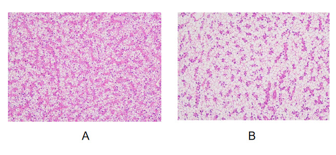
An LMBV infection model was established, and fresh tissues were processed into paraffin sections. Pathological alterations characteristic of viral injury were observed in liver tissue (Fig. 1). Extensive vacuolar degeneration occurred in hepatocytes, with numerous vacuoles of varying sizes distributed in the cytoplasm; hepatocytes exhibited swelling and indistinct cell boundaries. Hepatic necrosis and lysis were observed in multiple regions, manifested as karyopyknosis, karyorrhexis, and even complete dissolution and loss of cell nuclei. The hepatic cords were arranged disorderly, hepatic sinusoids were dilated and congested, and inflammatory cell infiltration was visible in partial stromal areas.
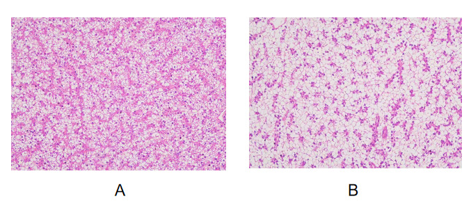
Compared with the virus-infected group, tissues from H. cordata-treated group exhibited alleviated pathological lesions, reduced inflammatory cell infiltration and less tissue damage, indicating that Houttuynia cordata could exert protective effects against virus-induced tissue injury.
3.2. Pathological Lesions of Intestinal Mucosa Induced by LMBV Infection
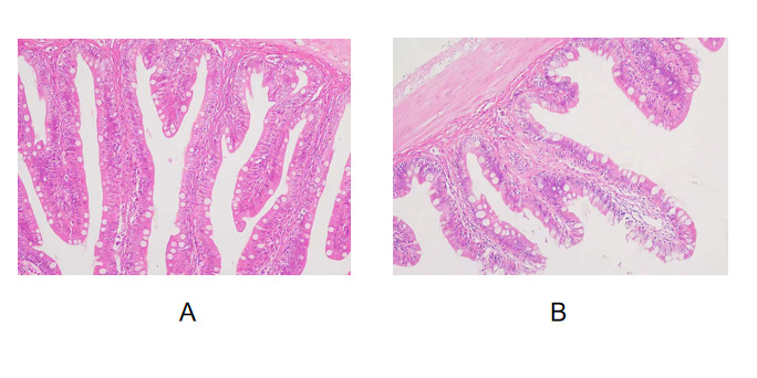
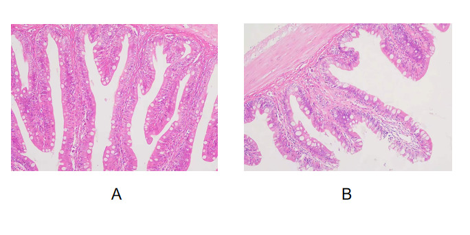
An LMBV infection model was established, and fresh intestinal tissues were prepared into paraffin sections. Obvious destructive pathological changes were detected in the intestinal mucosa (Fig. 2). Intestinal villi showed swelling, shortening, rupture and even focal shedding, with the overall structural integrity of villi destroyed. Mucosal epithelial cells exhibited degeneration and necrosis, and exfoliation and defects of epithelial layers were observed in partial regions. The lamina propria presented edema and loose structure accompanied by inflammatory cell infiltration; the number of goblet cells decreased with disordered distribution.
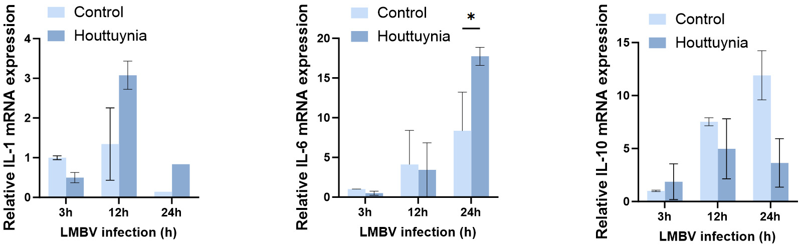
3.3. Houttuynia cordata Significantly Upregulates Transcriptional Expression of IL-6 under LMBV Infection
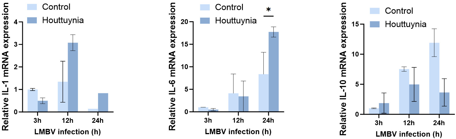
EPC cells were infected with LMBV and divided into two groups: one treated with Houttuynia cordata extract, and the other left untreated. Cells were harvested at 48 h post-infection to assess changes in IL family gene expression. The results showed that IL-6 levels were markedly upregulated relative to the control group, indicating that Houttuynia cordata effectively promotes IL-6 expression at 24 h following LMBV infection (Fig. 3).
3.4. Temporal Expression Profiles of Upstream Signaling Molecule TGF and Adaptor Protein TRAF6
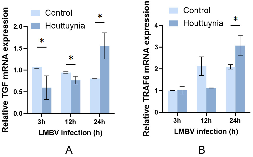
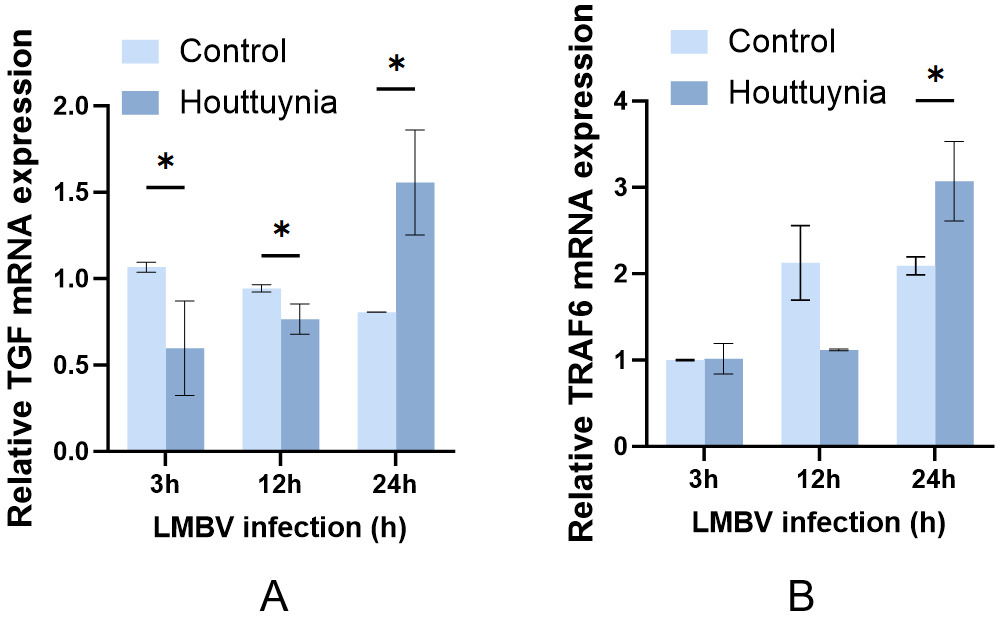
After verifying that H. cordata extract exerts immunomodulatory effects via IL-6, we further examined the temporal expression patterns of upstream signaling molecules and adaptor proteins to elucidate the molecular mechanism by which this extract promotes IL-6 expression at 24 h post-infection under viral stimulation. As illustrated in Figure 4, the transcriptional level of TGF in the control group remained relatively stable at basal levels throughout the entire infection cycle. In contrast, TGF transcription abundance in the H. cordata-treated group was lower than in the control group at the early and middle infection stages (3 h and 12 h), followed by a remarkable upregulation at the late infection stage (24 h).
3.5. Transcriptional Regulatory Effects of Core Kinases TAK1 and ERK
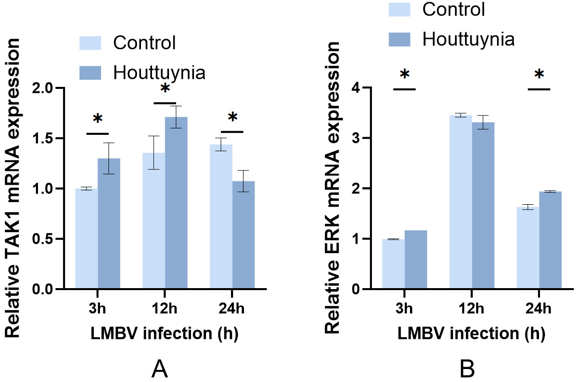
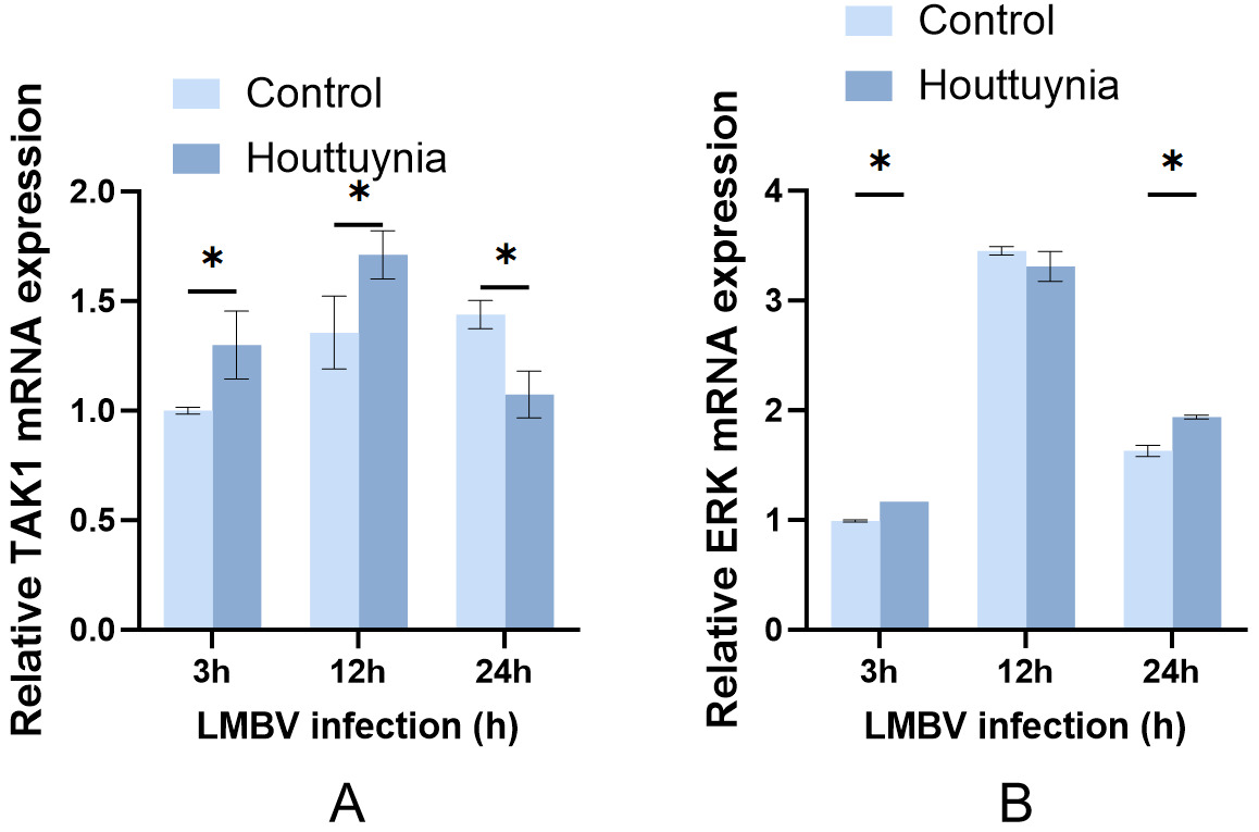
To further clarify how extract of H. cordata facilitates IL-6 secretion, we investigated the expression changes of TAK1 and ERK under extract treatment (Fig. 5). In the control group, TAK1 mRNA levels rose slowly throughout infection, consistent with the temporal activation pattern of upstream TRAF6. The transcriptional levels of TAK1 in the H. cordata-treated group were higher than those of the control at the early and middle infection stages (3 h, 12 h). At the late infection stage (24 h), TAK1 expression in the treatment group decreased to a level lower than the control, which prevents inflammatory damage caused by sustained excessive pathway activation. Meanwhile, ERK mRNA in both groups peaked at 12 h post-infection and declined at 24 h. The transcriptional levels of ERK in the treatment group were higher than in the control at the early (3 h) and late (24 h) infection stages, but comparable at the middle stage. Temporal regulation of ERK by H. cordata can synergistically modulate the intensity of inflammatory pathway activation. Collectively, H. cordata initiates TAK1-mediated signaling cascades in advance, accelerates the onset of immune responses, and promotes downstream transcriptional regulation.
3.6. Dynamic Activation Kinetics of the c-Fos–AP-1 Transcriptional Branch
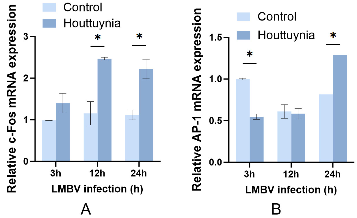
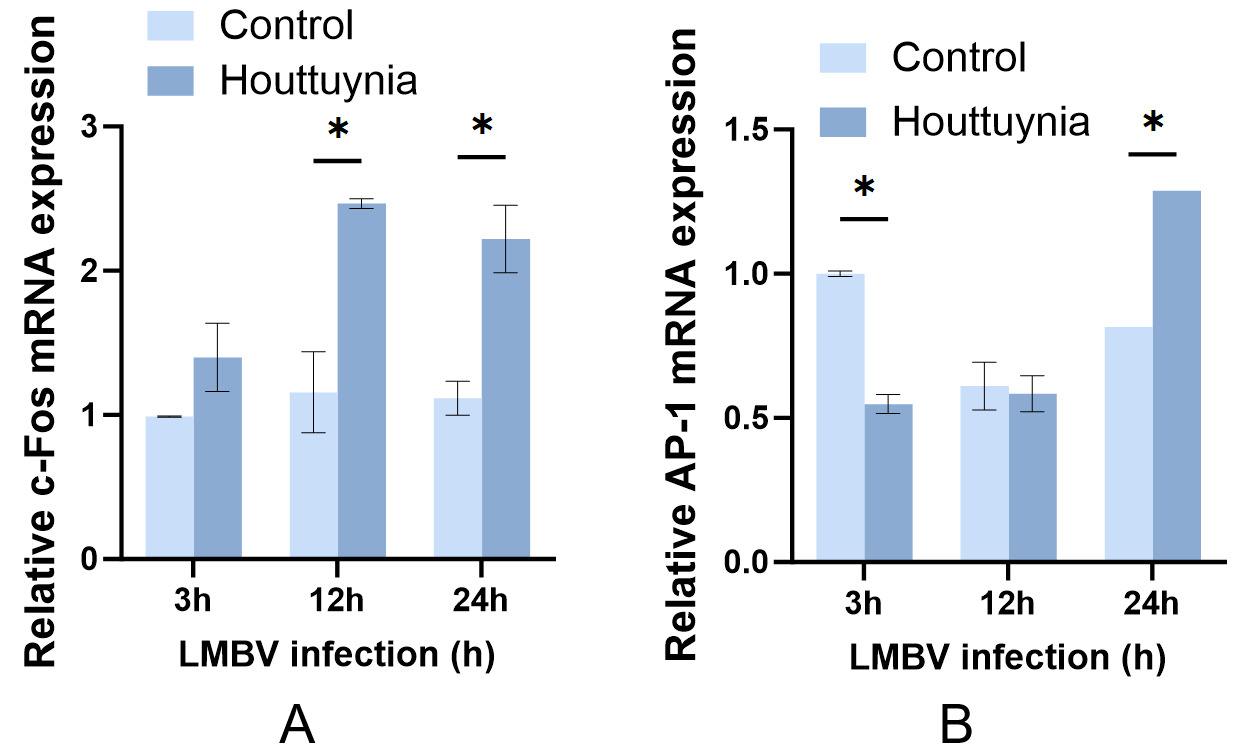
To further clarify how H. cordata extract promotes IL-6 secretion, we systematically investigated the terminal downstream branch of the TAK1-MAPK pathway. In the control group, c-Fos mRNA exhibited a gentle upward trend throughout infection (Fig. 6). By contrast, transcriptional levels of c-Fos in the H. cordata-treated group were markedly higher than those in the control at all three time points and continued to rise with prolonged infection time. AP-1 mRNA in the control group increased persistently along with LMBV infection and reached an obvious activation peak at 12 h (Fig. 6). AP-1 expression in the treatment group displayed a distinct delayed pattern: at the middle infection stage (12 h), AP-1 transcriptional levels were significantly lower than the control, indicating prominent suppression of pathway activation. At the late-infection stage (24 h), AP-1 expression in the treated group exceeded that in the control group and remained at high transcriptional activity. Combined with the expression profile of upstream c-Fos, these data reveal that H. cordata does not suppress the AP-1 pathway overall; instead, it remodels the temporal kinetics of its activation to blunt the inflammatory peak at mid-infection while sustaining pathway activity at late infection to preserve immune defense functions. Collectively, these results demonstrate that Houttuynia cordata persistently upregulates c-Fos expression at the transcriptional level, providing abundant subunit pools for subsequent AP-1 activation.
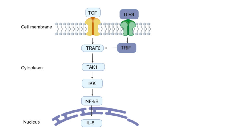
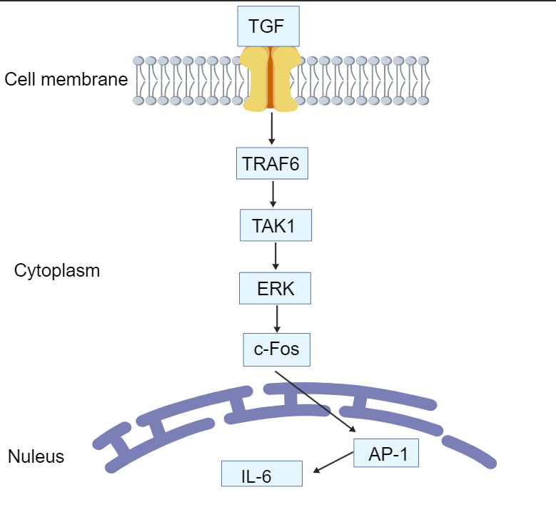
3.7. Houttuynia cordata Extract Mediates Inflammatory Signal Transduction via the TRAF6–TAK1–ERK–c-Fos–AP-1 Axis
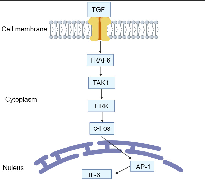
Combined with the temporal expression profiling data of each molecule, the complete signaling pathway through which Houttuynia cordata regulates the inflammatory response in fish upon LMBV infection can be summarized as follows (Fig. 7): TRAF6 is persistently upregulated throughout the entire course of LMBV infection, and H. cordata pretreatment can induce early activation of TAK1 to accelerate signal transduction. At the late stage of infection, H. cordata upregulates the expression of upstream TGF-β signaling components, thereby further amplifying the activity of the core TRAF6–TAK1 signaling axis. This cascade sequentially triggers sustained high expression of downstream c-Fos, markedly elevates the transcriptional level of the terminal transcription complex AP-1, and ultimately robustly induces the expression of pro-inflammatory cytokine IL-6. By temporally potentiating the host’s innate inflammatory defense response, H. cordata exerts protective effects against LMBV infection.
4. Discussion
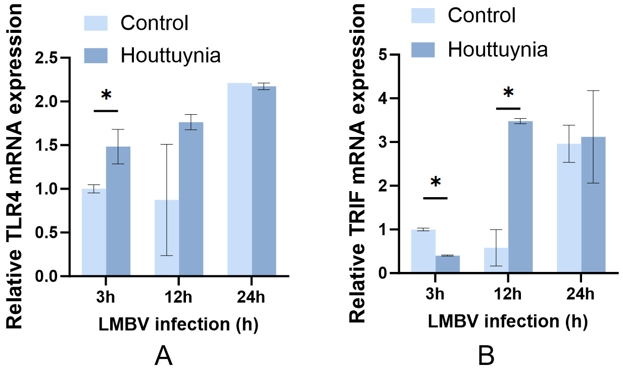
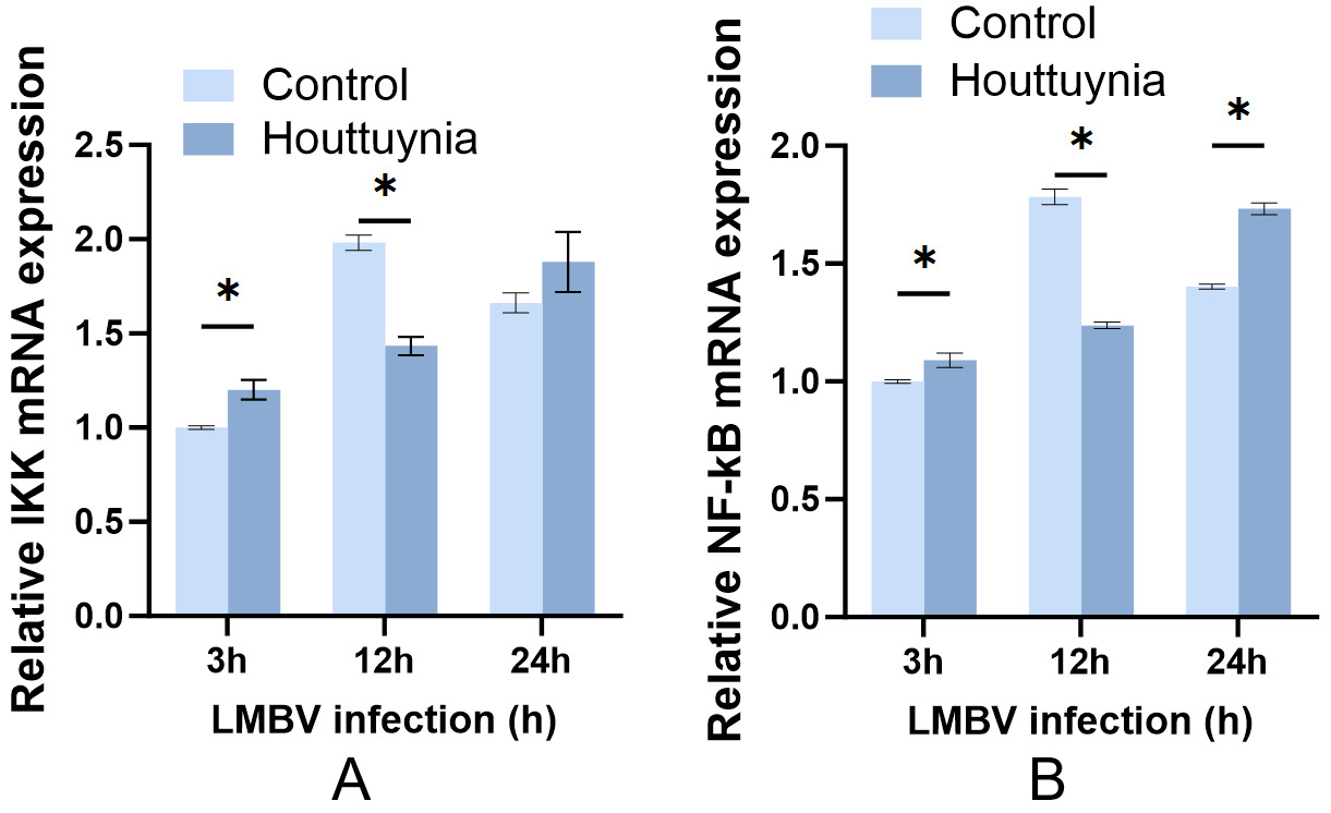
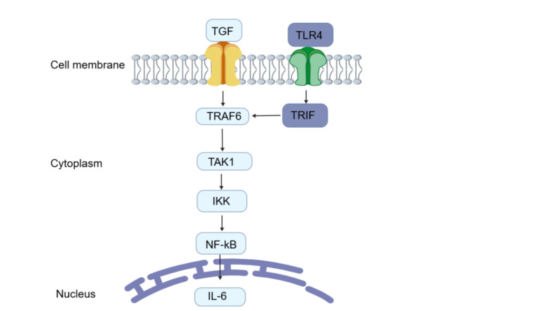
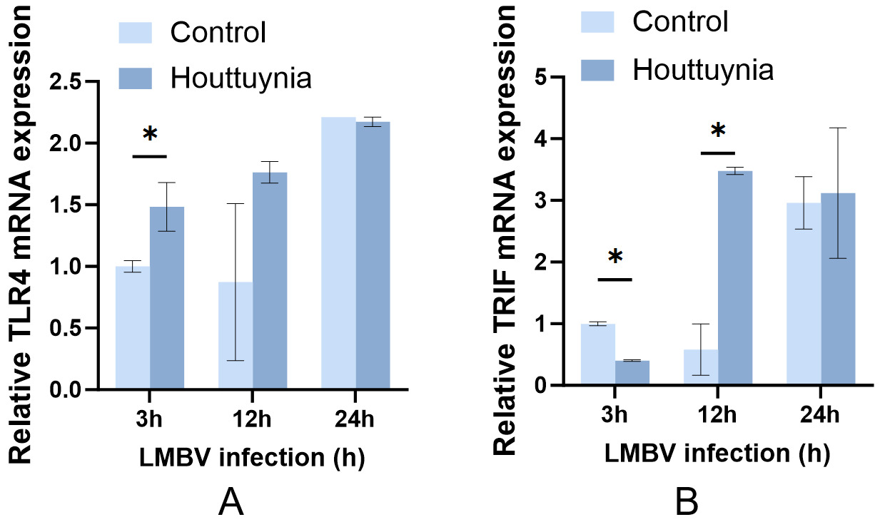
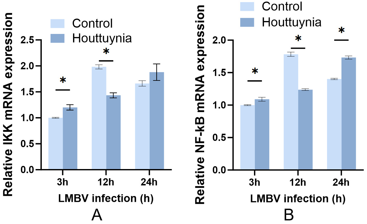
This study verified, using an LMBV-infected cell model, that the antiviral immunoregulatory effect of H. cordata does not rely on the TLR4-TRIF and TNF-TRAF6 branch signaling pathways (Supplemental Figure S1). Instead, it primarily regulates TAK1 via the TGF-TRAF6 signaling axis and preferentially activates the ERK/c-Fos/AP-1 cascade, thereby markedly driving high-level IL-6 expression at the late stage of LMBV infection to exert anti-LMBV immune regulation. No significant change in TRIF expression was observed after Houttuynia cordata treatment (Supplemental Figure S2), indicating that this herb initiates immune responses through a TLR4-dependent but TRIF-independent pathway. With no obvious activation of downstream IKK-NF-κB signaling (Supplemental Figure S3). Collectively, these results demonstrate that H. cordata facilitates IL-6 secretion via the TGF-AP-1 signaling cascade.
Analysis of hepatic and intestinal cells infected with LMBV revealed that LMBV infection leads to extensive multi-organ tissue damage in largemouth bass, presenting typical pathological characteristics of systemic viral infection.14 Pathological manifestations observed in this experiment are highly consistent with the pathogenic characteristics of viruses belonging to the genus Ranavirus. LMBV invades hepatocytes to replicate and proliferate, directly inducing parenchymal liver injury by disrupting cellular ultrastructure and triggering apoptosis and necrosis. As the core organ governing metabolism and immune regulation in fish, structural damage to the liver directly causes metabolic disorders and impaired immune function in the host, which constitutes a critical pathological basis for mortality following viral infection.15 Among these organs, the liver serves as the primary target organ for viral replication, exhibiting the most severe cellular degeneration and necrosis. The intestine constitutes the core component of the mucosal immune system in fish and acts as the first physical barrier against pathogen invasion in the host.16 Virus-induced destruction of intestinal mucosal structure not only impairs intestinal digestion and absorption functions, but also increases the permeability of the mucosal barrier, thereby triggering secondary intestinal flora translocation and local inflammatory cascades, which further exacerbate systemic viral dissemination and host tissue injury.17 Damage to the intestinal mucosal barrier further opens pathways for viral invasion and dissemination, and these two factors synergistically accelerate disease deterioration. Previous studies have confirmed that multi-organ tissue necrosis, organ failure, and systemic inflammatory responses triggered by LMBV infection represent the core pathological mechanisms underlying the high morbidity and mortality of infected fish.18 The baseline pathological characteristics identified in this study also provide a morphological reference standard for subsequent evaluations of the tissue-protective and antiviral effects of bioactive substances such as Houttuynia cordata.
Extracts of H. cordata sequentially activate the TGF-TRAF6-TAK1-AP-1-IL-6 inflammatory signaling axis to mediate host antiviral inflammatory responses, and this pathway represents one of the key mechanisms underlying innate immune defense against viral infection in fish.19 H. cordata does not merely exert a simple activating or inhibitory effect on this pathway; instead, it achieves bidirectional immunoregulation via temporal regulation at multiple hierarchical levels, ranging from upstream ligands and adaptor proteins to core kinases, transcription factors, and effector cytokines. In the early and middle stages of infection, it suppresses excessive pathway activation to alleviate tissue damage induced by inflammatory storms.20 At the late stage of infection, it upregulates pathway activity to enhance immune clearance and tissue repair capacity.21 Previous studies have confirmed that bioactive components such as flavonoids and polysaccharides in H. cordata exert immunomodulatory and anti-inflammatory activities by regulating the MAPK/AP-1 signaling pathway.22 The results of this experiment supplement the functional mechanism from the perspective of temporal regulation and provide experimental evidence at the level of molecular signaling pathways for the application of Houttuynia cordata in the prevention and control of viral diseases in aquaculture.
Within the TGF-TRAF6-TAK1-AP-1-IL-6 inflammatory signaling axis, TGF represents a family of multifunctional polypeptide cytokines that play central roles in physiological processes, including the regulation of immune homeostasis.23 In this experiment, TGF exhibited a temporal expression pattern with downregulation at the early stage (3–12 h) and significant upregulation at the late stage (24 h). Its upregulated expression level was higher than that of TNF, indicating that TGF serves as the primary initiating signal of this pathway. In the early phase of infection, TGF suppression prevents excessive inflammatory cellular damage, whereas upregulated TGF cooperates with TNF to activate downstream signals at the late infection stage. Such bidirectional regulation constitutes a distinctive feature of H. cordata when compared with commonly used aquaculture herbal medicines including Astragalus membranaceus and Abrus cantoniensis.24 TRAF6 is a crucial adaptor protein in innate immunity,TRAF6 expression is progressively upregulated with prolonged natural LMBV infection. The downstream hub kinase TAK1 exhibits a temporal expression pattern characterized by an elevation at 12 h, followed by a decline at 24 h. Its early upregulation initiates inflammatory cascades, whereas the late reduction relies on host negative feedback to constrain excessive inflammatory responses. Meanwhile, IKK-α expression remains unaltered, and the NF-κB pathway is not activated, indicating that TAK1 signaling modulated by Houttuynia cordata selectively biases toward the ERK/c-Fos/AP-1 branch. H. cordata persistently upregulates the c-Fos gene, which drives robust AP-1 elevation at 24 h and ultimately triggers burst-like overexpression of IL-6 at the late infection stage, with its expression level reaching 2.1-fold that of the control group. As a pivotal pro-inflammatory cytokine against ranavirus in fish, high IL-6 expression at late infection strengthens host antiviral capacity, whereas moderate suppression at early infection prevents inflammatory dysregulation. These findings provide a molecular interpretation of the functional mechanism underlying the use of Houttuynia cordata for LMBV disease control in aquaculture.
Most existing studies have focused on the pathways underlying the antibacterial and anti-mammalian viral activities of H. cordata,25 This study identified AP-1 as the core transcription factor responsible for the H. cordata-mediated upregulation of IL-6. Nevertheless, it should be noted that the crude extract of H. cordata is a complex mixture of multiple compounds. Thus, we cannot pinpoint which individual active component drives the observed cellular responses in our cell-based assay. Further work is needed to isolate and verify the bioactive monomer. Current research on herbal medicines against LMBV mainly centers on Astragalus membranaceus and Abrus cantoniensis. Astragalus membranaceus exerts direct antiviral and interferon-related effects to suppress the activity and replication of largemouth bass ranavirus. Abrus cantoniensis primarily functions via organ antioxidant repair; its ingredients such as astragalus polysaccharides significantly enhance non-specific immunity and repair damaged tissues in mandarin fish. To date, no reports have described the antagonistic effect of H. cordata against LMBV through the regulation of interleukin signaling pathways. The present work fully elucidates the TGF-TRAF6-TAK1-ERK-AP-1-IL-6 regulatory axis and fills the relevant research gap. In aquaculture production, there are no specific therapeutic drugs for LMBV, and existing vaccines exhibit limited protection efficacy. Since H. cordata can elevate IL-6 levels, it has promising potential for development as an aquaculture immune additive or vaccine adjuvant. Nevertheless, this study has several limitations. Although the steam-distilled aqueous extract of H. cordata exhibits clear immunomodulatory effects in vitro, this laboratory-prepared distillate is difficult to use for large-scale on-farm applications. In contrast, herbal powder is cheap and convenient for feed supplementation in practical aquaculture. Nevertheless, herbal powder suffers from inconsistent levels of volatile active substances, such as houttuynin. There remains a notable practical-application gap between laboratory steam-distilled extract and raw herbal powder, and further research is needed to balance biological efficacy, production cost and operability for real aquaculture deployment.
All experiments were performed solely using an in vitro cell model without in vivo LMBV challenge verification in largemouth bass. Crude extracts of H. cordata were used, making it impossible to pinpoint the bioactivity of individual active compounds. No concentration gradient for herbal extracts was established, resulting in a lack of dose-response relationships and an inability to identify optimal effective supplementation concentrations. Future research directions include conducting in vivo feeding and viral challenge trials, isolating monomeric compounds from Houttuynia cordata to identify key active ingredients, and investigating the feasibility of combining Houttuynia cordata with LMBV vaccines as immune adjuvants.
Acknowledgments
The authors gratefully acknowledge financial support from the Research Fund for the Doctoral Program of WHPU, grant number 2024RZ033, and the Project of Hubei Key Laboratory of Animal Nutrition and Feed Science (ANFS202510).
Author contributions
Writing – original draft: Qiao Wang (Equal), Qian Liu (Equal). Data curation: Rui Kong (Lead). Conceptualization: Haihui Liu (Lead). Resources: Guokang Yu (Lead). Writing – review & editing: Xianqin Hu (Equal), Chi Zhang (Equal).
Interest conflict statement
The authors have declared no conflict of interest.
Ethics Statement
This research strictly abides by the 3Rs (Replacement, Reduction, Refinement) ethical principles for laboratory animals. An in vitro EPC cell model was used to perform virus propagation, preliminary screening of drug concentrations, and fundamental molecular assays, thereby largely avoiding large-scale in vivo trials in live fish. The minimum effective sample size was determined based on pre-experimental data; multiple organs, including liver and intestine, were simultaneously harvested from each individual fish for multi-indicator histological analyses to maximize tissue utilization and minimize the total number of experimental fish. During tissue sampling, gentle capture with soft nylon nets, deep anesthesia using tricaine methanesulfonate (MS-222), and humane euthanasia via anesthetic overdose were implemented to refine all sampling procedures and substantially alleviate stress and pain inflicted on experimental fish.
Informed Consent Statement
All authors and institutions have confirmed this manuscript for publication.
Data availability
Data will be made available on request.