
Abbreviations
NB: Nutrient Broth
CFU: colony forming units
PCR: Polymerase Chain Reaction
C: Control group
BL: Low concentration of Bacillus subtilis BSP12 group (1×10⁷ CFU/g)
BH: High concentration of Bacillus subtilis BSP12 group (1×10⁸ CFU/g)
LGR: Length growth rate
WGR: Weight gain rate
SGR: Specific growth rate
SR: Survival rate
1. Introduction
European eel (Anguilla anguilla) is one of the 16 eel species and is widely cultured in the southeast coastal area of China, bringing significant economic benefits to farmers due to its delicious meat and high nutritional value.1 To maximize production efficiency, artificial eel aquaculture is mostly realized in land based and high-density manner, however, intensive eel aquaculture increases the risk of infectious disease outbreaks.2
Probiotics are a class of active microorganisms that are beneficial to the host by colonizing in host and improving the composition of the host’s intestinal flora.3,4 They have been recognized as environmentally friendly alternatives to antibiotics and have been shown to be effective in combating bacterial infections in aquaculture.5,6 Among probiotic candidates, Bacillus subtilis is one of the most extensively studied species in aquaculture. It has been reported to promote growth performance in various fish species, including striped catfish (Pangasius hypophthalmus),7 Nile tilapia (Oreochromis niloticus),8 and primarily through enhancing intestinal digestive enzyme activities such as amylase, lipase, and protease.7 Additionally, B. subtilis modulates host immune responses, as evidenced by increased IgM levels in rainbow trout (Oncorhynchus mykiss),9 enhanced peroxidase activity in mucus.10 Furthermore, host-associated B. subtilis strains can improve intestinal histomorphology in rainbow trout.11
On the other hand, the mineral research on aquaculture mainly focuses on the diet minerals supplementation.12 So far, the effects of B. subtilis from the aquacultural intestines source on important minerals of the carcass and their relations with host growth performance have not been elucidated. Mineral elements of K, Na, Ca, Mg, Zn, and Fe represent the predominant mineral fraction in European eel (Anguilla anguilla) muscle.13 Subsequent studies have confirmed that these elements as the major mineral constituents of eel flesh, with concentrations typically following the order K > Na > Ca > Mg > Zn > Fe.14 Beyond their quantitative dominance, these minerals are physiologically different: K and Na maintain osmotic balance and cellular ionic homeostasis,12 and Ca and Mg are essential for skeletal development, muscle contraction and nerve transmission.15 Zn serves as an indispensable trace element involved in immune response,16 and Fe is essential for oxygen transport.15 Therefore, the modulation of these minerals by probiotic supplementation could be related to eel growth performances, and even with the nutritional quality of farmed eel as a human food source.
Although the growth-promoting effects of dietary Bacillus in aquaculture have been well documented, its impacts on eel growth performance and muscle mineral deposition, and their relationships, remain poorly understood. To address this gap, we hypothesize that dietary supplementation with the autochthonous B. subtilis strain BSP12 would: (i) improve the growth performance of European eel, (ii) alter the content of important minerals in eel muscle. The correlation between eel growth performance and those modified minerals will also be explored.
2. Materials and Methods
2.1. European eel
In this experiment, 20 healthy European eels (average weight 310.67±1.73 g) for Bacillus isolation and safety testing experiment were collected from a fish company in Guangzhou of Guangdong province, China. Colonies were screened by Bacillus morphology (Fig. S1). The purified strain was stored in 40% glycerol at -80°C.
2.2. Antimicrobial activity of Bacillus strains
A total of 12 pathogenic strains and 5 Bacillus strains isolated from healthy eel intestines (Table S1) were cultured for 12 h to logarithmic phase, and adjusted to 1.0×10⁶ CFU/mL by 10-fold gradient dilution. The Oxford cup method was used, with chloramphenicol (50.0 μg/mL) as a positive control and sterile water as a negative control. Pathogens (100 μL, 1.0×10⁶ CFU/mL) were spread on ready NB solid medium. Five autoclaved Oxford cups (7 mm outer diameter) were placed on each plate and gently pressed to ensure the bottoms fit tightly. Each cup was added with 150 μL of Bacillus strains, chloramphenicol and sterile water, respectively. Plates were incubated at 30°C for 18 h, and inhibition diameters, measured as halo sizes, were recorded. All tests were performed in triplicate.
2.3. Antibiotic susceptibility test
Antibiotic susceptibility was determined by disk diffusion following CLSI (2024).17 Strains were incubated for 12 h and diluted to 1×10⁶ CFU/mL. One hundred fifty microliters were spread onto NB agar, and antibiotic discs were placed on the plates, which were then incubated at 37°C for 12 h. Inhibition zone diameters were measured. Strains were classified as resistant, intermediate, or sensitive.17 All tests were performed in triplicate.
2.4. Determination of optimal growth temperature, pH and bile tolerance
Optimal growth temperature. Strains were inoculated into 100 mL NB medium and incubated at 15, 20, 25, 30, 37 and 45°C with 180 rpm. Growth was measured at OD600 every 2 h using a spectrophotometer (UV759CRT, Shanghai Youke Instrument Company, Shanghai, China).
Optimal pH and bile tolerance. Strains were incubated at pH 2, 5, 7, 9 and 11, or bile salts (5‰, 20‰, 40‰, 60‰ and 80‰) at 37°C with 180 rpm.18 Growth was measured at OD600 every 2 h using a spectrophotometer.
2.5. Evaluation of extracellular enzyme activities
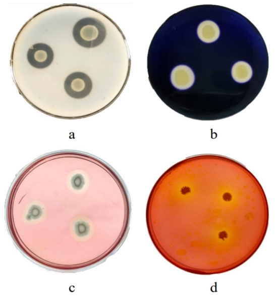
Candidate strains were spot seeded on protease, amylase, cellulase and lipase selective media,19 incubated at 37°C for 12 h, and examined for hydrolysis circles. Three parallels were set for each group.
2.6. Molecular identification by 16S rDNA
16S rRNA gene sequencing was used for identification. The 16S rRNA gene was amplified using primers 7F (5’-CAGAGTTTGATCCTGGCT-3’) and 1540R (5’-AGGAGGTGATCCAGCCGCA-3’) under the following conditions: 95°C for 5 min, 30 cycles of 94°C for 30 s, 57°C for 30 s, and 72°C for 90 s, with a final extension at 72°C for 10 min.20 PCR products were sequenced by Shanghai Bioengineering Co., Ltd. Sequences were analyzed by BLAST. The sequence was submitted to GenBank (OQ612706). Phylogenetic trees were constructed using MEGA 11.0.
2.7. Experiment design and biosafety test
2.7.1. Experiment design
810 eels (13.11±2.15 g, 23.55±2.07 cm) were obtained from Guangzhou, China, and they were acclimatized for two weeks before the experiment. Nine tanks (sized 200 cm length × 200 cm width × 50 cm heigh) were randomly divided into three groups (three replicate in each group), and 90 eels in every tank were randomly divided. Three as treatment group with a low concentration of B. subtilis (1.0 × 10⁷ CFU/g) mixed feed (BL), three as another treatment group with a high concentration of B. subtilis (1.0 × 10⁸ CFU/g) mixed feed (BH), the last three as a control group with an equal amount of sterile saline feed added (C). Rearing conditions were maintained as follows: water temperature 20±1°C, salinity 28.0±2.0 psu, pH 8.0±0.5, dissolved oxygen >8 mg/L, and 70% daily water exchange. Eels were fed at 8% body weight twice daily (07:00 and 19:00) for 28 days under 12L:12D photoperiod. Water quality parameters (temperature, salinity, pH and dissolved oxygen) were daily monitored in each tank after every water exchange. No significant differences were observed among the nine tanks or among the three treatment groups (p > 0.05). Throughout the experimental period, the water level in each tank was maintained at 30 cm.
2.7.2. Biosafety test
Before the formal experiment, 54 healthy European eels (Anguilla anguilla) from the same manufacturer were selected with an average weight of 11.58±1.85 (g) and an average length of 20.33±2.75 (cm) for the safety test of candidate strains. Eels were randomly divided into three groups (n=6 per tank, three tanks per group): low-dose (1×10⁷ CFU/mL), high-dose (1×10⁸ CFU/mL), and control (0.65% sterile saline).
Following Amenyogbe et al. (2021)21 eels were intraperitoneally injected (0.1 mL/fish) and monitored for 14 days for adverse signs and mortality. This route bypasses the gastrointestinal barrier to assess systemic virulence.22
2.7.3. Feed Preparation
The basal diet (fish meal 60%, soybean meal 19%, alpha starch 18%, fish oil 2%, vitamins and minerals 1%) contained 47.00% crude protein, 4.00% crude fat, 10.00% moisture, and 17.00% crude ash. It was ground and mixed with BSP12 suspension at 1.0×10⁷ CFU/g (BL) and 1.0×10⁸ CFU/g (BH). The control diet was prepared with an equal amount of sterile saline. All feeds were dried and stored at 4°C.
2.7.4. Sample processing and analysis
Eels were starved for 24 h before sampling. Growth was recorded on 0, 14 and 28 d. Six eels per tank (18 per group) were measured for weight and length. Three eels per tank (nine per group) were anesthetized with MS-222 for muscle collection in a biological safety cabinet. After execution, blood was removed and whole-body muscle was collected, mashed, labeled, and stored in liquid nitrogen at −80°C. Day 0 samples were mixed as Initial; days 14 and 28 samples were named C14, BL14, BH14, C28, BL28, and BH28.23
Growth performance. Growth performance was evaluated by weight gain rate (WGR), length growth rate (LGR), survival rate (SR) and specific growth rate (SGR).
Mineral analysis. Muscle samples (5 g) were ashed at 550°C, digested with HNO₃/HClO₄, dissolved in 0.5 mol/L HNO₃, and fixed in 25 mL volumetric flask. Mineral content was determined by atomic absorption spectrometry (PE900T, Platinum Elmer Co., Ltd., USA).
2.8. Statistical analysis
Normality and homogeneity of variance were examined using the Shapiro–Wilk test and Levene’s test, respectively. Differences among groups were evaluated by one-way ANOVA followed by LSD post-hoc test. Data are presented as mean ± standard error (SE). The relationships between mineral concentrations and growth parameters were assessed using Spearman’s rank correlation. For multiple correlation comparisons, p-values were adjusted using the Benjamini–Hochberg false discovery rate (FDR) method. Statistical significance was set at p < 0.05. All analyses were performed using IBM SPSS Statistics, version 27.0 (IBM Corp., Armonk, NY, USA).
3. Results
3.1. Morphological and antimicrobial characterization of isolated strains
Five Gram-positive strains were isolated from the intestinal tract of healthy European eel, named BSP1, BSP3, BSP4, BSP5 and BSP12. Five Bacillus strains showed antibacterial activity against 12 pathogens. Among of the five Bacillus strains, BSP12 had the highest inhibitory rate (91.67%) (Table S1). Based on morphological and antibacterial properties, BSP12 was selected for further experiments.
3.2. Antibiotic susceptibility test
BSP12 showed significant sensitivity to all nine antibiotics (Table S2). It was most sensitive to cephalothin (43.18±1.36 mm), followed by florfenicol, erythromycin, enrofloxacin, and chloramphenicol (>25.00 mm).
3.3. Growth characterization of the potential probiotic BSP12
3.3.1. Growth performance of the BSP12 at different temperatures, pH and different bile salt concentrations
The growth of BSP12 observed at 15°C, 20°C, 25°C, 30°C, 37°C and 45°C, with optimal growth at 37°C. (Fig. S2a).
Its growth was observed at pH 5, pH 7, and pH 9, with optimal growth at pH 7. OD600 increased with pH from 5 to 7 (p > 0.05), and decreased from 7 to 9 (p > 0.05). Growth was inhibited at pH <2 or >11 (Fig. S2b).
BSP12 grew at all bile salt concentrations (5‰, 20‰, 40‰, 60‰ and 80‰). OD600 values ranked: 80‰ < 60‰ < 40‰ < 20‰ < 5‰, indicating 5‰ as optimal (Fig. S2c).
3.3.2. Enzyme-producing properties of BSP12
As shown in Fig. 1, transparent circles were observed on protease medium, cellulase medium, and amylase medium; and colored circles were observed on lipase medium. It indicated that the BSP12 has the ability to produce protease, amylase, cellulase and lipase.
3.4. Safety analysis of BSP12 in animals
The safety assessment showed that European eels injected with Bacillus at concentrations of 1×10⁷ CFU/mL and 1×10⁸ CFU/mL were active and feeding well, and none exhibited pathological signs or disease symptoms, including mucus, lesions, edema, scale loss, or hemorrhage. During the biosafety test, all groups were 0% mortality rate, and the internal organs were well organized without lesions. The results indicated that no pathological signs, disease symptoms or mortalities were induced by BSP12.
3.5. Effect of B. subtilis BSP12 on the growth performance and survival of European eel
Final body weight, WGR, and SGR were significantly higher in BH than control (p < 0.05), improved by 18.08%, 222.94%, and 194.59%, respectively (Table 1). BL also showed elevated values (9.58%, 108.68%, and 97.30%), with significant differences for WGR and SGR (p < 0.05). Concerning survival rates, at the end of the experiment, compared with the control, those of 96.70%, BL, and BH were 100.00% and 100.00%, respectively, with no significant difference observed (p > 0.05). At the end of the experiment, concerning the temperature, salinity, pH, and DO, there were no significant differences among all groups (p > 0.05, data not shown)
3.6. Effect of B. subtilis BSP12 on minerals in eel muscle
As it can be seen from Table 2, at the end of the experiment, on day 28, three elemental contents (K, Mg, and Zn) were significantly increased (p < 0.05) in BH28 group, as compared to the C28 group, and there was no significant difference (p > 0.05) in other elemental contents. Concerning the BL28, only Zn elemental content was significantly increased (p < 0.05) in the BL28 group, compared to the C28, while all other elemental contents showed no significant change (p > 0.05).
3.7. Correlation between mineral content in eel muscle and growth performance
Spearman correlation showed BSP12 altered mineral-growth correlations (Table 3). In C, only Na correlated with WGR, LGR, and SGR (p < 0.05 or p < 0.01). In BL, K, Na, Mg, Zn, and Fe correlated with multiple growth indicators (p < 0.05 or p < 0.01), with Na correlating with all indicators. Mg and Zn correlated with final weight, WGR, and SGR (p < 0.05 or p < 0.01), but not in C.
It is noteworthy that the BH group showed higher correlation coefficients than the BL group. K and Na were significantly or extremely significantly positively correlated with all growth performance indicators (p < 0.05 or p < 0.01). Additionally, the correlation coefficients of Mg and Zn with WGR and SGR were higher in the BH group than in the C and BL groups, where the correlations of Mg and Zn with WGR and Mg with SGR reached an extremely significant level (p < 0.01), and the correlation of Zn with SGR reached a significant level (p < 0.05) (Table 3).
4. Discussion
In this study, BSP12 was selected for further experiments based on its morphological and antibacterial properties (Table S1 and Fig. S1). Moreover, an important prerequisite for the selection of probiotic strains is that they are not antibiotic resistant.24 In this study, nine antibiotics were tested, and the results showed that the candidate Bacillus strain BSP12 showed high sensitivity to all nine antibiotics (Table S2), suggesting that the strain may not carry resistance genes to aquatic antibiotics, and it had a good safety profile for environmental release.
Growth characteristics of BSP12 were determined to standardize conditions for extracellular enzyme activity assays. BSP12 grew at 15–45°C, with optimal growth at 37°C (Fig. S2a), consistent with Ghosh et al. (2002) for Bacillus strains from rohu intestine (35–37°C).25 BSP12 grew at pH 5–9, with optimal growth at pH 7, consistent with Li et al. (2020) for Bacillus CPA1-1 from Macrobrachium nipponense (pH 5–9) and Athulya et al. (2024) for Bacillus tropicus ACS1 from tilapia (pH 5–8).26,27 BSP12 tolerated bile salts at 5‰ to 80‰. In comparison, B. cereus from Nile tilapia intestine survived at 5‰,28 and Bacillus sp. KADR1 from rohu intestine tolerated 25‰, 50‰ and 75‰.29 The broader tolerance range of BSP12 (5‰ to 80‰) relative to these strains suggests enhanced gastrointestinal survival capacity in fish
It has been found that the addition of Bacillus to feed increased the activity of trypsin, amylase and lipase in the intestinal tract of Asian sea bass, and it also increased the body weight and specific growth rate of Asian sea bass.30 In addition, Liu et al. (2017) found that the addition of 1×10⁸ CFU/mL of Bacillus sp. to feed significantly increased the body weight,31 specific growth rate and digestive enzyme activities such as protease and amylase of tilapia (Oreochromis niloticus), while fish’s own endogenous digestive enzymes have difficulty in degrading, digesting and absorbing nutrients such as high proteins and fats in feeds. In this study, the candidate Bacillus strain BSP12 had strong protease, amylase, cellulase, and lipase activities (Fig. 1), indicating that the addition of Bacillus BSP12 in fish feed could improve nutrients digestion and absorption in the intestinal tracts of fish.
Probiotic Bacillus is nonpathogenic, has no side effects, and is highly safe; safety testing of probiotics to ensure their safety is one of the important prerequisites for their application in the eel production process.32 In this study, animal safety tests showed that BSP12 was nonpathogenic to the European eels at concentrations of 1×10⁷ CFU/mL and 1×10⁸ CFU/mL with injection.
In the present study, after 28 days of feeding experiment, survival rates were evenly equal in BL and BH groups, and final body weight, WGR, and SGR were significantly increased in BH as compared to the C group (p < 0.05) (Table 1). This indicated that the application of a higher dose (1×10⁸ CFU/g) of B. subtilis BSP12 could significantly improve the growth performance of European eel. These results were supported by Olowe et al. (2024) whose results showed that the final body weight, WGR, and SGR of Japanese eel (Anguilla japonica) were significantly increased by adding either 106 CFU/g or 107 CFU/g of B. subtilis to the basal diet (p < 0.05).33 According to previous reports, this improvement may be attributed to the increased activity of digestive enzymes such as protease, amylase and lipase qualitative by B. subtilis.34 Therefore, the improvement in growth performance of European eels observed in the present study may be related with the protease, amylase, cellulase and lipase produced by this strain (Table 1 and Fig. 1).
All aquatic animals require minerals to perform their vital physiological and biochemical functions and to maintain their normal life processes.12 Fewer studies have examined the effects of Bacillus as a microbial agent on the mineral content of eel carcass. In this study, results showed (Table 2) that Bacillus had different regulatory effects on macro-minerals (K, Na, Ca, and Mg) and trace minerals (Zn and Fe) in the muscle of the European eel.
In the present study, results showed that, compared with the control group, during the entire experimental period, Ca content in the muscle of eels in BH28 increased but was not significant (p > 0.05), whereas Mg content increased significantly (p < 0.05) (Table 2). It has been reported that probiotics improve Mg bioavailability through various mechanisms, including the production of short-chain fatty acids and the regulation of the intestinal microbiota.35
In this study, at the end of the experiment, compared to the C28, K in BH28 was significantly increased in muscle (p < 0.05) (Table 2). Kurchaeva et al. (2020) reported that feeding rabbits (Oryctolagus cuniculus) with a diet supplemented with 0.5% of the symbiotic additive “Pro-Stor” (containing Bacillus subtilis, Bacillus licheniformis and lactic acid bacteria) for 42 days resulted in a higher muscle K content in the experimental group (356.0 mg) than in the control group (348.0 mg).36 The present results are consistent with this finding.
Our results also showed that BSP12 treatment positively affected trace minerals (Zn and Fe). Zn content was significantly increased in both BL28 and BH28 groups, compared with the control group (p < 0.05). Zn content in BH28 was elevated by 13.33% (Table 2) compared with C28. Fe content was also increased in BL and BH, but the difference was not significant (p > 0.05). Benamirouche et al. (2020) reported that broilers fed Pediococcus acidilactici, Saccharomyces cerevisiae, and Yucca schidigera extract for 50 days showed significantly increased Zn and Fe contents in muscle (p < 0.05).37 Although they could not exclude a synergistic effect between the probiotic and Yucca schidigera extract, they attributed the increased muscle mineral deposition primarily to probiotic metabolites. In the present study, this result was supported by their conclusion that probiotics can enhance Zn and Fe deposition in muscle tissues without reliance on exogenous mineral sources or phytogenic additives. BSP12 supplementation significantly increased the contents of K, Mg and Zn in eel muscle (Table 2), suggesting that this probiotic strain promotes mineral deposition in host muscle tissue.
Concerning the correlation between muscle mineral content and growth performance, the Spearman correlation analysis results are shown in Table 3. In the C group, only Na correlated significantly with growth indicators, while in the BL and BH groups, multiple minerals (K, Na, Mg, Zn, and Fe) showed significant correlations with growth performance, with the correlation coefficients generally higher in the BH group. These results indicate that after BSP12 improved host growth performance, the minerals deposited in the muscle showed a stronger correlation with growth performance. The host’s growth status could even be assessed by examining the correlation between muscle minerals and growth performance. Previous studies have shown that the mineral content in muscle of farmed animals changed with their growth status. Xie et al. (2023) studied the content of essential mineral elements in the muscle tissues of different-sized Oncorhynchus mykiss. Initially, this content increased as the fish size increased, but then decreased.38 They speculated that the changes in mineral element content might be related to vigorous growth.
Interestingly, in this study, we noted that at the end of experiment, Zn content in test (both BL28 and BH28 groups) was all significantly increased in eel muscle (p < 0.05), and the results showed that this increased content of Zn was significantly correlated with SGR and WGR of eel (Table 2 and Table 3). We also noted that at the end of experiment, the significant increase of Mg content in BH of eel muscle had similarly positive correlation with eel SGR and WGR (p < 0.01) (Table 2 and Table 3). This could be speculated that BSP12 might enhance intestinal Mg absorption as suggested by Varvara and Vodnar (2024),35 and another possible mechanism might involve probiotics increasing the bioavailability of Mg deposition in host tissues by producing short-chain fatty acids (SCFAs) and regulating the intestinal flora as proposed by Scholz-Ahrens et al. (2007).39 Those interpretations are also consistent with previous studies, demonstrating that dietary Mg supplementation promoted muscle fiber development and improved growth performance (WGR, SGR) in Megalobrama amblycephala (Bao et al., 2022) and enhanced tissue mineral deposition and weight gain in Eriocheir sinensis (Chen et al., 2026).40,41 However, direct evidence for this mechanism in this study requires further investigation in our future research.
Overall, due to the BSP12 supplementation, the correlations between multiple minerals and growth indicators were enhanced, suggesting that some mineral deposition was closely associated with eel growth performance. This enhanced correlation may contribute to improving growth performance in the food eel.
5. Conclusion
B. subtilis BSP12 from the host intestinal tract showed antagonistic effects against 11 pathogenic indicator bacteria, with broad-spectrum inhibition; adaptability to temperature, pH, and bile salts; and production of protease, amylase, cellulase, and lipase. It was sensitive to nine antibiotics, indicating safety for environmental release. BSP12 supplementation in European eel rearing: (i) it significantly improved growth performance (final body weight, WGR, and SGR) (p < 0.05), (ii) it significantly increased mineral deposition, such as Zn and Mg. The increase of those minerals was significantly correlated with WGR and SGR. These findings reveal the relationship between BSP12-mediated mineral deposition and growth performance in the food eel.
Acknowledgments
This research was supported the Natural Sciences Fund of China (Grant No. 40776091), High-level Personnel Start-up Fund of Foshan University (gg07017), and Public Health Service Improvement Fund of Foshan (2018AB003701). We thank Mrs. Manfeng Lai, Mr. Muzhi Guo, and Mr. Zixian Guo for their help with part of the statistical work.
CRediT - Authors’ Contribution
Conceptualization: Yanbiao Guo (Equal), Siyi Chen (Equal). Methodology: Yanbiao Guo (Equal), Siyi Chen (Equal). Formal Analysis: Yanbiao Guo (Equal), Jintao Lǚ (Equal), Lingxiao Li (Equal), Jingyin Liu (Equal), Bin Jiang (Equal), Siyi Chen (Equal), Xinyou Lin (Equal), Yuqi Li (Equal). Investigation: Yanbiao Guo (Equal), Jintao Lǚ (Equal), Lingxiao Li (Equal), Jingyin Liu (Equal), Bin Jiang (Equal), Siyi Chen (Equal), Xinyou Lin (Equal), Yuqi Li (Equal). Writing – original draft: Yanbiao Guo (Equal), Jintao Lǚ (Equal). Writing – review & editing: Yanbiao Guo (Equal), Jintao Lǚ (Equal), Jingyin Liu (Equal), Bin Jiang (Equal), Haifu Zhang (Equal), Siyi Chen (Equal), Xinyou Lin (Equal), Yuqi Li (Equal). Funding acquisition: Yanbiao Guo (Lead).
Conflict of interest
Authors declare no conflict of interest.
Ethical Conduct Approval – IACUC
The experiments involving Anguilla anguilla in this study were conducted in strict accordance with the Management Rules of Laboratory Animals (Chinese Order No. 676 of the State Council, revised 1 March 2017) and were approved by the Animal Care and Use Committee at Foshan University.
Informed Consent Statement
All authors and institutions have confirmed this manuscript for publication.
Data Availability Statement
All are available upon reasonable request.