
_and_kegg_pathway_enrichment_(b)_of_degs_between_wildtype_*f._columnare*_g_4.tiff)
1. Introduction
Flavobacterium columnare, a Gram-negative bacterium, is the causative agent of columnaris disease in a wide diversity of freshwater fish, including wild and farmed fish species.1 Since its initial identification in the Mississippi River in the United States of America,2 the causative pathogen has been continuously the focus of fish disease research in relation with genetic variety, epidemiology, immune response, possible treatment and vaccine development (e.g. Kumru et al.3; LaFrentz et al.4; Kayansamruaj et al.5; Abdelrahman et al.6; de Abreu Reis Ferreira et al.7; Fraslin et al.8; Xiao et al.9; Jin et al.10; Sivaranjan et al.11). With the development of the gene deletion strategy,12 type IX secretion system (T9SS), a unique secretion system present in the phylum Bacteroidetes,13,14 has been revealed as the virulence required for the pathogenicity of F. columnare.15,16 Recently, peptidase genes, including two peptidase-encoding genes (C6N29_11545 and C6N29_11550) and tspA, which encodes a predicted tail-specific protease, have been confirmed as virulence factors of F. columnare using a deletion strategy or by disrupting T9SS.17
In Gram-negative bacteria, another secretion system, T6SS, which functions in competition and colonization over resources, has been widely reported,18,19 and T6SS can be classified into four distinct subtypes, namely T6SSi to T6SSiv.20,21 In the Bacteroidetes, the presence of T6SSiii has been reported,3,22 with its first report in F. johnsoniae.23 However, in T6SSiii, some components, such as TssA, TssJ, TssL, and TssM were not reported, but they are functional in T6SSi, and some unique components of T6SSiii, TssN, TssO, and TssP were hypothesized to compensate functionally for the missing proteins.23 Recently, the assembly of five components, TssNQOPR was experimentally validated through protein-protein interaction.22 In a recent bioinformatic analysis on species in the Flavobacterium, a complete composition of T6SS, or the lack of a few components, such as tssG, tssN, tssQ in T6SS have been identified in different species in the genus.3 However, as a member of the Bacteroidetes, F. columnare has been reported to possess partial T6SS-encoding genes, but any attempt to understand the possible function of its T6SS and its effector proteins has so far remained poorly characterized.3,22,24
It is known that TssB, a core structural protein of the T6SS, plays a critical role in system assembly and function.25 The present study was thus designed to characterize the possible function of tssB in F. columnare. The gene deletion mutant and complementation strains were then established. The colony morphology, growth rate, antibacterial competition assay, and transcriptome sequencing were performed to examine the characteristics of this ΔtssB and to test if tssB is functional as a component of T6SS. The reduction in growth rate and in antibacterial competition against Aeromonas hydrophila F4 was then observed for ΔtssB, which may imply the functional presence of T6SS in F. columnare.
2. Materials and methods
2.1. Bacterial strains, plasmids
Flavobacterium columnare G4 was isolated previously and has been maintained for research in laboratory,12,26 and was cultured in Shieh liquid or solid medium at 28 ℃.27 Aeromonas hydrophila, isolated previously in the institute, and Escherichia coli were cultured in Luria-Bertani (LB) medium at 28 °C and 37 °C, respectively. Antibiotics used were based selectively on the resistance of plasmids or strains, and were purchased (Sangon Biotech), with 1 µg/mL tobramycin and 10 µg/mL tetracycline for F. columnare, and 100 µg/mL ampicillin for E. coli.
All bacterial strains and plasmids used in this study are listed in Table 1.
2.2. Bioinformatics analysis
Homologous genes were identified through local BLAST searches against the F. columnare G4 genome (unpublished data) using BioEdit software. Nucleotide and amino acid sequences of target genes were analyzed via online BLAST platform (NCBI BLAST: https://blast.ncbi.nlm.nih.gov/Blast.cgi; UniProt BLAST: https://www.uniprot.org/blast). Protein domain architecture and functional annotations were performed using the Pfam database (http://pfam.xfam.org/). Signal peptide sequences and cleavage sites were predicted using SignalP 6.0 (https://services.healthtech.dtu.dk/services/SignalP-6.0/), while transmembrane domains were identified through Deep TMHMM 1.0 (https://services.healthtech.dtu.dk/services/DeepTMHMM-1.0/). Multiple sequence alignments of nucleic acid and protein sequences were generated using Clustal Omega (https://www.ebi.ac.uk/Tools/msa/clustalo/). Neighbor-joining (NJ) phylogenetic tree was generated using amino acid sequences with a bootstrap value of 10,000 times in MEGA 11 software.
2.3. Construction of ΔtssB deletion mutant and tssB+ and tssB+tssC+ complementary strains
Gene deletion mutant of F. columnare was constructed as previously reported.12,15 The knockout plasmid pMS75-tssB was constructed as described in Table 1 and subsequently transformed into E. coli S17-1 λpir competent cells to serve as the donor strain for conjugation. The donor strain and the recipient F. columnare G4 were harvested separately through centrifugation at 5,000 × g for 10 min at 25 °C, followed by two washes with 1 mL Shieh liquid medium, before being resuspended in 100 μL Shieh medium. The two strains were mixed, thoroughly vortexed, and spotted onto a 0.45 μm nitrocellulose filter membrane (NC membrane) (Millipore) placed at the center of a Shieh agar plate. After static incubation at 28 °C for 24 h, the conjugative bacterial lawn was streaked onto Shieh selection plates supplemented with 1 μg/mL tobramycin and 10 μg/mL tetracycline. The plates were incubated at 28 °C until single colonies appeared, yielding the first homologous recombination products. A single colony of the obtained recombinants was inoculated into 100 μL Shieh liquid medium and cultured with shaking for 48 h. The culture was then plated onto Shieh plates containing 5% sucrose for counter-selection. Following incubation at 28 °C until single colonies emerged, strains that underwent the second homologous recombination were obtained. Double-plate screening (Shieh plate vs Shieh plate supplemented with 5 μg/mL tetracycline, incubated at 28 °C for 12 h) was performed to identify tetracycline-sensitive clones. Finally, PCR was conducted using the out-flanking verification primers tssBTF/tssBTR to confirm the mutant strain. The successfully generated mutant was designated as ΔtssB.
Complementation strains of F. columnare were also constructed using a strategy as previously described.12,15 The complementation plasmids pCP23-tssB and pCP23-tssBtssC were constructed as described in Table 1. These plasmids were then separately transformed into E. coli S17-1 λpir competent cells, which served as the donor strains for conjugation. The ΔtssB deletion mutant was used as the recipient strain. For conjugation, 1 mL each of donor and recipient cultures (OD600 = 0.5) was collected and centrifuged at 5,000 × g for 10 min at room temperature. The cell pellets were washed twice by resuspension in 1 mL Shieh medium, followed by centrifugation. Finally, the donor and recipient cells were resuspended in 100 μL Shieh medium, thoroughly mixed, and spotted onto an NC membrane placed at the center of a Shieh agar plate. After incubation at 28 °C for 24 h, the bacterial lawn was spread onto Shieh selection plates supplemented with 1 μg/mL tobramycin and 10 μg/mL tetracycline. The plates were incubated at 28 °C until single colonies appeared. Single colonies were picked and inoculated into Shieh liquid medium containing 5 μg/mL tetracycline for further cultivation. The resulting complementation strains were designated as tssB+ strain and tssB+tssC+ strain, respectively.
All the primers used in this study were synthesized by Sangon Biotech Co., Ltd. (Shanghai, China), and are shown in Table 2.
2.4. Western blotting analysis
Protein expression profile of various F. columnare strains were analyzed using sodium dodecyl sulfate-polyacrylamide gel electrophoresis (SDS-PAGE) and Western blotting (WB), following the previous protocol.12 Specifically, the wildtype strain G4, ΔtssB deletion mutant, tssB+ complementary strain, tssB+tssC+ complementary strain were separately inoculated into Shieh liquid medium (supplemented with 5 μg/mL tetracycline for complementary strains) and cultured under shaking until the logarithmic growth phase (OD600 = 0.5). One milliliter aliquot of each culture was collected, and bacterial cells were separated from supernatants by centrifugation at 5000 × g for 10 min at 4 °C. The cell pellets were resuspended in 100 μL 1 × SDS loading buffer, while the supernatants were filtered through 0.22 μm membrane (Millipore) to remove residual cells. Subsequently, whole-cell proteins and extracellular proteins were resolved by SDS-PAGE and transferred onto polyvinylidene difluoride (PVDF) membranes (Millipore), which was blocked at room temperature for 1 h in tris-buffered saline (TBS) containing 5% skimmed milk. The membrane was then incubated with anti-TssB polyclonal antibody diluted 1:500 in TBS with 1% skimmed milk for 1 h at room temperature. The anti-TssB polyclonal antibody was generated in rats with the entire protein, MLSNHGIGGNEVPLDANEAISEIPQNRTIIAQKLTAESPVKPELVEGLTTIEKVFDHFKPEIKVDFENLDGSTKMENLNFKNLGDFGVKGITQQSNFLTGLETEKDQYQKIIKQLKSNKILKGALEDPDAKKALLDSLQSLIKELEENK by a company (Wuhan Dai’an Biotechnology Co.). Then, the membrane was washed four times, each for 5 min, with TBST (TBS containing 0.1% Tween 20). Goat anti-rat IgG-HRP diluted at 1:5000 was added for 30 min incubation at room temperature, followed by four washes with TBST. Finally, chemiluminescent detection was performed, and images were obtained using a Bio-Rad ChemiDoc™ MP Imaging System.
2.5. Growth curve determination and colony morphology
The wildtype strain G4 of F. columnare and its ΔtssB deletion mutant were streaked onto Shieh solid plates and incubated statically at 28 °C for 24 h. Single colonies were then inoculated into Shieh liquid medium and cultured with shaking at 28 °C and 180 rpm until the OD600 reached 0.4. The cultures were sub-cultured at a 1:40 ratio into fresh medium (400 mL in an Erlenmeyer flask), thoroughly mixed, and 2 mL suspension was immediately collected to measure the initial OD600 (recorded as 0 h). The flasks were incubated in a shaker at 28 °C (180 rpm), and the OD600 was measured every hour until the experiment was terminated during a ten hour culture. Each experiment was performed in triplicate, and the mean values were used for analysis.
Aliquots of early-log-phase cultures of the wild-type strain G4, ΔtssB mutant, were evenly spread onto Shieh agar plates and incubated statically at 28 °C until single colonies formed. Colony morphology was photographed using a Leica microscope (Germany).
2.6. Antibacterial effect of ΔtssB mutant
The wildtype strain G4 of F. columnare, ΔtssB deletion mutant, tssB+ single-complemented strain, tssB+tssC+ double-complemented strain, and the competitor wildtype strain A. hydrophila F4 were cultured to an OD600 of 0.4. Subsequently, 2 μL of each F. columnare strains, and A. hydrophila F4, or a 1:1 (v/v) mixture of F. columnare and A. hydrophila F4 was spotted onto Shieh agar plates. The plates were incubated at 28 °C for 8 h, and afterward phenotypic observations were performed.
Following incubation, bacterial lawns were scraped from the plates, serially diluted, and plated for colony counting. The antibacterial effect of different F. columnare strains against A. hydrophila F4 was evaluated by comparing the colony-forming units (CFU) of the control group (A. hydrophila F4 alone) and the experimental group (A. hydrophila F4 co-cultured with F. columnare strains).
2.7. Transcriptome analysis of F. columnare wildtype strain and ΔtssB deletion mutant
The wildtype strain G4 of F. columnare and its isogenic ΔtssB mutant were streak-inoculated onto Shieh agar plates and cultured at 28 °C for 24 h. Single colonies were incubated at 28 °C with 180 rpm until reaching OD₆₀₀ of 0.4. Bacterial cells were harvested and total RNA was extracted. RNA integrity and quantity were rigorously examined as being 9.50, 9.80, and 9.80 for three samples of the wildtype strain, and being 9.80, 9.80, and 9.80 for three samples of the ΔtssB mutant using an Agilent 2100 Bioanalyzer (Agilent Technologies, CA, USA). Following quality verification, a strand-specific library was constructed according to Parkhomchuk et al.28 and validated using the Agilent 2100 Bioanalyzer. Qualified libraries were pooled based on effective concentration and target sequencing depth, followed by Illumina sequencing. The transcriptomic sequencing experiments were conducted in triplicates. Raw reads underwent quality control and filtering of low-quality sequences using fastp with default parameters. Clean reads were aligned to the F. columnare G4 reference genome (unpublished data) using Bowtie2.29 Transcriptome assembly and analysis were performed using Rockhopper.
Differential expressed genes (DEGs) analysis between the two groups were performed using the DESeq2 R package (version 1.20.0). Genes identified by DESeq2 with an adjusted P-value (padj) < 0.05 were designated as DEGs.30 Gene Ontology (GO) enrichment analysis of DEGs was performed using the GOseq R package. Statistical enrichment of DEGs in Kyoto Encyclopedia of Genes and Genomes (KEGG) pathways was analyzed using KOBAS software. DEGs were ultimately validated by quantitative real-time PCR (qRT-PCR).
2.8. Quantitative real-time PCR
For validation of differentially expressed genes, total RNA was extracted separately from F. columnare strains G4 and ΔtssB mutant using the RNeasy® Mini Kit (Qiagen, Cat. No. 74104) according to the manufacturer’s protocol. Extracted RNA was immediately reverse-transcribed into cDNA following DNase I treatment with gDNA Eraser (Takara, Japan), and cDNA synthesis was conducted using the PrimeScript™ RT Reagent Kit (Takara, Japan) in a 20 μL reaction system: 10 μL DNase-treated RNA, 1.0 μL PrimeScript RT Enzyme Mix I, 1.0 μL RT Primer Mix, and 4.0 μL 5× PrimeScript Buffer 2, with RNase-Free H₂O to final volume. Reactions started at 37 °C for 15 min, followed by 85 °C for 5 s.
The qRT-PCR was performed using iQ™ SYBR Green Supermix (Bio-Rad) on a CFX96™ Real-Time System (Bio-Rad) under following cycling conditions: 94 °C for 3 min; 45 cycles of 94 °C for 10 s, 54–60 °C for 20 s, 72 °C for 30 s, 80 °C for 5 s; followed by melting curve analysis (65–95 °C, 0.5 °C increments, 1 s/read). Reactions (20 μL) contained: 10 μL iQ™ SYBR Green Supermix (2×), 1.0 μL cDNA, 1.0 μL each forward/reverse primer (10 μM), and 7.0 μL H₂O. Gene expression changes were calculated using 2−ΔΔCt method, and normalized to 16S rDNA.
2.9. Statistical analysis
To compare the difference in growth curves between F. columnare G4 strain and ΔtssB mutant, two-sample t-test for area under the growth curve (AUC) comparison were employed as previously reported.31 Competition assays among two bacterial species were analyzed using a two-tailed Student’s t-test, with *, **, ***, **** indicating P < 0.05, 0.01, 0.001, 0.0001.
3. Results
3.1. Sequence characters of tssB
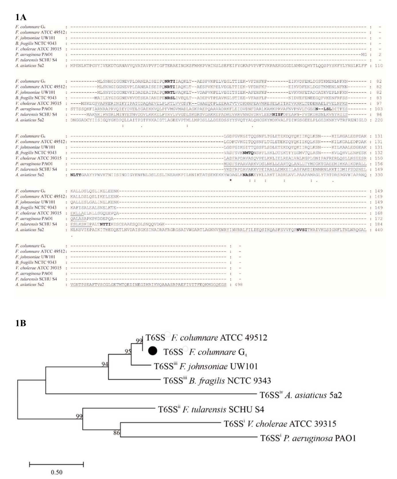
The tssB gene identified in F. columnare strain G4 (GenBank accession no: PV745264) contains a 450 bp open reading frame (ORF) encoding 149 amino acids (aa), with a predicted molecular mass of 16.59 kDa. The TssB protein was predicted to have no signal peptide, no transmembrane structure, and no conserved domain, but to be localized in bacterial cytoplasm (Fig. 1A). The amino acid sequence identity of TssB from F. columnare strain G4 has 100% identity to FCOL_09740 (GenBank: AEW86757.1) of F. columnare ATCC 49512, which is predicted to be a small subunit of the contractile sheath of T6SS. The TssB has sequence identities of 75.2%, 48.7%, 20.4%, 20.1%, 16.3%, and 6.3% with its homologs in Flavobacterium johnsoniae, Bacteroides fragilis, Vibrio cholerae, Pseudomonas aeruginosa, Francisella tularensis, and Amoebophilus asiaticus, respectively. Phylogenetic analysis indicated that TssB in F. columnare G4 was clustered closely with the molecule in F. columnare ATCC 49512, and then with F. johnsoniae, a species known to possess T6SSiii (Fig. 1B).
_and_phylogenetic_analysis_(b)_of_predicted_tssb_amino_acid.png)
3.2. Construction of ΔtssB mutant and complementation strains
To investigate the function of tssB, the deletion mutant, ΔtssB was constructed in the wildtype F. columnare G4, along with a tssB single-gene complementation strain, tssB+, and a tssB+tssC+ double-gene complementation strain, tssB+tssC+, as TssB+ and TssC are elements forming a complex together in T6SS.32,33
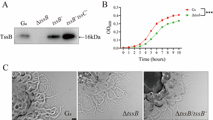
The whole-cell proteins of these four strains were analyzed using Western blotting with a polyclonal antibody against the TssB+ protein (Fig. 2A). In the ΔtssB mutant, a band corresponding to the TssB+ protein was not detected, in contrast to the obvious presence of TssB+ in the wild-type F. columnare G4. In tssB+ single-, and tssB+tssC+ double-complementation strains, a single band with similar molecular weight as in the wildtype strain was observed (Fig. 2A).
__growth_curve_(b)_and_colony_morphology_(c)_in_wildtype__mutant.png)
3.3. Colony morphology, growth and transcriptome analysis of ΔtssB mutant
The wildtype G4, ΔtssB mutant, and tssB+ all showed the typical well-developed rhizoid structure of colonies (Fig. 2B), similarly, as observed under a Leica microscope. The G4 strain, ΔtssB mutant, tssB+ all exhibited similar colony morphology characteristic of a typical spreading growth pattern with dry central elevation, thin edges, and well-developed rhizoids (Fig. 2C). These results indicate that the deletion of tssB gene had no effect on morphological feature of F. columnare colonies.
Turbidimetric measurement was employed to monitor continuously the optical density variation of wildtype G4 strain and ΔtssB mutant at different time points in order to examine the growth kinetic characteristics of the two strains (Fig. 2B). The wildtype G4 strain had a significantly higher growth rate that ΔtssB mutant, indicating that the deletion of tssB gene substantially impairs bacterial growth (P < 0.001).
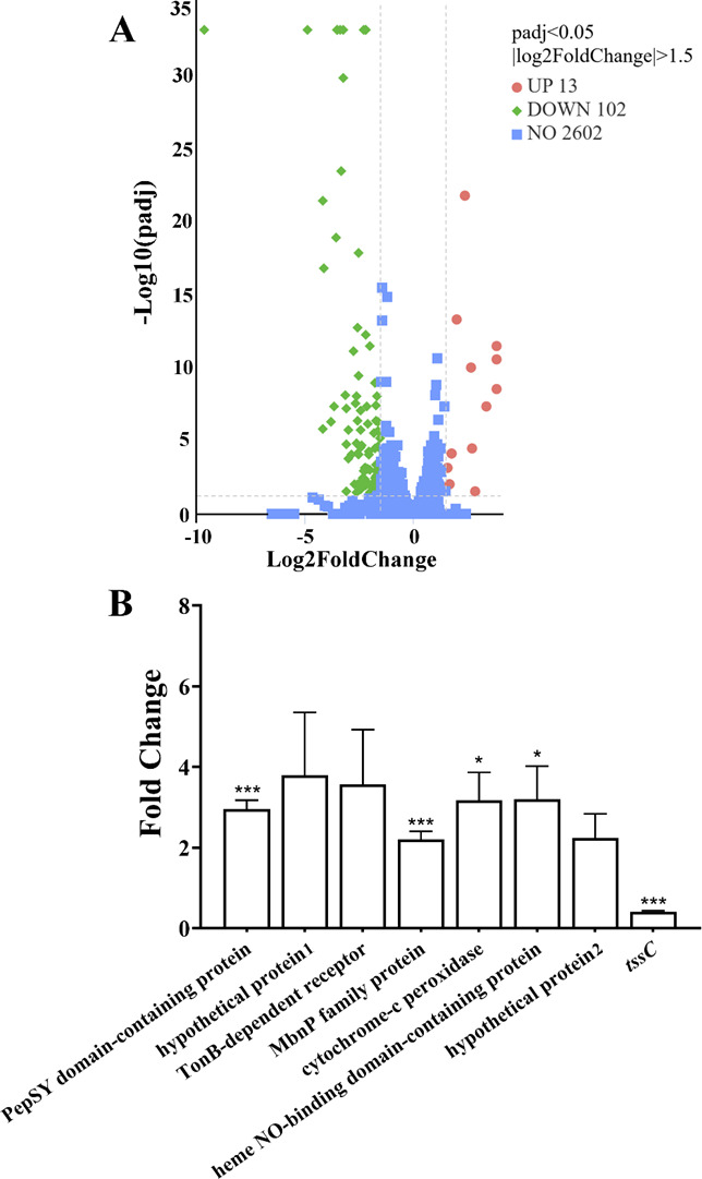
To understand the possible effect of tssB gene, transcriptome analysis was performed comparatively in ΔtssB mutant and the wildtype G4 strain. The analysis of differentially expressed genes (DEGs; P-value (padj) < 0.05) revealed a total of 115 genes, with 13 upregulated and 102 downregulated genes (Supplementary Table 1; Fig. 3A). DEGs exhibiting a fold change > 2.83 included 13 upregulated genes, such as TonB-dependent receptor, MbnP family protein, and cytochrome-c peroxidase, and 102 downregulated genes included Sigma-54-dependent transcriptional regulator, zinc metalloprotease, MarR family winged helix-turn-helix transcriptional regulator, DUF4291 domain-containing protein, and tssC (Supplementary Table 1). DEGs were verified by qRT-PCR, among which PepSY domain-containing protein, TonB-dependent receptor, MbnP family protein, and cytochrome-c peroxidase were significantly upregulated, but tssC expression was downregulated (Fig. 3B).
_between_*f._columnare*_g_4__and_the_*tssb*.png)
GO functional enrichment analysis indicated that the DEGs were significantly enriched in the Cellular Component term, membrane, and heme binding (Fig. 4A). KEGG pathway enrichment analysis demonstrated significant enrichment of DEGs in the nitrogen metabolism pathway (Fig. 4B).
_and_kegg_pathway_enrichment_(b)_of_degs_between_wildtype_*f._columnare*_g_4.tiff)
3.4. The loss of antibacterial effect of ΔtssB against A. hydrophila
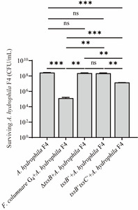
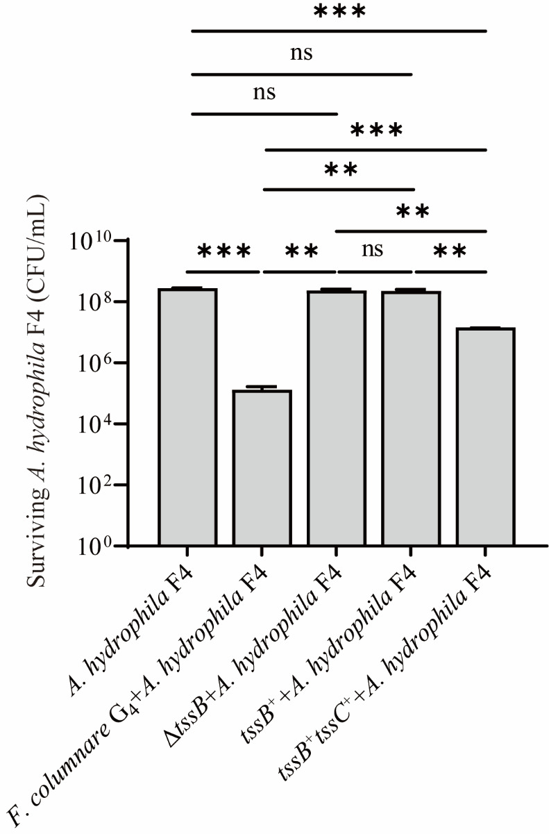
A wildtype A. hydrophila F4 strain was employed to examine the possible effect of ΔtssB mutant in combinations of comparative coculture (Fig. 5). It is obvious that when two wildtypes of A. hydrophila F4 and F. columnare G4 were cocultured together, a significant reduction in bacterial concentration of A. hydrophila F4 was observed when compared with the single culture of A. hydrophila F4 alone (P < 0.001). The ΔtssB deletion mutant showed no such antibacterial effect, as no change in A. hydrophila F4 concentration was observed when cocultured (P > 0.05). The antibacterial effect of single complementation strain, tssB+ was not observed (P > 0.05); but the double complementation strain, tssB+tssC+, had significant antibacterial impact as shown by the lower concentration of A. hydrophila F4 (P < 0.001), although the concentration level was higher than the effect caused by wildtype G4 (Fig. 5). These results thus indicated that the deletion of tssB resulted in the loss of antibacterial effect of F. columnare.
4. Discussion
In this study, ΔtssB, the deletion mutant of the tssB gene encoding a component of the contractile tail of T6SS3,23,25 was established in F. columnare G4. The ΔtssB strain showed similar colony morphology as its wildtype with rhizoids, but had a significantly lower growth rate than the wildtype. It is obvious that the antibacterial effect of ΔtssB on growth, defined as the concentration of A. hydrophila F4, was reduced significantly when compared with wildtype F. columnare G4, indicating the function of T6SS in F. columnare G4.
It has been suggested that the rhizoid colony and the non-rhizoid colony of F. columnare may be related to their virulence in infecting fish.7,34–36 Kunttu et al.34 reported that F. columnare strains with rhizoids always had high virulence. Li et al.35 reported the gene expression profiles of two strains, G4 and G18, of F. columnare with rhizoid and non-rhizoid colonies, respectively. In addition, different genomovars of F. columanre may be related to virulence,36 and a recent identification of virulent F. columanre was also reported to have rhizoid colony morphology.7 F. columnare strains have been reported with large genetic diversity, and T9SS has been identified as the virulence factor for F. columnare, which is also related to bacterial gliding mobility for colonizing host tissues and for colony morphology.37,38 On the other hand, the other two species, F. johnsoniae and F. psychrophilum in the same genus, are also reported with rhizoid colony shapes for their virulent strains.34,39,40 However, the similar rhizoid colony morphology between ΔtssB and the wildtype may indicate that tssB is not related to the formation of rhizoid colony in F. columnare.
In a comparative genomic analysis, virulent strains of F. columnar were found to contain several spike proteins of T6SS, the valine-glycine repeat G (VgrG).24 T6SS, which is widely present in Gram-negative bacteria, is typically recognized as an antibacterial weapon involved in interbacterial competition.21 As noted earlier, partial components of the T6SS have been identified in F. columnare in two previous reports.3,24 In F. johnsoniae, the T6SS was initially classified as T6SSiii because it differs from previously reported T6SSi and T6SSii in the absence of TssA, TssL, TssM, and TssJ.23 The T6SS in F. columnare has been identified as T6SSiii,3,24 as supported by the phylogenetic relationship between TssB in F. columnare and F. johnsoniae in the present study. With the availability of gene manipulation technique,12 the mutant, ΔtssB, was found to show significantly reduced growth rate, and also a defect in bacterial competitiveness, indicating at least a certain degree of functional loss of T6SS in the mutant. However, it should be emphasized that the exact gene composition of T6SS in F. columnare needs to be decoded and its role in bacterial competition requires further investigation. Whether the deficiency of tssB in F. columnare is related to the bacterial virulence also requires further in vivo investigation.
To further investigate the mechanism underlying the differential bacterial competitiveness between the wildtype F. columnare G4 and its ΔtssB deletion mutant, transcriptome sequencing revealed 13 upregulated and 102 downregulated genes. Although the possible function of these genes has not been examined in F. columnare, some of these genes, as reported in other bacteria, may indicate their possible relationship with T6SS. Durán et al.41 reported that in Pseudomonas fluorescens F113, FleQ, a member of the NtrC family can activate the expression of the F1-T6SS and F3-T6SS gene clusters by sensing c-di-GMP signals and nitrogen source status. This activation enhances interbacterial competitive ability (killing other bacterial strains) and rhizosphere colonization efficiency. In Burkholderia pseudomallei, the inactivation of tctR, a MarR family transcriptional regulator, resulted in an increase in hcp2-lacZ transcriptional fusion expression, indicating that TctR acts as a repressor of the T6SS-2 gene cluster.42 In Yersinia pseudotuberculosis, HpaR, another MarR family transcriptional regulator, can positively regulate the expression of T6SS4 in response to oxidative stress.43 MarR family regulators are typically involved in the regulation of virulence factor synthesis and aromatic compound catabolism,44 and the observed downregulation of MarR regulators may reflect their role in normal metabolism, thereby accounting for the lower growth rate of ΔtssB in the present study. Whether the downregulated sigma-54-dependent transcriptional regulator, which belongs to the NtrC family, and the MarR family winged-helix-turn-helix transcriptional regulators function as transcriptional regulators for F. columnare requires further investigation. In addition, the DUF4291 domain was reported to show high homology with ART-like effector proteins in pathogenic Legionella species,45 but whether the DUF4291 domain in F. columnare is an effector of T6SS also requires further investigation.
A few other downregulated genes may provide a hypothetical basis for explaining the lower growth rate of the ΔtssB mutant in the present study. The deletion of tssB and downregulation of tssC in the mutant might have resulted in the disruption of the T6SS sheath, as TssB and TssC are two components of the T6SS sheath.21 It may be possible that the loss of the T6SS sheath and probably the T6SS organelle may have an influence on the bacterial membrane, and on the growth rate of the ΔtssB mutant. On the other hand, the research on genes enriched in heme-binding function is rather limited in F. columnare, but available literature may indicate that T6SS is probably related to heme-binding and heme transportation. In Burkholderia thailandensis, the outer membrane heme transporter, HmuR was reported to interact with TseZ, a proteinaceous zincophore secreted through T6SS for zinc acquisition.46 In Pseudomonas aeruginosa, the oxygen-binding hemerythrin, Mhr, is secreted as an effector of the H2-T6SS and interacts with cbb3-type cytochrome c oxidase subunits, thereby influencing intracellular NADH/NAD+ levels.47 Collectively, these processes may influence bacterial growth. In the present study, the function of two upregulated genes, cytochrome-c peroxidase and heme NO-binding domain-containing protein, and some downregulated genes, such as hmpA, and cytochrome c, which were enriched in heme-binding, is unknown in F. columnare and requires further investigation.
In the present study, the downregulated genes enriched in nitrogen metabolism as revealed in KEGG include cytochrome c, cbb3-type cytochrome c oxidase subunit I, nirK, nitrate reductase, MFS transporter, some of which may have a relation with T6SS or with nitrogen metabolism as reported in other bacteria. As stated above, cbb3-type cytochrome c oxidase may have a relation with T6SS in Pseudomonas aeruginosa.47 In B. pseudomallei, the downregulation of T6SS is related to the reduction of nitrate reductase operons.48 The nirK is involved in bacterial denitrification.49 However, the exact mechanism of these genes enriched in nitrogen metabolism should be of interest for future research in F. columnare.
In summary, the mutant, ΔtssB, with the deletion of the tssB gene encoding a contractile tail component of T6SS showed reduced growth rate and reduced competitiveness when compared with the wildtype, indicating the functional presence of T6SS in F. columnare. The exact composition of the T6SS in F. columnare, its transcriptional regulation, and its effector proteins should be of interest for further investigation.
Acknowledgements
The research was financially supported by a grant from the National Natural Science Foundation of China (31772892), and the China Agriculture Research System of MOF and MARA (CARS-46). P. Nie received financial support from a special top talent plan "One Thing One Decision (Yishi Yiyi) ([2018]27) and “First Class Fishery Discipline” Programme ([2018]8, [2020]3) in Shandong Province, China.
Authors’ Contribution - CRediT
Conceptualization: Liu Yang (Equal), Nan Li (Equal). Data curation: Liu Yang (Equal), Nan Li (Equal). Formal Analysis: Liu Yang (Equal), Yu H. Deng (Equal). Investigation: Liu Yang (Equal), Nan Li (Equal). Methodology: Liu Yang (Equal), Nan Li (Equal). Software: Liu Yang (Equal), Bo Li (Equal), Yu H. Deng (Equal). Validation: Liu Yang (Equal), Nan Li (Equal). Writing – original draft: Liu Yang (Equal), Nan Li (Equal), Nan Li (Equal), Bo Li (Equal). Funding acquisition: Nan Li (Equal), Pin Nie (Equal). Project administration: Nan Li (Equal), Pin Nie (Equal). Resources: Nan Li (Lead). Supervision: Nan Li (Equal), Pin Nie (Equal). Visualization: Nan Li (Lead). Writing – review & editing: Bo Li (Equal), Pin Nie (Equal).
Ethical Conduct Approval – IACUC
All animal experimental procedures were approved with permission number 20240910 from the School of Marine Science and Engineering, Qingdao Agricultural University, and conducted in strict accordance with its guidelines for the care and use of laboratory animals.
Informed Consent Statement
All authors and institutions have confirmed this manuscript for publication.
Data Availability Statement
All are available upon reasonable request.