
Introduction
Global aquaculture has reached an all-time high, surpassing capture fisheries in aquatic animal production for the first time and consolidating its position as the fastest-growing animal food production sector worldwide.1 According to FAO reports, aquaculture production reached a historic record of 134.1 million tonnes, providing more than half of the fish destined for direct human consumption.2 Within this growth, the Nile tilapia (Oreochromis niloticus) has established itself as a cornerstone species for food security and applied biotechnology, owing to its hardiness, rapid growth, high survival rates, and the high protein quality of its fillets.1,2
In Latin America, tilapia production continues to trend upward, exceeding 4.5 million tonnes.2 Mexico has reaffirmed its position as a strategic producer with high potential, reporting a production of 102,128 tonnes of tilapia in 2024, becoming the most relevant freshwater species for the national aquaculture sector.3 This development is driven by optimal environmental conditions and increasing domestic market demand; however, the sustainability of intensive systems faces critical profitability challenges.4 In this context, feed stands out as the most expensive input, accounting for up to 60% of total production costs, primarily due to the price volatility and supply of fishmeal.5
Given the supply insecurity of animal-derived ingredients due to volatility, research efforts in the aquaculture sector have focused on finding more sustainable plant-based protein sources.4,6 The genus Amaranthus (amaranth) stands out among these alternatives due to its essential amino acid profile, which is rich in lysine and sulfur-containing amino acids, surpassing that of most commercial cereals and legumes.7
However, the biological utilization of plant sources is generally limited by antinutritional factors that reduce digestibility and affect the metabolism of aquatic organisms.8 The most common heat-resistant antinutrients in amaranth seeds include saponins and phytic acid, compounds capable of chelating essential minerals and inhibiting the activity of endogenous digestive enzymes, thereby decreasing protein and dry matter digestibility coefficients.7 Consequently, biotechnological processes have been developed to mitigate these compounds and increase nutrient bioavailability.6,9,10
Among these processes, enzymatic hydrolysis is a highly efficient alternative that selectively cleaves peptide bonds, thereby fragmenting polypeptide chains into low-molecular-weight peptides.11 The result of this process is not only an improvement in nutrient bioavailability and bioaccessibility but also the potential enhancement of the ingredient’s organoleptic properties, acting as a feeding attractant.8
Enzyme selection is a critical determinant, as commercial proteases exhibit different catalytic mechanisms. Alcalase® is a high-biotechnological-effectiveness endopeptidase; its ability to cleave the polypeptide chain internally allows it to break down amaranth globulins and albumins into low-molecular-weight peptides more efficiently than exopeptidases that act only at the chain ends.12 This structural breakdown is crucial for facilitating nutrient utilization in aquaculture. Studies comparing Alcalase® with enzymes containing exopeptidase activity (such as Flavourzyme®)13 demonstrate that while exopeptidases excel at liberating free amino acids, the internal cleavage of Alcalase® yields higher degrees of hydrolysis (DH) and an enriched profile of low molecular weight bioactive peptides11,14 DH is defined as the percentage of peptide bonds cleaved relative to the total peptide bonds in the proteins substrate, typically quantified on a trichloroacetic acid (TCA) soluble nitrogen basis.
For the optimization of these enzymatic processes, methodologies from previously studied plant matrices are commonly adapted, such as the standardization of operating conditions for legumes15: pH 8.0, 120 min, 55 °C, and a 4% enzyme/substrate ratio. This methodology for vegetables aligns with the ranges recommended in literature for various amaranth fractions and isolates, such as temperatures of 45 - 65 °C,16 pH 7.5 - 9.5,17 reaction times of 60 - 240 min,18 and enzyme/substrate ratios of 2 - 6% m/m.19
Nevertheless, there is a critical need to evaluate reaction kinetics specifically for whole amaranth seeds, considering the broad relationship reported between hydrolysates with high Degree of Hydrolysis (DH).20 Thus, evaluating hydrolysis time as a control variable is essential to optimize and achieve a maximized DH while maintaining effective degradation of the cellular matrix without compromising the ingredient’s nutritional value.21
Reviewing the background of amaranth use in aquaculture, most studies have been limited to evaluating the inclusion of raw amaranth seed meal, as in common carp (Cyprinus carpio),22 or protein concentrates from amaranth leaves in Nile tilapia (Oreochromis niloticus),23 without subjecting the whole matrix to biotechnological processes such as prior enzymatic hydrolysis. Conversely, studies on other plant matrices, such as soy,24 and on amaranth isolates,25 suggest that using hydrolysates with controlled, maximized DH is an efficient strategy to improve protein solubility and mitigate antinutritional components.
Despite known operating ranges for Alcalase® in plant systems, there is a critical knowledge gap regarding how the production of a protein hydrolysate from whole amaranth seeds, processed under a reaction time optimized to achieve an optimal DH, affects antinutrients and the subsequent in vivo digestive response of Nile tilapia. To address this, a standard 30% fishmeal substitution level was chosen as the optimal benchmark for plant proteins inclusion, ensuring diets’ palatability while avoiding nutritional imbalances that could alter normal digestive tract function during digestibility evaluations
Therefore, the present study aimed to obtain a protein hydrolysate from amaranth seeds (Amaranthus hypochondriacus) with an optimized degree of hydrolysis, using the enzyme Alcalase, to screen for antinutritional factors (saponins, phytic acid, condensed tannins, and trypsin inhibitors), as well as its impact on the apparent dry matter digestibility (ADMD) and apparent protein digestibility (APD) of amaranth.
Materials and Methods
Raw Material Preparation
Whole amaranth (Amaranthus hypochondriacus, var. Nutrisol) seeds were used in this study. The seeds were ground using an electric mill (Thomas-Wiley, Mod. 4) and passed an 80-mesh sieve (0.180 mm) to ensure uniform particle size. The resulting meal was then stored at 4 °C for subsequent biotechnological processing and chemical analysis.
Proximate analysis of the ingredient
The proximate composition of the amaranth meal was determined in triplicate at the Laboratory of Bioprocesses and Functional Foods of the Univerdad Autónoma de Sinaloa, following official methods26: moisture (method 930.15), crude protein (method 954.01, using a 5.85 conversion factor specific for amaranth), lipids (method 920.39), ash (942.05), and crude fiber (962.06). Nitrogen-free extract (NFE) was determined by difference.
Enzymatic Hydrolysis Process
To obtain the protein hydrolysate, the commercial enzyme Alcalase® 2.4 L (Sigma-Aldrich; catalog no. P5380, specified as ≥2.4 AU/g), derived from Bacillus licheniformis, was used. The process was carried out using the pH-stat method,27 following previously described hydrolysis conditions,15 with minor modifications: a substrate/water ratio of 1:10 (W/v), an enzyme/substrate ratio of 4% (v/w based on the total pure protein content of the substrate), a temperature of 55 °C, and pH 8.0. Upon completion of the optimized reaction time, the enzyme was inactivated via thermal shock at 85 °C for 15 min. The hydrolysate was then dehydrated using low-temperature hot air drying (40 °C) to preserve its biological value.
Degree of hydrolysis (DH%) determination
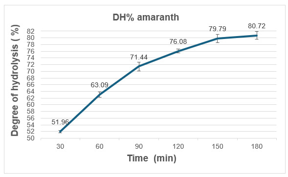
The kinetics of enzymatic hydrolysis were monitored to determine the optimal reaction time for maximum peptide bond cleavage. Aliquots (250 µL) were withdrawn from the reaction mixture at fixed intervals (30, 60, 90, 120, 150, and 180 min). Each aliquot was immediately quenched by adding an equal volume of 20% (v/v) trichloroacetic acid (TCA) to precipitate non-hydrolyzed protein, according to the acid precipitation method.28 Samples were then centrifuged at 10,000 × g for 15 min at 4 °C. Nitrogen content in the supernatant and total sample nitrogen were determined via the micro-Kjeldahl method.26 The degree of hydrolysis (DH %) was calculated using the following equation29: DH (%) = (TCA-soluble nitrogen/Total nitrogen) × 100. The protein content was determined using the following calculation: Protein (%) = ((V HCl x N HCl) x atomic weight of N / 1000 x sample weight) x FC. Where V HCl is the volume of HCl consumed (mL), N HCl is the normality of HCl, and FC is the conversion for amaranth (5.85).
Electrophoretic profile
To evaluate the molecular weight profile and the degradation of amaranth storage proteins during hydrolysis, sodium dodecyl sulfate-polyacrylamide gel electrophoresis (SDS-PAGE) was performed.29 Samples were diluted in Laemmli sample buffer (Bio-Rad Laboratories) supplemented with 5% β-mercaptoethanol and heated at 95 °C for 5 min prior to loading onto the gels.
Samples were separated using 15% polyacrylamide discontinuous gels or precast MINIPROTEAN® gels (Bio-Rad). A dual Color molecular weight standard (Bio-Rad) was used as a marker. Electrophoresis was performed at a constant voltage of 110 V for 120 min in Tris-Glycine- SDS buffer. Gels were stained overnight with Coomassie Blue R-250 and subsequently destained for 2 hours. Protein was visualized and digitized using the Gel Doc™ XR+ imaging system (Bio-Rad).
Antinutritional factors analysis
The concentration of antinutritional factors was evaluated using standardized methodologies: phytic acid,30 was determined by the bauxite/sulfosalicylate colorimetric method; saponins,31 were quantified using the vanillin-sulfuric acid oxidation method; condensed tannins,32 were measured via the vanillin-HCl assay; and trypsin inhibitors33 were determined based on the degree of bovine trypsin inhibition using the synthetic substrate BAPNA (N-benzoyl-DL-arginine-p-nitroanilide).
In vivo apparent digestibility
Three isoproteinic (35%) and isolipidic (10%) diets were formulated using a Microsoft Excel spreadsheet based on the proximate composition of the ingredients: a reference diet (RD) and two experimental diets with 30% substitution by raw amaranth meal (RAM) and hydrolyzed amaranth meal (HAM) (Table 1). Nile tilapias (initial body weight of 20 ± 3g) were distributed in a completely randomized design with three treatments and three replicates per treatment (n = 3; total of 9 tanks). Fish were stocked at a density of 30 fish per experimental tank. The bioassay was conducted in a continuous-flow system using 200-L conical-bottom tanks (Guelph type) equipped with continuous aeration. Water quality parameters were maintained with dissolved oxygen at 6.09 ± 0.44 mg/L; water temperature at 27.8 ± 0.5 °C; pH at 7.5 ± 0.2; total ammonia nitrogen 0.03 ± 0.01 mg/L and nitrite 0.1 ± 0.03 mg/L under a 12:12 h light: dark photoperiod.
Fish were acclimated to the experimental facilities and diets for 5 days prior to the start of date collection. Following acclimation, the digestibility bioassay was conducted over a total experimental period of 45 days. During this period, feed was offered to apparent satiation twice a day (08:00 and 15:00 h). Independently, fecal collection for digestibility determination was conducted during a separate 30-day window within this experimental period. To mitigate the effects of nutrient leaching into water, fresh feces were collected directly from the bottom cone of the tanks using a plastic container 30 to 45 minutes after each feeding event. The collected feces were gently rinsed with distilled water and immediately stored a -40 °C. At the end of the collection period, feces samples from each tank were pooled, lyophilized, and analyzed to determine the content of chromium oxide (Cr₂O₃) and proteins using the acid digestion method.34 For Ingredients, the apparent digestibility of dry matter (ADM) and protein (ADP) were calculated using standard equations for aquaculture nutrition studies35:
ADM = [(100 x ADC of tested diet) – ((100 - % tested ingredient) x ADC of reference diet))] / % tested ingredient
ADP = [(100 x ADP of tested diet x % protein in reference diets) – ((100 - % tested ingredient) x APD of reference diet x %protein of reference diet)] / (% tested ingredient x % protein in the tested ingredient)
Where ADC is the apparent dry matter digestibility (100 -100 x ((%Cr2O3 in diet) / (% Cr2O3 in feces)) and ADP is the apparent digestibility of protein (100 – 100 x ((% Cr2O3 in diet) / (% protein in diet)-1 x (% protein in feces / % Cr2O3 in feces)).
Statistical analysis
Statistical analysis was performed using STATISTICA® v.7.0. Data were first checked for normality and homogeneity of variance. A one-way ANOVA (α < 0.05) was then conducted to identify significant differences, followed by Tukey’s post-hoc test to categorize the treatments based on their statistical differences.
Results
The proximate chemical composition of the amaranth seed meal revealed the highest content of crude protein, followed by lipids, crude fiber, and nitrogen-free extract (NFE) (Table 2).
The degree of hydrolysis (DH) of amaranth seed proteins varied significantly among treatments (P < 0.05) (Figure 1, Table 3). Higher DH values, indicative of greater proteolytic activity and release of amino groups at 150 and 180 min with no significant difference (P > 0.05) between these two times points.
_.png)
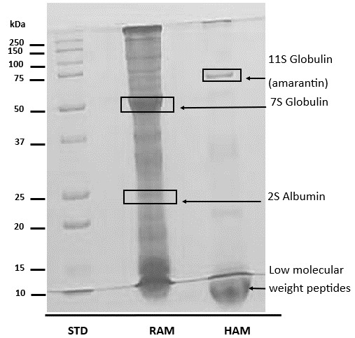
The effect of enzymatic hydrolysis with Alcalase® 2.4L on amaranth seed proteins was evaluated by SDS-PAGE (Figure 2). The electrophoretic profile revealed notable differences between RAM and HAM. While RAM was dominated by high-molecular-weight bands, HAM exhibited a significant shift toward lower-molecular-weight bands.
._h.png)
The hydrolysis process significantly reduced (P < 0.05) de concentrations of saponins and phytic acid by 35.95% and 16.79%, respectively, compared to RAM (Table 4). Regarding trypsin inhibitors, activity was detected in HAM, whereas these levels were non-detectable (ND) in RAM.
The hydrolysis process significantly increased (P < 0.05) the digestibility of the ingredient’s dry matter and protein, by 21.81% and 15.85%, respectively (Table 5). The HAH treatment showed superior performance, achieving protein digestibility greater than 90%.
Discussion
Proximate analysis of the ingredient
The proximal analysis of the amaranth meal revealed an outstanding nutritional profile for a plant-based matrix, highlighting its high crude protein and lipid content. These values are consistent with the ranges reported by Grundy et al.,7 who observed protein variations between 12 - 22% and lipid variations between 6 – 13% in various amaranth varieties. These results confirm that the variety used in this study maintains the biological and nutritional quality standards for the cultivated species. In the context of aquafeed formulation, amaranth seed has become a priority research subject because its protein levels significantly exceed of conventional cereals commonly used in the industry, such as rice (5.6%), maize (9.4%), wheat (12.8%), and oats (15.8%).36
From the perspective of Nile tilapia nutritional physiology, the contribution of lipids and NFE can represent a crucial bivalent energy source. Amaranth lipids, rich in unsaturated fatty acids such as oleic and linoleic acid, together with branched starch fractions that compose the NFE, facilitate the so-called protein-sparing effect, a concept widely documented in teleost physiology.37,38 This physiological mechanism allows tilapia juveniles to utilize the available carbohydrates and lipids to meet their metabolic energy demands and basal maintenance, redirecting the amino acids from the dietary proteins fraction exclusively toward muscle tissue synthesis and somatic growth.39 However, despite this favorable proximal profile, the direct utilization of raw amaranth meal is often limited by the structural complexity of its storage proteins and the latent presence of antinutritional factors,17,25 which justifies the need to implement biotechnological modification processes such as the enzymatic hydrolysis evaluated in this research.
Degree of hydrolysis (DH%)
Shorter hydrolysis times contributed to lower DH values due to reduced enzymatic activity25 Therefore, the hydrolysis conditions previously described,15 with a duration of 120 min, were maximized to 150 min for the amaranth seed substrate for other amaranth varieties,25 indicating that increased time improves hydrolysis yield, similarly to observations in soybean,24 and functional characterization studies with amaranth isolate,25 suggest that the use of hydrolysates with a controlled and maximized degree of hydrolysis can be an efficient strategy to improve protein solubility and also mitigate antinutritional components.
Electrophoretic profile
Enzymatic hydrolysis with Alcalase® 2.4L induced a drastic structural modification in the protein matrix of the amaranth seed (A. Hypochondriacus). The complete degradation of the bands corresponding to high-molecular-weight proteins, such as amarantin (75 kDa) and dense albumins (37–50 kDa), into peptide fractions markedly lower than 10 kDa confirms the high catalytic efficiency of this endopeptidase toward the internal peptide bonds of amaranth.
This transition toward a low molecular weight profile is consistent with what has been reported for amaranth bioactive peptides through extrusion and germination processes,40,41 which demonstrated that proteolysis with Alcalase® efficiently disrupts the compact globular core of amaranth. From a physiological perspective, the reduction in molecular weight not only increases the solubility of the ingredient but also potentially releases cryptic peptide sequences with antioxidant and antimicrobial biological activity.25
The generation of this peptide profile below 10 KDa from sources such as amaranth offers a significant metabolic advantage for tilapia, as these components, being pre-digested in the form of oligopeptides, peptides, and tripeptides, bypass the need for exhaustive gastric and pancreatic hydrolysis, thereby reducing the cellular energy expenditure required for endogenous enzyme secretion.37 Furthermore, at the physiological level, this lower molecular weight peptide profile may enhance nutrient bioavailability and bioaccessibility for aquatic organisms compared to the intact macromolecules of RAM.37,39
Antinutritional factors
A relevant finding was the significant reduction in saponins and phytic acid in HAM, mitigating interference with mineral and nutrient bioavailability and bioaccessibility, which are recurrent in RAM.
When comparing the saponin results of the present study with specialized pseudocereal literature,40–42 it can be demonstrated that the thermal and leaching treatments during enzymatic hydrolysis can destabilize the hydrophobic interactions of saponins with amaranth cell membrane proteins, facilitating their removal.
At the physiological level in tilapia, saponins are amphipathic compounds that act as natural surfactants and, at high concentrations, can interact with the cholesterol in the plasma membrane of enterocytes, causing permeabilization and even cellular lysis of the intestinal epithelium.43 By rescuing the saponin load in the diet through HAM production, it is possible to prevent microvillar shedding in the fish’s foregut, safeguarding the integrity of the mucosal barrier and optimizing the net surface area available for nutrient absorption.
Phytic acid is a chelating agent that reduces protein digestibility in aquatic organisms; therefore, the use of biotechnological processing is necessary to achieve levels compatible with animal metabolism.44 The reduction observed in this study may suggest that the globular core of amaranth exerts a protective effect on phytates, limiting more severe dephosphorylation. Although it represents a moderate reduction, its physiological impact is highly relevant to the mineral homeostasis of tilapia. Free phytic acid possesses a highly reactive polyanionic structure that chelates essential divalent cations in the intestinal lumen, forming insoluble phytates that precipitate and are excreted, inducing metabolic deficiencies and increasing reactive phosphorus levels in aquaculture effluents.45
Furthermore, at the physiological level, phytate electrostatically binds to the terminal lysine and arginine residues of proteins and endogenous enzymes, creating ternary complexes that mechanically block the catalytic site of proteases.43,44 Therefore, the reduction of phytic acid achieved in HAM can decrease the formation of insoluble aggregates, keeping protein fractions hydrophilic and available for the action of digestive enzymes.
Additionally, the absence of detectable condensed tannins (ND) via the vanillin-HCl method rules out issues associated with the indiscriminate precipitation of endogenous digestive enzymes or astringency that could compromise feed palatability.44
On the other hand, the detection of trypsin inhibitory activity in HAM, compared with its non-detectable (ND) state in RAM, does not result from de novo synthesis of inhibitory compounds but rather from a critical increase in their structural bioaccessibility.
The native trypsin inhibitors of amaranth are a seed defense mechanism against injury and herbivores, primarily belonging to the Potato I family. They are encapsulated or integrated into insoluble hydrophobic aggregates along with storage globulins and glutelin’s, which prevents their efficient extraction during conventional analytical assays.46,47 As reported in the literature, low activity levels (17–27 TIU/g) in amaranth seeds47 result from the compaction of storage proteins, which act as a physical shield that masks the activity of these inhibitors in in vitro assays. Furthermore, the targeted hydrolysis with Alcalase® in this study selectively disrupted these macro-aggregates and unfolded the compact globular structure, thereby releasing these stable fractions and enabling their solubilization and detection in the analyzed extract.
Additionally, it has been documented that extensive proteolysis generates short, cryptic peptide fragments rich in amino acids that can act as structural mimics or competitors within the active site of the bovine enzyme used in the spectrophotometric assay, falsely suggesting an increase in inhibitory units.48 However, the residual presence of trypsin inhibitors in HAM did not represent a physiological limitation, given that the high digestibility (>90%) indicates that their concentration remained at levels insufficient to compromise in vivo digestibility in diets for O. niloticus juveniles.
Apparent digestibility
The superiority in the apparent digestibility coefficients (ADC) of HAM reinforces the efficacy of enzymatic pre-digestion compared to RAM. The results in the present study can be explained by the enzymatic activity of Alcalase®, its affinity for the substrate under study, and its resistance to gastrointestinal digestion, thereby allowing the absorption of free amino acids and low-molecular-weight peptides.40,41
Likewise, these results can also be explained by the reduced levels of antinutrients observed in this study, which favored the increase in digestibility coefficients in diets for tilapia juveniles, as verified in studies on beans,49 chickpeas,50 jackfruit,10 and mesquite.51,52
The increase in ingredient digestibility, exceeding 92% in HAM, experimentally demonstrates that enzymatic pre-digestion compensates for the digestive limitations associated with the use of raw plant meals in aquaculture.
Physiologically, HAM’s success in terms of digestibility lies in the synchronization of intestinal absorption kinetics. Raw plant proteins, such as those in RAM, possess highly compact tertiary and quaternary structures, resulting in a significant fraction of nitrogen reaching the hindgut without being hydrolyzed. In contrast, HAM peptides come into immediate contact with intestinal brush-border peptidases, resulting in a coordinated flow of absorption without saturating epithelial active transport sites, thereby drastically increasing the apparent coefficient.53
The improvement in digestibility is consistent with previous studies on the replacement of fishmeal with untreated amaranth seed meal (Amaranthus cruentus), which reached up to 200 g/kg in carp,22 and up to 800 g/kg using amaranth leaf protein concentrate in tilapia.23 The tolerance limit is strictly conditioned by the level of ingredient processing and the species evaluated, for instance, the protein concentration in amaranth leaves eliminates structural barriers such as fiber and phytic acid that the whole grain generally retains.22 Furthermore, amaranth hydrolysate may represent an efficient alternative for the formulation of low-cost, high-nutritional-value aquafeeds.
This study shows that HAM is not merely a replacement for fishmeal but a functional ingredient that optimizes the nutritional utilization of tilapia. However, we emphasize that these digestibility coefficients reflect the 30% inclusion level; further research should investigate how these dynamics shift at higher inclusion rates or under varying metabolic demands.
Acknowledgments
The authors thank the National Council of Humanities, Sciences, and Technologies (CONAHCYT) for their support. Special thanks are extended to the Autonomous University of Sinaloa (UAS), the Autonomous University of Nayarit (UAN), and the National School of Fisheries Engineering for their institutional support.
Authors’ Contribution per CRediT
Formal Analysis: Dayse G. Silva Mendes (Equal), Luís M. Sánchez Magaña (Equal), Mario A. Gómez Favela (Equal). Investigation: Dayse G. Silva Mendes (Equal), Luís M. Sánchez Magaña (Equal), Mario A. Gómez Favela (Equal). Writing – original draft: Dayse G. Silva Mendes (Lead). Writing – review & editing: Edith O. Cuevas Rodríguez (Equal), Luís M. Sánchez Magaña (Equal), Francisco J. Valdez González (Equal). Supervision: Edith O. Cuevas Rodríguez (Equal), Francisco J. Valdez González (Equal). Conceptualization: Oscar I. Zavala Leal (Lead). Methodology: Oscar I. Zavala Leal (Equal), Breidy L. Cuevas Rodríguez (Equal), Juan C. Bautista Covarrubias (Equal). Funding acquisition: Francisco J. Valdez González (Lead). Resources: Francisco J. Valdez González (Lead).
Competing of Interest – COPE
The authors declare that they have no conflicts of interest for the publication of this manuscript.
Ethical Conduct Approval – IACUC
This research was authorized by the University Bioethics committee of the Autonomous University of Nayarit
Informed Consent Statement
All authors and institutions have confirmed this manuscript for publication.
Data Availability Statement
All are available upon reasonable request.