
_bacterial_c.tiff)
Introduction
The global aquaculture industry has expanded rapidly to meet rising seafood demand, leading to intensified farming practices and increased stocking densities.1 However, diseases frequently occur in intensive aquaculture systems, and although antibiotics provide short-term relief, they exacerbate antibiotic resistance and environmental pollution, creating an urgent need for sustainable alternatives.2 Consequently, identifying sustainable alternatives to antibiotics has become essential for the long-term viability of aquaculture.
Probiotics have gained prominence as viable substitutes for antibiotics and are widely utilized in aquaculture.3 Among probiotic candidates, lactic acid bacteria, particularly L. acidophilus AC, are extensively studied for their beneficial effects.4 Live L. acidophilus AC has been shown to enhance growth, boost immunity, and increase disease resistance in various aquatic species.5 Similarly, heat-inactivated L. acidophilus AC exhibits potentiality in promoting growth, regulating immune function, and improving gut health.6 For example, Zhang et al.7 reported that heat-inactivated lactic acid bacteria significantly enhanced growth performance and disease resistance in tilapia (Oreochromis niloticus), while Ren et al.8 observed improved intestinal health in Pacific white shrimp (Litopenaeus vannamei) through modulation of gut microbiota and metabolites.
The large yellow croaker (Larimichthys crocea) is a marine aquaculture species of significant economic value along China’s southeastern coast; however, the development of aquaculture has long been constrained by issues such as slow growth and frequent disease outbreaks. Although many studies have demonstrated that heat-inactivated L. acidophilus AC promotes growth in aquatic animals, few studies have investigated whether the addition of heat-inactivated L. acidophilus AC to L. crocea feed has distinct probiotic effects. Furthermore, research on the mechanism of heat-inactivated L. acidophilus AC remains insufficient. Against this backdrop, this study evaluated the effects of heat-inactivated L. acidophilus AC on the growth performance of juvenile L. crocea, and through the integration of metabolomics and microbial community analysis to elucidate the mechanisms. The results are expected to provide practical guidance for the application of heat-inactivated L. acidophilus AC in L. crocea aquaculture and promote a shift toward sustainable, antibiotic-free aquaculture practices.
Materials and Methods
Preparation of Heat-inactivated L. acidophilus AC Suspension and Experimental Diets
L. acidophilus AC, previously isolated from the intestine of L. crocea,9 was confirmed to exhibit probiotic properties in our laboratory. Hemolysis tests verified its lack of pathogenicity. Glycerol-preserved L. acidophilus AC was revived, and single colonies were inoculated into Lactobacillus-selective broth. Cultures were incubated at 37°C for 48 hours in a constant-temperature shaker. Bacterial concentration was measured as optical density (OD) at 600 nm and adjusted to 1×10¹⁰ CFU/g and 1×10¹¹ CFU/g for the experimental groups. The suspensions were heat-inactivated in a 70°C water bath for 10 minutes. Inactivation was confirmed using the dilution plate method: aliquots of the heat-treated suspension were spread onto MRS agar plates (three replicates per group) and incubated anaerobically at 37°C in an anaerobic jar. Plates were inspected for colony growth at 12, 24, and 36 hours to ensure complete inactivation.
Experimental diets were prepared by uniformly spraying the heat-inactivated L. acidophilus AC suspension onto a basal diet (Jianma brand L. crocea compound feed: crude protein ≥47%, crude fat ≥5%). The control diet (NC group) was sprayed with an equivalent volume of phosphate-buffered saline (PBS). Diets were dried at 37°C in an oven, coated with a 10% sodium alginate solution for stability, and stored at 4°C. Fresh diets were prepared every three days to maintain the quality.
Experimental Animal Husbandry Management
The feeding trial was conducted at the Changzhi Island marine aquaculture base (Zhoushan Peninsula Aquaculture Co., Ltd.). Juvenile fish were acclimated in seawater cages for two weeks, fed the basal diet. Nine cages (1m × 1m × 1m) were each stocked with 30 healthy fish (average initial weight: 102.8 ± 3.2 g). These cages were randomly assigned to three dietary treatments, resulting in three replicate cages per treatment. The treatments were: (1) NC group (basal diet + PBS), (2) ABI1 group (basal diet + 1×10¹⁰ CFU/g heat-inactivated L. acidophilus AC), and (3) ABI2 group (basal diet + 1×10¹¹ CFU/g heat-inactivated L. acidophilus AC). Fish were housed in 1×1×1 m cages and fed twice daily (05:00 and 17:00) at approximately 4% of body weight for 56-d. Water quality was as follows: temperature 18.1–25.9°C, salinity 26–30‰, nitrite <0.005 mg/L, ammonia nitrogen <0.2 mg/L, and dissolved oxygen >6.5 mg/L.
Sample Collection
Following the 56-d trial, fish were fasted for 24 h. The number, body length and weight in each group was recorded for subsequent growth indexes analysis. Twelve fish were randomly selected from each cage, and three of them were used for the whole body composition analysis, three fish were dissected to obtain muscle, liver and viscera that used to calculate the muscle body composition analysis, the hepatosomatic and the viscerosomatic indexes. In addition, six fish were dissected, and the intestinal tissues from three of them were fixed in 4% paraformaldehyde for histological examination. The remaining three intestines were placed in sterile cryovials and frozen in liquid nitrogen for intestinal microbiome and metabolome analysis.
Growth Parameter Analysis
Growth parameters were calculated using the following equations.
Weight Gain Rate (WGR, %) = 100 × (Wt – W0) / W₀
Specific Growth Rate (SGR, %/d) = 100 × (ln Wt - ln W0) / t
Condition Factor (CF, g/cm3) = 100 × Wt / L3
Hepatosomatic Index (HSI, %) = 100 × Wh / Wt
Viscerosomatic Index (VSI, %) = 100 × Wv / Wt
Where: W0 = initial body weight (g), Wt = final body weight (g), L = body length (cm), Wh = liver weight (g), Wv = visceral weight (g), and t = experimental duration (days).
Intestinal Histological Analysis
Fixed intestinal samples were processed by Hangzhou Haoke Biotechnology Co., Ltd. for paraffin embedding and hematoxylin-eosin (HE) staining. Sections were scanned using a digital whole-slide pathology scanner (KF-FL-020) and analyzed with Visiopharm software to quantify, villus length and goblet cell numbers.
Intestinal Microbiota Analysis
Total DNA was extracted from intestinal samples using the MP FastDNA Spin Kit (MP Biomedical, USA). The V3–V4 region of the 16S rRNA gene was amplified with primers 338F (5’-ACTCCTACGGGGAGGCAGCA-3’) and 806R (5’-GGACTACHVGGGTWTCTAAT-3’). PCR products were purified, quantified, and sequenced on the Illumina NovaSeq 6000 platform. Raw reads were filtered, trimmed, and merged to generate operational taxonomic units (OTUs) for diversity, differential abundance, and correlation analyses.
Intestinal Non-targeted Metabolomics Analysis
Intestinal samples stored at -80 °C were sent to Beijing Biomarker Technologies Co., Ltd. for non-targeted metabolomics analysis. The liquid chromatography-mass spectrometry (LC-MS) system included the Waters Acquity I-Class PLUS ultra-high-performance liquid chromatography tandem system and the Waters Xevo G2-XS QTof high-resolution mass spectrometer. Dual-channel data acquisition was performed using MassLynx V4.2. Peak extraction and alignment were performed using Progenesis QI software. Data identification utilized the online METLIN database, public databases, and Biomarker’s internally constructed library via Progenesis QI software. Theoretical fragmentation identification was also conducted to ensure that the deviation of parent ion mass numbers was less than 100 ppm and the deviation of fragment ion mass numbers was <50 ppm. Data analysis was performed using the Biomarker Cloud platform (http://www.biocloud.net).
Statistical Analysis
Data were analyzed using one-way analysis of variance (ANOVA) followed by Duncan’s multiple range test in SPSS 27.0. Results are expressed as mean ± standard deviation, with statistical significance set at P < 0.05.
Results
Effects of Heat-inactivated L. acidophilus AC on the Growth Performance of Juvenile L. crocea
As presented in Table 1, the WGR and SGR increased with the higher concentrations of heat-inactivated L. acidophilus AC, though these differences were not statistically significant compared to the NC group (P > 0.05). Notably, the CF in the ABI1 group was significantly higher than that in the NC group (P < 0.05). No significant differences were observed in the HSI or VSI across all groups (P > 0.05).
Effects of Heat-inactivated L. acidophilus AC on Intestinal Tissue of Juvenile L. crocea
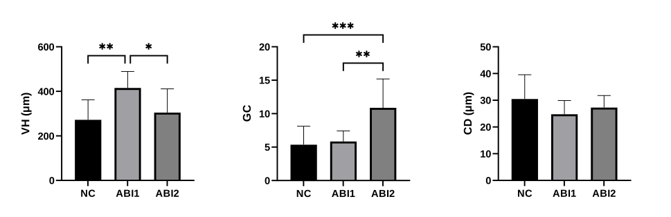
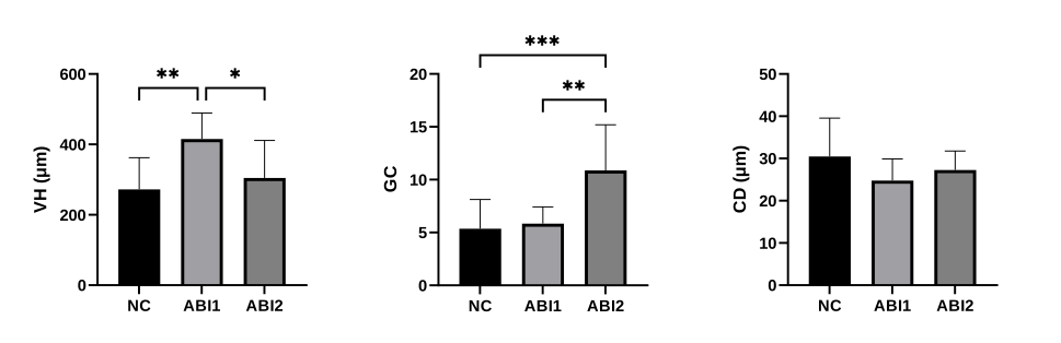
Quantitative analysis (Fig. 1) confirmed that compared to the NC group, the villus length was significantly greater in the ABI1 group (P < 0.05), while the number of goblet cells per villus was significantly higher in the ABI2 group (P < 0.05).
Effects of Heat-inactivated L. acidophilus AC on the Intestinal Microbiota of Juvenile L. crocea
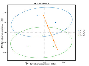
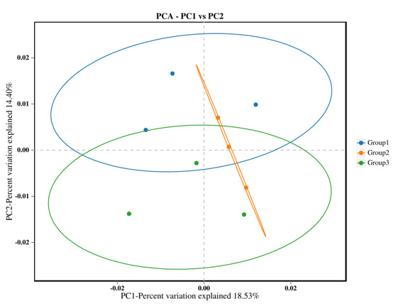
Alpha diversity analysis (Table 2) showed no significant differences in Chao1, ACE, or Shannon indices among the NC, ABI1, and ABI2 groups (P > 0.05). However, the Simpson index was significantly lower in the ABI2 group (P < 0.05), indicating a decrease in microbial diversity. Principal Component Analysis (PCA, Fig. 2) revealed differences in microbial community structures in the ABI1 and ABI2 groups compared to the NC group.
_bacterial_c.tiff)
At the phylum level (Fig. 3), the dominant intestinal microbiota in L. crocea included Proteobacteria, Bacteroidota, Actinobacteriota, Acidobacteriota, Chloroflexi, and Firmicutes. With increasing concentrations of heat-inactivated L. acidophilus AC, Actinobacteriota abundance increased, while Firmicutes abundance decreased. The relative abundance of Gemmatimonadota was significantly higher in the ABI1 group (P < 0.05). At the genus level (Fig. 3), dominant genera included unclassified_Bacteria, unclassified_Micrococcaceae, unclassified_Gemmatimonadaceae, Sphingomonas, unclassified_Vicinamibacterales, and unclassified_Microscillaceae. Higher concentrations of heat-inactivated L. acidophilus AC were associated with increased abundance of unclassified_Micrococcaceae, Sphingomonas, unclassified_Microscillaceae, unclassified_Vicinamibacteraceae, Devosia, and unclassified_Sphingomonadaceae, while unclassified_Vicinamibacterales decreased.
Effects of Heat-inactivated L. acidophilus AC on the Intestinal Metabolites of Juvenile L. crocea
Differential Metabolite Analysis
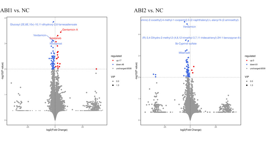
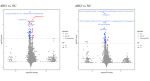
From the volcano plots, where each data point corresponds to a specific metabolite. The horizontal coordinate (x-axis) represents the fold change in expression levels of metabolites between different groups (log2 Fold Change), and the vertical coordinate (y-axis) represents the level of statistical significance (-log10 P-value). The size of the points is positively correlated with the variable importance in projection (VIP) value of the OPLS-DA model, meaning that the larger the point, the stronger the discriminating ability of the metabolite in the model, and the higher the reliability of the screening results. Blue marks indicate down-regulated differentially expressed metabolites, red marks indicate up-regulated differentially expressed metabolites, and gray represents metabolites that were detected but no significantly different. To further highlight key metabolites, the five metabolites with the highest significance based on P-values were labeled in the figure.
Volcano plots (Fig. 4) demonstrated the significant metabolic shifts. Compared to the NC group, the ABI1 group exhibited 17 significantly upregulated and 48 significantly downregulated metabolites. Key upregulated metabolites included Gentamicin A and Seletalisib, while Glucosyl (2E,6E,10x)-10,11-dihydroxy-2,6-farnesadienoate, Verdamicin, and Nocloprost were notably downregulated. In the ABI2 group, 3 metabolites were significantly upregulated and 61 downregulated, with Verdamicin, (R)-3,4-Dihydro-2-methyl-2-(4,8,12-trimethyl-3,7,11-tridecatrienyl)-2H-1-benzopyran-6-ol, and 5b-Cyprinol sulfate among the most downregulated.
Differential Metabolite Fold Change Analysis
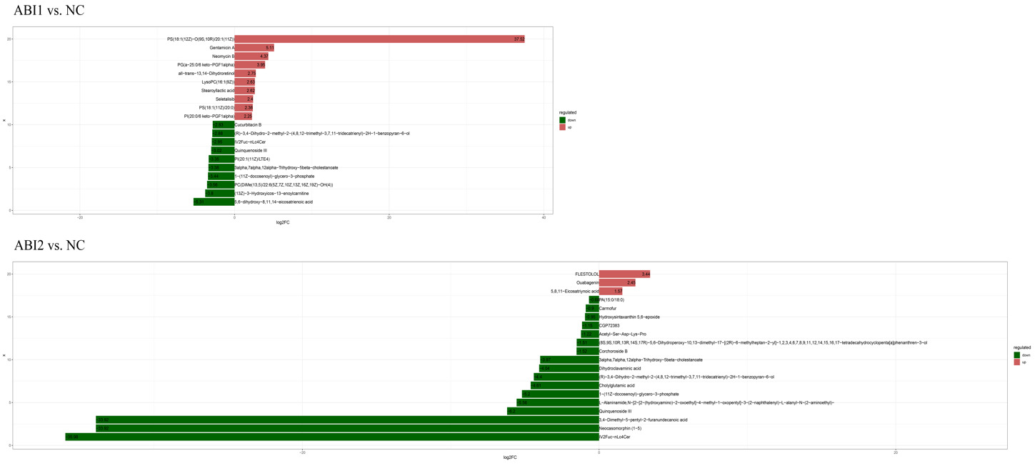
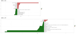
Fold change analysis (Fig. 5) showed that in the ABI1 group, PS(18:1(12Z)-O(9S,10R)/20:1(11Z)) exhibited the greatest upregulation (37.52-fold), while 5,6-dihydroxy-8,11,14-eicosatrienoic acid showed the largest downregulation (5.31-fold). In the ABI2 group, FLESTOLOL was the most upregulated (3.44-fold) metabolite, and IV2Fuc-nLc4Cer was the most downregulated (35.98-fold) metabolite.
KEGG Functional Annotation and Enrichment Analysis
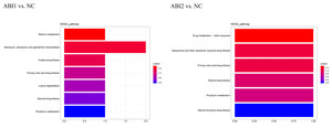
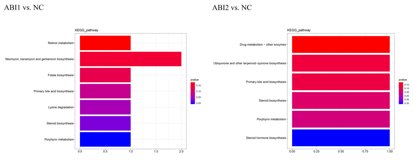
The KEGG pathway enrichment analysis of differential metabolites, as shown in Fig. 6, indicates that the differential metabolites between the ABI1 and NC groups were enriched in retinol metabolism, neomycin, kanamycin and gentamicin biosynthesis, folate biosynthesis, primary bile acid biosynthesis, lysine degradation, steroid biosynthesis, and porphyrin metabolism. At the same time, the differential metabolites between the ABI2 and NC groups were enriched in drug metabolism-other enzyme systems, ubiquinone and other terpenoid-quinone biosynthesis, primary bile acid biosynthesis, steroid biosynthesis, porphyrin metabolism, and steroid hormone biosynthesis. It can be seen that the differential metabolites of both experimental groups compared to the control group were enriched in three pathways: primary bile acid biosynthesis, steroid biosynthesis, and porphyrin metabolism.
Discussion
Effects of Heat-inactivated L. acidophilus AC on the Growth Performance and Intestinal Tissue Structure of Juvenile L. crocea
The morphological structure of the intestines directly influences fish health and growth by affecting digestion and absorption, making it a key factor in improving growth performance in aquaculture. Intestinal villus length, and number of goblet cells are key structural indicators that influence fish growth and digestive function. Longer intestinal villi increase the surface area available for nutrient absorption, thereby significantly improving growth rates and feed conversion efficiencies.10,11 Intestinal goblet cells secrete mucus to maintain mucosal integrity and protect intestinal tissues from environmental and microbial stressors.12,13 In this study, feeding a diet supplemented with heat-inactivated L. acidophilus AC significantly improved intestinal morphology, as evidenced by increased villus length and goblet cell count in the ABI1 group. These changes suggest enhanced intestinal structural development and barrier function. At the genus level, the abundance of Sphingomonas in the experimental groups was slightly higher than that in the control group. Previous studies have shown that supplementation with Sphingomonas can improve intestinal morphology (by increasing the VH/CD ratio in the ileum) and increase the abundance of beneficial bacteria,14 such as Lactobacillus, that produce short-chain fatty acids (SCFAs). SCFAs promote epithelial cell proliferation and maintain barrier integrity.15 Although SCFA levels were not directly measured, the improvements in intestinal structure and enhanced barrier function observed in this study may be associated with this bacterial strain.
The CF in Group ABI1 increased significantly. This index is positively correlated with crude fat content and reflects fish growth and health status,16 suggesting that heat-inactivated L. acidophilus AC may indirectly improve fattening by influencing fat digestion and absorption. In this study, although weight gain and specific growth rate showed an upward trend with increasing supplementation levels, the differences were not significant. Therefore, heat-inactivated L. acidophilus AC does not appear to directly enhance growth performance but rather mediates these effects indirectly by improving CF and tissue structure and enhancing intestinal health.
Effects of Heat-inactivated L. acidophilus AC on the Intestinal Microbiota of Juvenile L. crocea
The results showed that heat-inactivated L. acidophilus AC did not significantly affect the Chao1 index, ACE index, and Shannon index of the intestinal microbiota of L. crocea, but it did significantly affect the Simpson index. The Chao1 and ACE indices mainly reflect species richness, and there were no significant differences in these indicators among the groups, indicating that heat-inactivated L. acidophilus AC treatment did not change the total number of microbial species in the intestine. The Simpson’s index reflects community evenness; a lower value indicates lower diversity.4 Although this index was significantly lower in the ABI2 group than in the NC group, the difference was minimal, suggesting that high-concentration heat-inactivated L. acidophilus AC only slightly affected microbial community evenness and did not cause significant changes in community structure; however, it may still play a positive role in maintaining the balance of the gut microbiota.
The intestinal microbiota of fish plays a critical role in digestion, immunity, disease resistance, growth, and development.17 In this study, Proteobacteria was the dominant phylum in both the control and experimental groups, which is consistent with previous studies on marine fish.18 Furthermore, in the intestines of L. crocea supplemented with heat-inactivated L. acidophilus AC, the relative abundance of Actinobacteria showed an increasing trend. Since microorganisms belonging to this phylum have been shown to enhance disease resistance in fish and maintain microbial balance, an increase in their abundance may be beneficial to the host.19As mentioned earlier, at the genus level, Sphingomonas may enhance intestinal barrier function in fish by supporting intestinal epithelial development and microbial interactions; therefore, an increase in its relative abundance is also beneficial to the host.
Effects of Heat-inactivated L. acidophilus AC on the Intestinal Metabolites of Juvenile L. crocea
Metabolomic analysis revealed significant treatment-specific metabolic changes in the ABI1 group, with Seletalisib and various phospholipid metabolites (including PS(18:1(12Z)−O(9S,10R)/20:1(11Z)), PI(20:0/6 keto−PGF1alpha), PS(18:1(11Z)/20:0) and LysoPC) showing significant upregulation. As components of the cell membrane, these phospholipids play a key role in maintaining cell integrity and signal transduction.20–22 Seletalisib has been shown to enhance disease resistance in zebrafish.23 5,6-dihydroxy-8,11,14-eicosatrienoic acid, an eicosanoid, is a member of the eicosanoid family and plays a role in inflammatory and immune responses. Therefore, we conclude that the upregulation of seletalisib and various phospholipid molecules in the ABI1 group reflects enhanced membrane integrity, improved signal transduction, and increased resistance to disease, whereas 5,6-dihydroxy-8,11,14-eicosatrienoic acid (an eicosanoid) indicates lower levels of inflammation. These metabolic characteristics are consistent with the improvements in intestinal function.
In the ABI2 group, 5b-Cyprinol, 3alpha,7alpha,12alpha-Trihydroxy-5beta-cholestanoate 1-(11Z-docosenoyl)-glycero-3-phosphate, and Cholylglutamic acid were significantly downregulated metabolites. As bile salts or bile acid derivatives, they are directly involved in metabolic processes such as lipid digestion and absorption.24–27 5,8,11-Eicosatrienoic acid is a polyunsaturated fatty acid analog involved in the regulation of inflammation, influencing cell membrane fluidity, and lipid metabolism in fish.28 Its significant upregulation may enhance the inflammatory response. Overall, the results from the ABI2 group may indicate that lipid metabolism is affected at higher supplementation levels, which could negatively affect growth performance and health status.
In KEGG pathways, compared with the NC group, the differential metabolites in both the ABI1 and ABI2 groups were enriched in three common pathways: primary bile acid biosynthesis, steroid biosynthesis, and porphyrin metabolism pathways. Primary bile acids are essential for the digestion and absorption of lipids,27 steroids, which maintain cellular structural stability as membrane components,29 and porphyrins, which are involved in redox reactions and energy metabolism37. These changes in metabolic pathways suggest that heat-inactivated Lactobacillus acidophilus AC primarily exerts its effects by regulating metabolism and homeostasis rather than by directly promoting growth performance.
Conclusion
This study demonstrates that adding L. acidophilus AC, heat-inactivated at an appropriate concentration, to the diet significantly improves the intestinal morphology, intestinal health, and metabolic profile of juvenile L. crocea. Adding 1010 CFU/g of heat-inactivated L. acidophilus AC produced the most favorable response, particularly in terms of CF, intestinal structure, gut health, and improved metabolism.When the dosage was increased to 1011 CFU/g, no further beneficial effects were observed, and negative effects, such as reduced metabolic efficiency and enhanced inflammatory responses emerged, indicating that the dose-response relationship for this intervention has plateaued or even begun to decline.This finding provides scientific evidence for the precise application of heat-inactivated probiotics in aquaculture.
Acknowledgements
This work was supported by the National Natural Science Foundation of China (42176119).
Article Type
Laboratory-Based Studies.
Funding Statement
The study was supported by the National Natural Science Foundation of China (42176119).
Conflict of Interest Statement
The authors declare that they have no known competing financial interests or personal relationships that could have appeared to influence the work reported in this paper.
CRediT Authorship Contribution Statement
Writing – original draft: Linfeng Huang (Lead). Writing – review & editing: Linfeng Huang (Lead). Supervision: Jiahang Xu (Lead). Investigation: Zhiqiang Ran (Lead). Visualization: Shanshan Wang (Lead). Formal Analysis: Haoyu Qiu (Lead). Software: Chenzhi Tao (Lead). Methodology: Ping Wang (Lead). Funding acquisition: Ping Wang (Lead).
Attestation Statements
Data related to any of the subjects in the study has not been published previously. All study and manuscript data will be made available to the journal editors upon request before and/or after manuscript publication for review or query. This study was designed and reported in accordance with the ARRIVE 2.0 guidelines for animal research, and the corresponding checklist was completed and submitted.
Data Sharing Statement
Data will be made available on request.