
Introduction
The rapid expansion of intensive aquaculture has been pivotal in meeting the global demand for aquatic products. However, the prevalent high-density, high-input production model chronically subjects farmed fish to multiple stressors, such as oxidative stress, dysregulated lipid metabolism, and exposure to chemical toxicants. This constant stress has led to a high prevalence of non-infectious hepatopathies, primarily characterized by hepatocyte damage, posing a major bottleneck to the industry’s sustainable and healthy development 1. The common carp (Cyprinus carpio L.) represents the most commercially important freshwater aquaculture species in China. Functioning as a central hub for metabolism, detoxification, and immune regulation, the liver is pivotal for maintaining organismal homeostasis in fish. In aquaculture practice, dietary factors such as high-fat feeds, oxidized lipids, or mycotoxin contaminated readily induce hepatic steatosis, inflammation, and necrosis, which collectively compromise growth performance, disease resistance, and survival.2 Therefore, elucidating the molecular mechanisms underlying hepatocyte damage and developing effective nutritional interventions are crucial for advancing aquatic animal nutrition and health research.
The exploration of plant extracts as aquafeed additives has gained considerable momentum, driven by their inherent safety, natural origin, and multi-target properties. Curcumin, a natural polyphenol derived from turmeric (Curcuma longa) rhizomes, is well established in mammalian models to possess potent antioxidant, anti-inflammatory, and lipid metabolism modulating activities. It has shown particular efficacy in protecting against chemical-induced and metabolic liver injuries.3 In aquaculture, curcumin supplementation has been reported to enhance growth performance, boost systemic antioxidant defenses, and mitigate histopathological liver damage in common carp.4 However, existing research in aquatic species has largely remained at the phenotypic level, focusing on whole-organism or tissue level observations and conventional physiological and biochemical analyses. Consequently, the subcellular targets of curcumin and its regulatory effects on key signaling pathways remain poorly understood. This knowledge gap impedes a mechanistic understanding of its hepatoprotective action and limits the rationale for its precise dosage and application in aquafeeds.
Mitochondria, the central organelles governing cellular energy metabolism and apoptosis, are pivotal for maintaining hepatocyte homeostasis. Hepatic injuries triggered by diverse stressors are frequently linked to mitochondrial dysfunction, which manifests as loss of membrane potential, impaired ATP production, disrupted dynamics, and defective mitophagy.5 Consequently, preserving mitochondrial integrity both structural and functional has emerged as a promising therapeutic strategy for hepatoprotection. In mammalian systems, curcumin has been shown to enhance mitochondrial biogenesis and function by modulating pathways like Nrf2/ARE and AMPK/PGC-1α. However, whether it functions via similar mechanisms in aquatic species, specifically in common carp hepatocytes, remains unclear and lacks systematic investigation.
To address this, an in vitro model was established using primary common carp hepatocytes, wherein injury was induced by carbon tetrachloride (CCl₄) and concomitant oxidative stress. This model enabled the systematic evaluation of curcumin’s effects on cell viability, redox status, and mitochondrial function. The investigation focused on multiple facets of mitochondrial biology including dynamics, biogenesis, autophagy, and apoptotic signaling to elucidate the molecular mechanisms through which curcumin confers hepatoprotection by targeting mitochondria. A primary innovation of this research is the specific focus on mitochondria as a subcellular target in common carp, revealing an organelle-targeted mechanism for curcumin-induced hepatoprotection. The study extended beyond assessing fundamental parameters like mitochondrial membrane potential and ROS levels to integrate an analysis of curcumin’s regulatory effects on apoptosis-related genes (caspase-3, caspase-9, and bcl-2) and key mitophagy markers (PINK1 and Parkin). Thus, this work provides a theoretical foundation for the precise application of curcumin as a mitochondrial modulator in aquafeeds and offers novel perspectives for nutritional strategies aimed at enhancing hepatic health in farmed fish.
Materials and methods
Cells and Culture. Primary hepatocytes from common carp (Cyprinus carpio) were isolated following a modified protocol based on the method described by Yin et al.6 Briefly, healthy juvenile carp (body weight 50 ± 5 g, obtained from a local fishery) were anesthetized with MS-222 (100 mg/L). Livers were aseptically excised in a laminar flow hood. A two-step in situ perfusion method was employed: first, the liver was perfused for 5–8 minutes with calcium- and magnesium-free Hank’s Balanced Salt Solution (HBSS, containing 0.5 mM EGTA; Thermo Fisher) to remove blood; subsequently, digestion was performed for 10–15 minutes using standard DMEM/F12 medium supplemented with 10% fetal bovine serum (FBS), 100 U/mL penicillin, 100 μg/mL streptomycin, and 0.05% type IV collagenase (Sigma). The resulting cell suspension was filtered through a 100-μm nylon mesh and centrifuged at 800 × g for 5 minutes. The cell pellet was washed twice with phosphate-buffered saline (PBS) and resuspended in complete medium. Cells were seeded onto culture plates pre-coated with 0.01% poly-L-lysine (Sigma) and maintained at 28°C in a humidified incubator with 5% CO₂ (Thermo Scientific, Forma Series II). After 24 hours, the medium was replaced to remove non-adherent cells. All subsequent experiments were performed using adherent hepatocytes in good condition, typically cultured for 48–72 hours.
Experimental Design. An oxidative hepatocyte injury model was established using carbon tetrachloride (CCl₄). Preliminary dose-response assays (data not shown) determined that a 24-hour exposure to 10 mM CCl₄ (Sigma-Aldrich) induced a consistent reduction in cell viability to approximately 50% of the control level, accompanied by marked oxidative stress (e.g., increased ROS and MDA), and this concentration was therefore selected for model induction. Subsequently, a dose-response screening of curcumin (0.5–10 μM) was performed on CCl₄-injured carp hepatocytes. Based on the restoration of cell viability and reduction of oxidative stress markers, 2.5 μM curcumin was identified as the optimal protective concentration (data not shown).
The experiment comprised four groups (Control group, CCl₄ group, 2.5 μM curcumin group and 10 μM curcumin group). For each group, three independent replicate experiments were conducted, and within each experiment, three parallel wells (technical replicates) were assessed. The Control group was incubated in standard DMEM/F12 medium for 8 hours. To induce injury, cells in the remaining groups were first exposed to DMEM/F12 medium containing 10 mM CCl₄ for 4 hours. After this incubation, the medium was replaced. Cells in the CCl₄ group then received fresh DMEM/F12 medium for an additional 4 hours. To evaluate the therapeutic effects of curcumin, cells in the intervention groups received DMEM/F12 medium supplemented with either 2.5 μM curcumin or 10 μM curcumin for the subsequent 4-hour recovery period.
Major Reagents and Instruments. Key reagents: Mitochondria Isolation Kit (Sigma-Aldrich); ATP Assay Kit (Beyotime); Reactive Oxygen Species Assay Kit (DCFH-DA, Beyotime); Trizol Reagent for total RNA extraction (Invitrogen); PrimeScript™ RT Reagent Kit for reverse transcription (TaKaRa); TB Green® Premix Ex Taq™ II for real-time quantitative PCR (TaKaRa); Activity Assay Kits for mitochondrial respiratory chain complexes I, II, III, citrate synthase (CS), and succinate dehydrogenase (SDH) (Nanjing Jiancheng Bioengineering Institute).
Major instruments: Inverted fluorescence microscope (Nikon Eclipse Ti2); Real-time quantitative PCR system (Bio-Rad CFX96 Touch); Multifunction microplate reader (Thermo Scientific Multiskan GO); Refrigerated benchtop centrifuge (Eppendorf Centrifuge 5424 R).
Measurement of Mitochondrial Metabolic Enzyme Activities. Following treatments, mitochondrial proteins were extracted using a commercial Mitochondria Isolation Kit. Protein concentrations were determined with the Bicinchoninic Acid (BCA) Assay (Beyotime). The activities of respiratory chain complexes I (Co I), II (Co II), III (Co III), citrate synthase (CS), and succinate dehydrogenase (SDH) were measured using corresponding activity assay kits according to the manufacturers’ protocols, with absorbance or fluorescence readings acquired on a multifunction microplate reader.
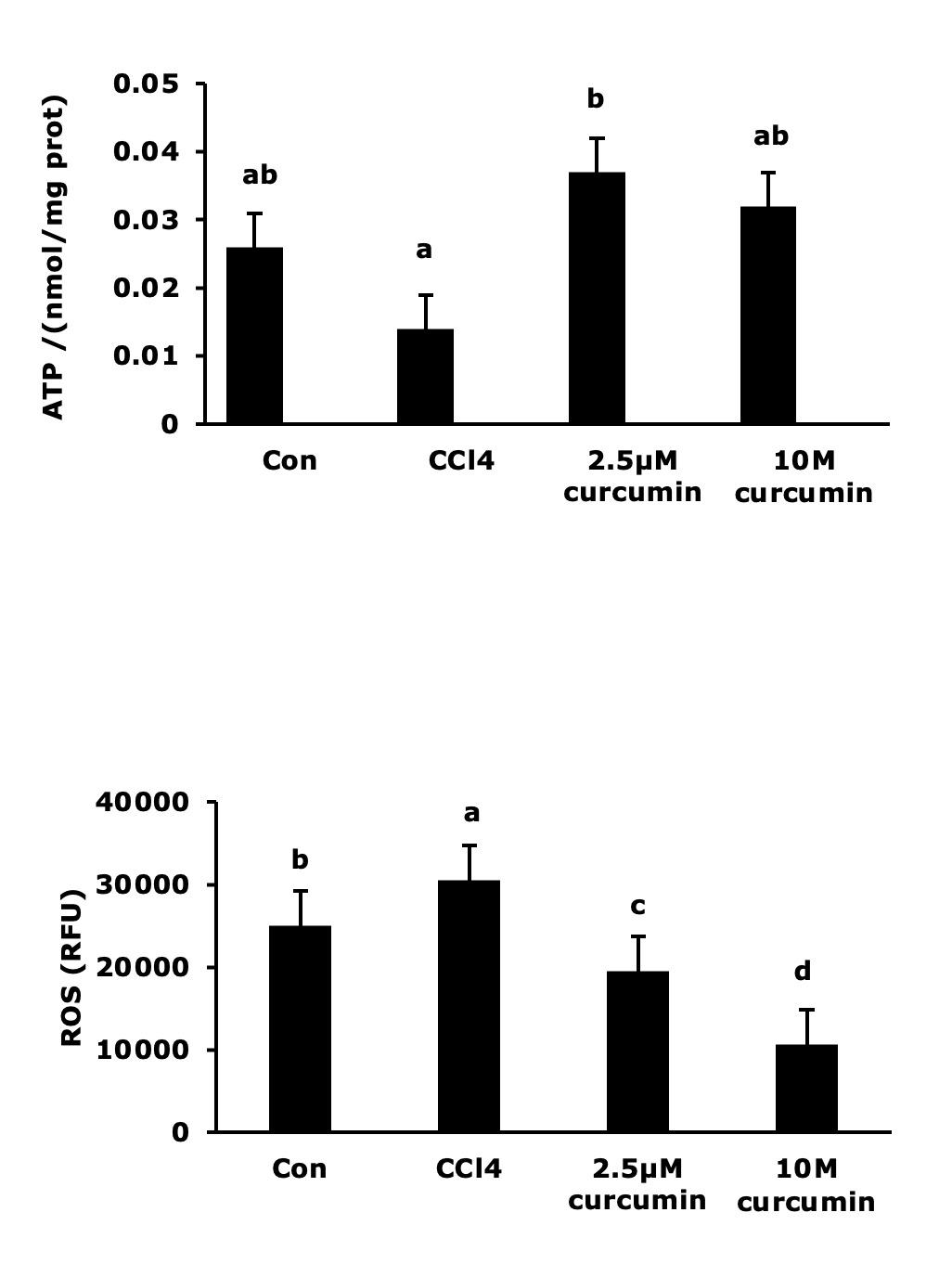
Determination of ATP Content and Intracellular ROS Levels. ATP content was quantified using an ATP assay kit based on the luciferin-luciferase chemiluminescence method. Cell lysates were collected, and luminescence (relative light units, RLU) was measured on a microplate reader. ATP concentration (nmol/mg protein) was calculated using a standard curve. Intracellular ROS levels were assessed using the fluorescent probe 2’,7’-dichlorodihydrofluorescein diacetate (DCFH-DA). After treatment, cells were incubated with 10 μM DCFH-DA in the dark for 30 minutes, washed with PBS, and then observed under a fluorescence microscope for image acquisition. Fluorescence intensity (relative fluorescence units, RFU) for quantitative analysis was simultaneously measured using a microplate reader.
Quantitative Real-Time PCR (qRT-PCR) Analysis. Total RNA was extracted from cells using Trizol reagent. RNA concentration and purity were assessed by spectrophotometry, with A260/A280 ratios ranging from 1.8 to 2.0. One microgram of RNA was used for genomic DNA removal and subsequent cDNA synthesis via reverse transcription. Gene-specific primers were designed based on common carp (Cyprinus carpio) sequences retrieved from NCBI, using Primer Premier 5.0 software. All primers (sequences listed in Table 1) were synthesized by Sangon Biotech (Shanghai). qRT-PCR was performed on a Bio-Rad CFX96 system using TB Green® Premix Ex Taq™ II, under the following cycling conditions: initial denaturation at 95°C for 30 s, followed by 40 cycles of 95°C for 5 s and 60°C for 30 s. The β-actin gene was used as an internal reference. Relative mRNA expression levels of target genes were calculated using the 2−ΔΔCt method. All samples were analyzed in triplicate.
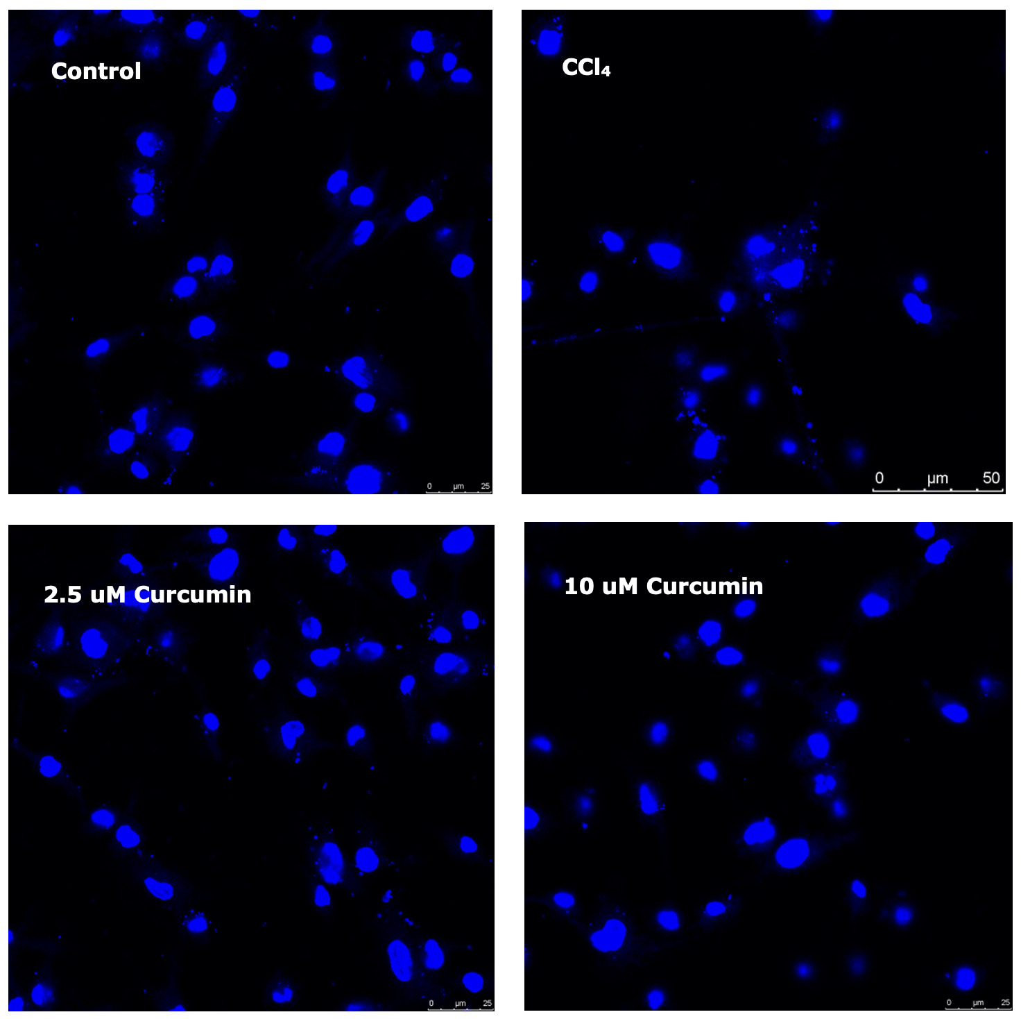
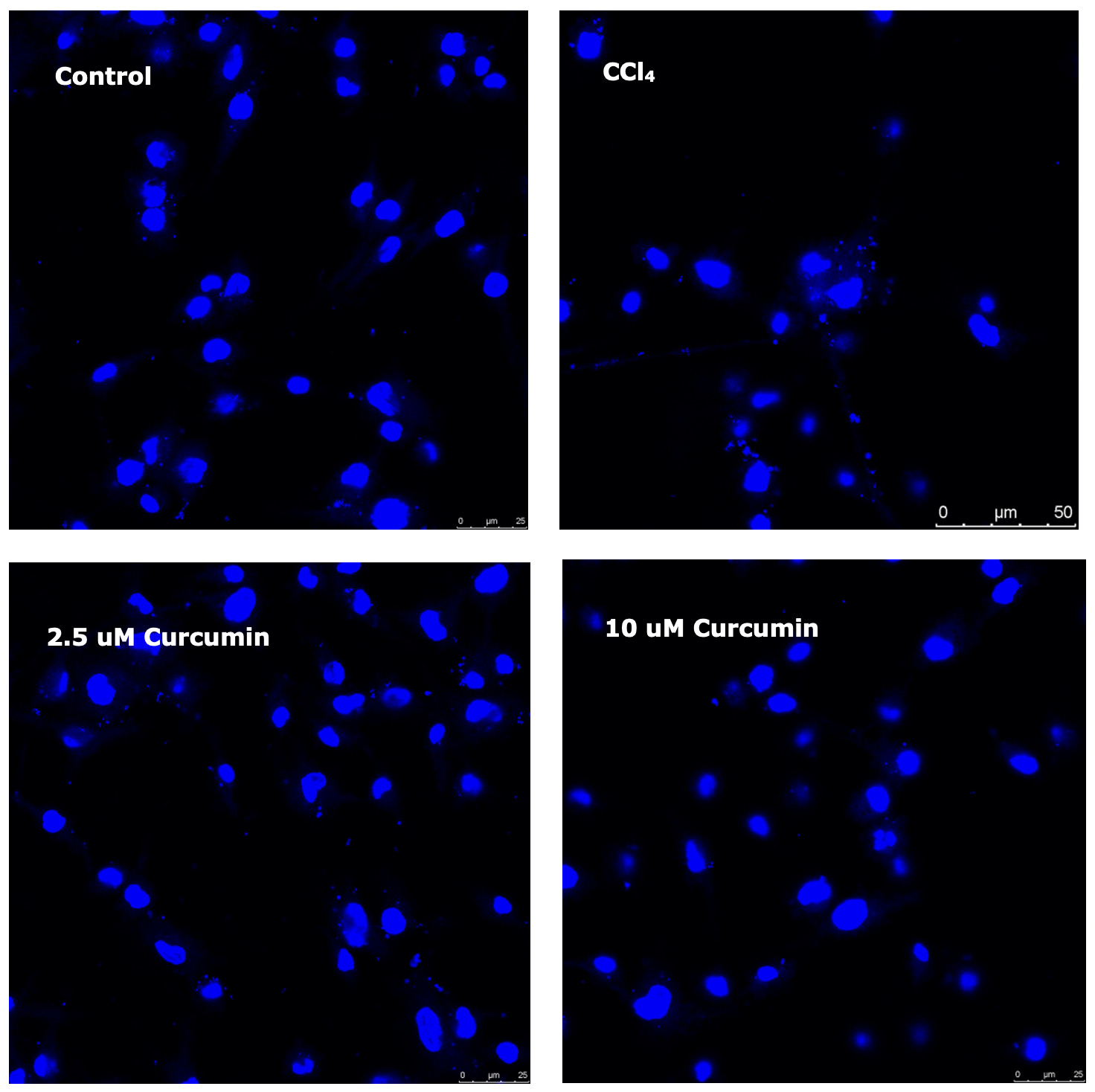
Apoptosis assay. Cell apoptosis was assessed using Hoechst 33258 staining. After fixation with 4% paraformaldehyde, cells were stained with 5 μg/mL Hoechst 33258 for 10 minutes and observed under a fluorescence microscope. Apoptotic cells were identified by characteristic nuclear condensation and fragmentation.
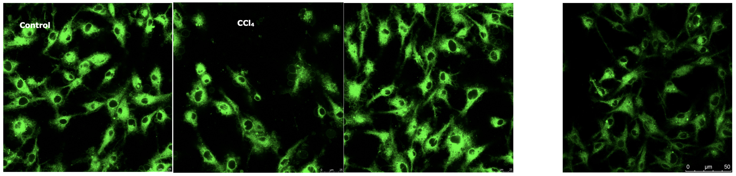
Mitochondrial morphology and content. Mitochondria were visualized in live cells using MitoTracker Green FM. Cells were incubated with 100 nM MitoTracker Green FM for 30 minutes, washed, and immediately imaged under a fluorescence microscope. The average fluorescence intensity was quantified using ImageJ software as an indicator of relative mitochondrial mass.
Data Processing and Statistical Analysis. All data are presented as the mean ± standard deviation (Mean ± SD) derived from nine independent biological replicates (n=3×3). Statistical analyses were performed using SPSS 26.0 software. Differences among groups were assessed by one-way analysis of variance (ANOVA). When ANOVA indicated a significant effect (P < 0.05), Duncan’s multiple range test was applied for post-hoc pairwise comparisons. The significance level was set at P < 0.05.
Results
Effects of Curcumin on Mitochondrial Metabolic Function in CCl₄-Induced Carp Hepatocytes. As summarized in Table 2, CCl₄ exposure significantly inhibited the activity of key mitochondrial respiratory chain enzymes in carp hepatocytes. Specifically, compared to the control, activities of complex I (Co I), complex III (Co III), and citrate synthase (CS) were reduced by 11.0%, 53.0%, and 47.3%, respectively, in CCl₄-treated cells (P < 0.05). Curcumin intervention induced a concentration-dependent recovery of mitochondrial metabolic function. Notably, in cells co-treated with CCl₄ and 2.5 μM curcumin, Co I activity was elevated by 124.5% relative to the CCl₄ group, surpassing even the control level (P < 0.05). Meanwhile, Co III and CS activities were fully restored to levels comparable to the control (P > 0.05). In contrast, while 10 μM curcumin restored Co I and Co III activities to control levels, it only partially rescued CS activity. Moreover, succinate dehydrogenase (SDH) activity remained suppressed at a level similar to the CCl₄ group (P < 0.05). No significant alterations in complex II (Co II) activity were detected across treatment groups (P > 0.05).
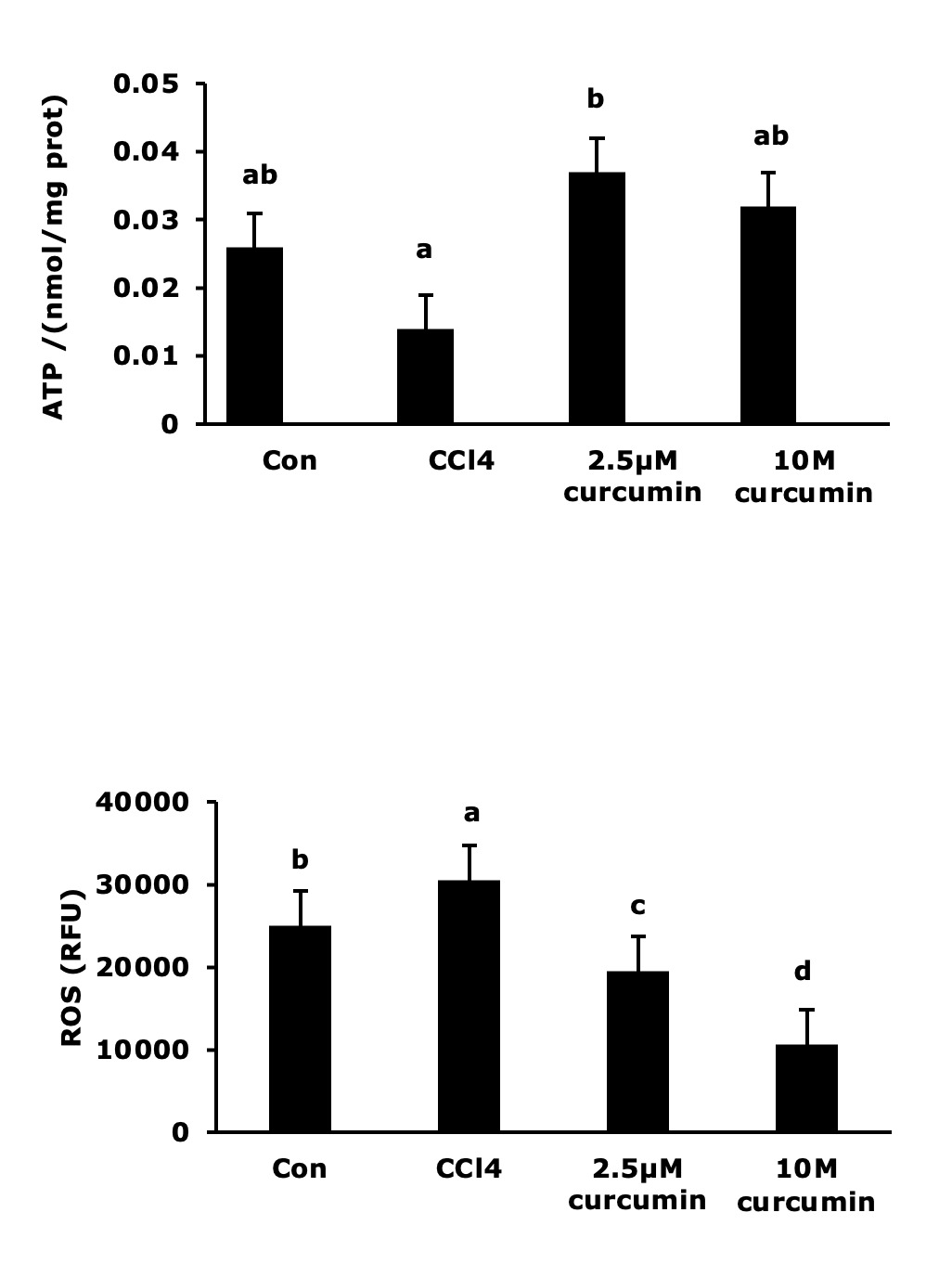
Consistent with these metabolic disruptions, CCl₄ severely compromised both energy homeostasis and redox balance (Fig. 1). CCl₄-treated cells exhibited a significant decrease in intracellular ATP content alongside a marked increase in reactive oxygen species (ROS) levels compared to controls (P < 0.05). Curcumin treatment effectively reversed these alterations. Notably, 2.5 μM curcumin not only restored ATP content to a level above that of the CCl₄ group but also reduced ROS below the baseline control level. Despite demonstrating the most potent direct antioxidant effect, as evidenced by the lowest ROS levels, 10 μM curcumin was only as effective as the 2.5 μM dose in restoring ATP synthesis.
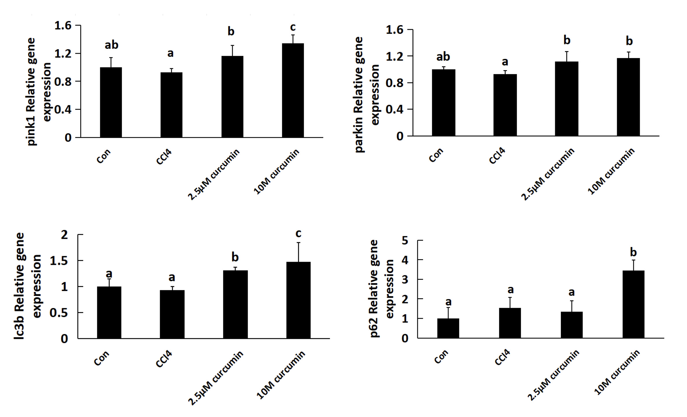
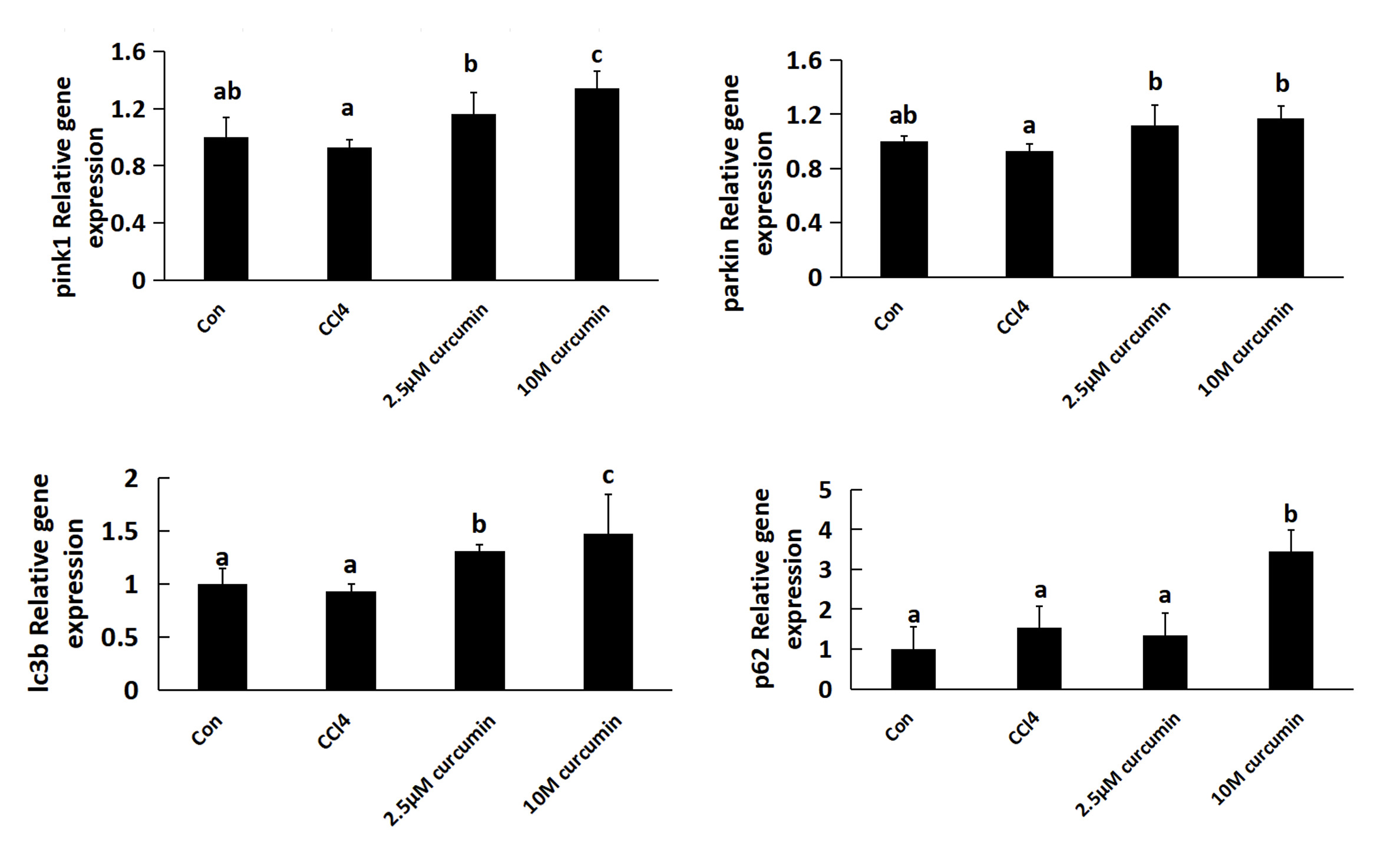
Effect of Curcumin on Mitophagy in CCl₄-Induced Carp Hepatocytes. CCl₄ exposure and curcumin treatment significantly altered the expression of key mitophagy-related genes (Fig. 2). CCl₄ treatment induced an upward (though not statistically significant) trend in the mRNA levels of PINK1, Parkin, LC3B, and the autophagy substrate p62 compared to the control (P > 0.05). Curcumin treatment induced a dose-dependent upregulation of these genes. Specifically, 10 μM curcumin significantly elevated the expression of PINK1, Parkin, and LC3B relative to the CCl₄ group (P < 0.05). Notably, p62 expression in the 10 μM curcumin group exhibited abnormal accumulation, reaching approximately 3.4-fold the control level and significantly exceeding levels in all other groups (P < 0.05). In contrast, 2.5 μM curcumin exerted a more moderate upregulating effect on these genes.
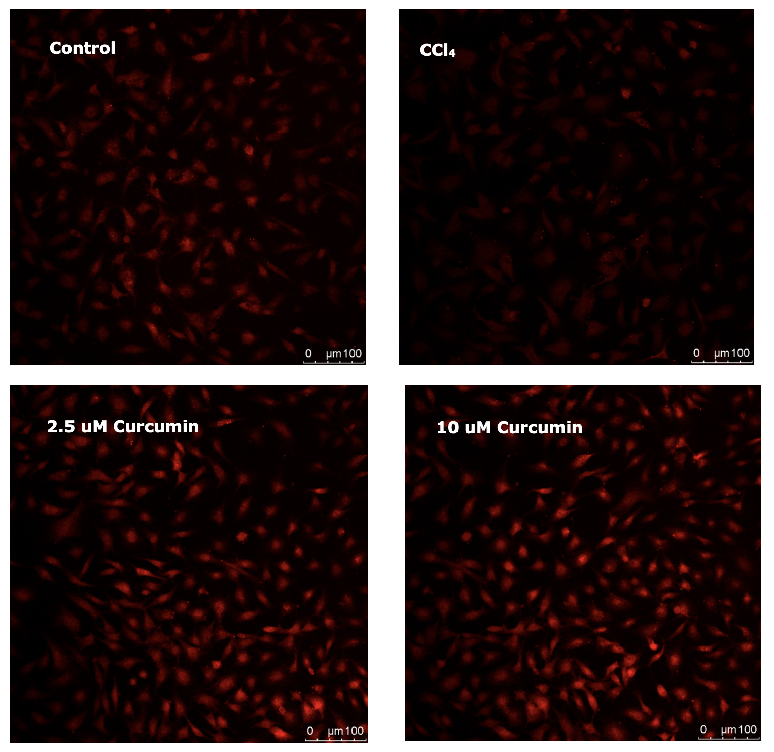
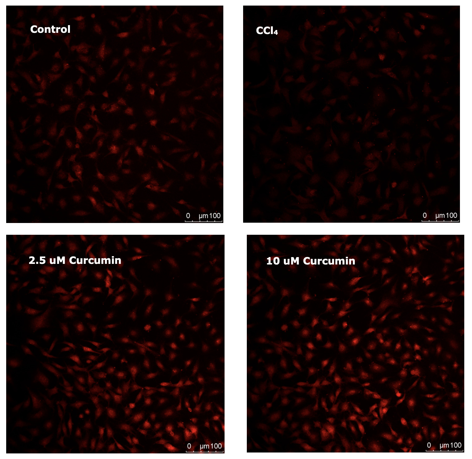
Fluorescence staining of mitochondrial autophagosomes provided complementary morphological evidence (Fig. 3). The number of mitophagosomes was reduced in CCl₄-treated cells compared to controls, indicating impaired autophagic flux. Curcumin treatment increased mitophagosome numbers, with the more pronounced effect observed at the 10 μM concentration. This morphological finding is consistent with the pronounced upregulation of mitophagy-related genes observed at this higher curcumin concentration.
Effect of Curcumin on Apoptosis in CCl₄-Exposed Carp Hepatocytes. CCl₄ treatment significantly altered the expression of key apoptosis-related genes (Table 3). Specifically, mRNA levels of the pro-apoptotic genes caspase-9 and caspase-3 were markedly upregulated in CCl₄-treated cells (P < 0.05), with caspase-3 expression increased approximately 2.5-fold relative to the control. Conversely, expression of the anti-apoptotic gene bcl-2 was significantly downregulated (P < 0.05). Curcumin treatment reversed these alterations in a dose-dependent manner. Treatment with 2.5 μM curcumin restored caspase-3 and caspase-9 expression to control levels and maintained bcl-2 expression. Although 10 μM curcumin also reduced caspase-3 expression, the effect was less pronounced than with 2.5 μM curcumin, and it was less effective at sustaining bcl-2 expression.
Morphological assessment of apoptosis corroborated the gene expression findings (Fig. 4). Nuclear staining revealed typical apoptotic morphology in CCl₄-treated cells, including chromatin condensation, nuclear fragmentation, and pyknosis. In contrast, curcumin-treated cells displayed intact, uniformly stained nuclei, comparable to controls, indicating effective inhibition of apoptosis.
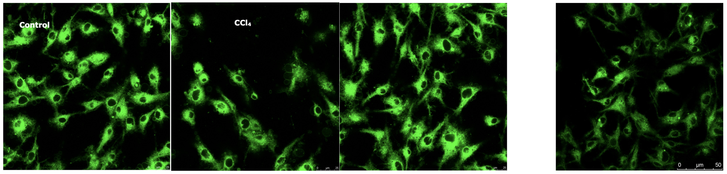
Mitochondrial fluorescence intensity was markedly reduced in the CCl₄ group (Fig. 5), suggesting a loss of mitochondrial mass or impaired membrane integrity. Both control and curcumin-treated cells exhibited robust mitochondrial fluorescence, indicating that curcumin treatment aids in preserving mitochondrial mass and integrity. This preservation of mitochondria is consistent with the observed attenuation of mitochondrial apoptosis.
Discussion
Mitochondria are central regulators of cellular energy metabolism and redox homeostasis, and their functional integrity is crucial for hepatic stress adaptation and metabolic balance in aquatic animals.7 Here, we demonstrate that CCl₄ exposure significantly inhibits the activity of Co I and Co III, as well as CS, in carp hepatocytes. This inhibition resulted in depleted ATP content and elevated ROS levels. These findings are consistent with the classic pathological hallmarks of CCl₄ toxicity: an “energy crisis” and an “oxidative burst,” stemming from electron transport chain blockade and impaired tricarboxylic acid (TCA) cycle flux.8 Mechanistically, inhibition of Co I and Co III activities promotes electron leakage and excessive ROS production. Concurrently, suppression of CS-the TCA cycle rate-limiting enzyme-directly impedes the energy synthesis pathway.9 Collectively, these disruptions converge to form the core mechanism underlying CCl₄-induced hepatocyte injury.
Curcumin exerted a distinct, concentration-dependent effect on mitochondrial recovery. At 2.5 μM, curcumin not only significantly enhanced Co I activity above the CCl₄-treated level but also fully restored the activities of Co III, CS, and SDH. This coordinated restoration led to increased ATP production and a reduction of ROS below baseline control levels. This synergistic restoration of bioenergetics and redox homeostasis aligns with the effects of curcumin reported in mammalian hepatocytes 10. In contrast, while 10 μM curcumin exhibited superior direct antioxidant capacity, it was markedly less effective than the 2.5 μM dose at restoring CS activity and ATP synthesis. This dichotomy suggests a mechanistic shift: lower concentrations primarily facilitate mitochondrial metabolic repair, whereas higher concentrations exert a predominant direct antioxidant effect. This concentration-dependent mechanistic shift, previously unreported in aquatic species, offers a novel framework for defining the optimal therapeutic window of curcumin in aquaculture.
While studies have established the antioxidant and immunomodulatory roles of curcumin in aquatic species,11,12 its systematic regulation of key mitochondrial metabolic enzymes in fish hepatocytes is poorly understood. Notably, the optimal protective concentration identified here (2.5 μM) is lower than the 5 μM typically effective in rat hepatocytes 10, highlighting a species-specific difference. This divergence likely stems from species-specific differences in basal mitochondrial metabolic rates and curcumin metabolism,13 underscoring the necessity for species-tailored dosing in aquafeed formulations. From an applied perspective, the efficacy of 2.5 μM curcumin in restoring mitochondrial function provides key experimental support for its development as a sustainable aquafeed supplement. Its use could enhance stock resilience to environmental stressors, aligning with industry goals for sustainable aquaculture. In summary, a low dose of curcumin (2.5 μM) synergistically ameliorates bioenergetic and redox dysfunction in damaged hepatocytes through coordinated restoration of mitochondrial metabolic enzyme activities. In contrast, the primary effect of higher concentrations is potent, direct antioxidant action.
Mitophagy, the selective autophagy of damaged mitochondria primarily mediated by the PINK1/Parkin pathway, is essential for maintaining mitochondrial quality control and cellular homeostasis. In this pathway, LC3B is incorporated into the expanding autophagosomal membrane, whereas p62 serves as an autophagy receptor and substrate. The steady-state level of p62 is a key indicator of autophagic flux efficiency. Furthermore, quantification of autophagosome number and morphology provides direct morphological evidence of mitophagic activity.14,15 In this study, CCl₄ treatment induced an upward trend in mRNA levels of PINK1, Parkin, LC3B, and p62, yet significantly reduced the number of mitochondrial autophagosomes. This dissociation suggests that CCl₄-induced damage fails to translate gene expression signals into functional mitophagy. This indicates a probable block in the progression from mitophagy initiation to completion, a phenomenon consistent with reports of impaired autophagic flux in CCl₄-treated zebrafish liver.16 It is thus plausible that impaired activation or execution of mitophagy represents a species-specific response to this toxicant in fish hepatocytes.17 Furthermore, 10 μM curcumin robustly upregulated PINK1, Parkin, and LC3B expression, but also caused a pronounced accumulation of p62 (approximately 3.4-fold over controls). Together with the observed increase in mitophagosome number, these data indicate that high-concentration curcumin potently initiates mitophagy. However, the concomitant p62 accumulation implies an obstruction in autophagic flux, likely at the stage of autophagosome-lysosome fusion or lysosomal degradation.18 In contrast, 2.5 μM curcumin moderately elevated the expression of these mitophagy-related genes without inducing p62 accumulation, and also increased mitophagosome numbers. This pattern indicates that 2.5 μM curcumin supports a complete and functional mitophagic cycle, enabling efficient clearance of damaged mitochondria.
Mechanistically, the observed discordance in the CCl₄ group elevated mitophagy-related gene expression coupled with reduced mitophagosome numbers suggests a concurrent disruption of both autophagic initiation and flux. We propose that CCl₄, while triggering transcriptional upregulation, induces severe oxidative stress that compromises critical downstream steps such as autophagosome maturation or lysosomal function thereby arresting functional mitophagy.18 The dose-dependent effects of curcumin underscore the complex and finely-tuned regulation of mitophagy by natural compounds in fish. At a high dose (10 μM), curcumin appears to obstruct autophagic flux, resulting in the accumulation of damaged mitochondria. This finding integrates coherently with the observed poor restoration of CS and SDH activity by high-concentration curcumin, revealing a cross-pathway mechanism that underpins its biphasic action: impaired mitochondrial clearance via blocked autophagy exacerbates metabolic dysfunction. Diverging from reports in mammalian systems, 10 μM curcumin did not confer a net protective benefit in our model. Instead, by impairing autophagic flux, it likely exacerbated cellular dysfunction. This species discrepancy may arise from differences in lysosomal enzyme regulation or autophagic signaling between fish and mammals.17
Dysregulation of apoptosis, a programmed cell death pathway often initiated by mitochondrial damage, which exacerbates hepatic injury and metabolic dysfunction in aquatic animals.19 A critical regulatory node is the balance between pro-apoptotic factors (e.g., caspase-9, caspase-3) and anti-apoptotic proteins (e.g., Bcl-2). In fish hepatocytes, the mitochondrial (intrinsic) apoptotic pathway is predominant. Here, caspase-9 acts as an initiator, cleaving and activating the effector caspase-3 to execute the apoptotic program. The Bcl-2 protein family, which includes anti-apoptotic (e.g., Bcl-2) and pro-apoptotic (e.g., Bax) members, governs this process by regulating mitochondrial outer membrane permeabilization (MOMP), the pivotal event controlling cytochrome c release.20,21 CCl₄ treatment significantly upregulated caspase-3 and caspase-9 expression and downregulated bcl-2 in carp hepatocytes. Consistent with these molecular changes, Hoechst 33342 staining revealed nuclear fragmentation, and MitoTracker Green FM staining indicated loss of mitochondrial mass. Together, these data demonstrate that CCl₄ triggers the intrinsic apoptotic pathway via mitochondrial destabilization. This aligns with reports of CCl₄-induced apoptosis in hepatocytes of other cyprinids, such as grass carp.22 The underlying mechanism likely involves oxidative stress and bioenergetic failure resulting from mitochondrial damage.
Curcumin exerted a concentration-dependent inhibition of apoptosis. At 2.5 μM, it significantly suppressed CCl₄-induced caspase-3 elevation, restored caspase-9 to baseline, and sustained bcl-2 expression. This coordinated gene regulation, together with the observed preservation of nuclear integrity and mitochondrial mass, indicates that low-concentration curcumin effectively blocks the intrinsic apoptotic cascade. This anti-apoptotic action is functionally integrated with curcumin’s concurrent restoration of mitochondrial metabolism and autophagic flux, forming a coherent cytoprotective network. Mechanistically, this synergy can be explained by curcumin’s dual role in preserving mitochondrial health and promoting the clearance of damaged organelles, which collectively limits cytochrome c release and subsequent caspase activation. This integrated mechanism resonates with the synergistic interplay between mitochondrial protection and apoptosis suppression reported in other teleost models.23 In contrast, 10 μM curcumin was less effective, producing a weaker downregulation of caspase-3 and only marginal restoration of bcl-2 expression. This diminished efficacy is likely a direct consequence of the impaired autophagic flux and resulting accumulation of dysfunctional mitochondria caused by the high curcumin dose. Notably, bax expression did not differ significantly among groups, indicating that the intrinsic apoptotic pathway in CCl4-injured carp hepatocytes may be primarily regulated through bcl-2 rather than Bax. The significant downregulation of bcl-2 by CCl4 and its restoration by curcumin, coupled with unchanged bax, suggest that curcumin exerts its anti-apoptotic effect mainly by upregulating Bcl-2 expression.
The anti-apoptotic properties of curcumin in aquatic species are well-documented. For example, it protects Litopenaeus vannamei intestinal cells by downregulating caspase-324 and reduces hepatocyte apoptosis in blunt snout bream (Megalobrama amblycephala) 25. However, its coordinated regulation of the intrinsic apoptotic pathway alongside mitochondrial function and autophagy in fish hepatocytes remains unexplored. Notably, the anti-apoptotic effect of curcumin in our model appears confined to the intrinsic pathway, whereas in mammalian hepatocytes it often involves modulation of both intrinsic and extrinsic pathways.26 This divergence underscores fundamental species-specific differences in apoptotic signaling.21 In summary, curcumin governs apoptosis in CCl₄-injured carp hepatocytes in a concentration-dependent manner. A low dose (2.5 μM) elicits comprehensive protection by preserving mitochondrial integrity and sustaining autophagic flux, thereby blocking the intrinsic apoptotic cascade. Conversely, a high dose (10 μM) compromises this protection, largely due to its disruptive effect on autophagic flux.
Conclusion
In conclusion, this study demonstrates a dose-dependent hepatoprotective effect of curcumin against CCl₄-induced injury in common carp hepatocytes. A low dose (2.5 μM) confers comprehensive cytoprotection through synergistic restoration of mitochondrial bioenergetics, maintenance of autophagic flux, and inhibition of the intrinsic apoptosis pathway. In contrast, a high dose (10 μM) acts primarily as a potent antioxidant but fails to coordinately restore metabolic and autophagic functions. These findings decipher the organelle-specific mechanism of curcumin and establish a precise dosage rationale for its use as a mitochondria-targeted aquafeed supplement. This work provides a mechanistic foundation for advancing hepatic health and implementing precision nutrition strategies in aquaculture.
Acknowledgements
This work was supported by Natural Science Foundation of Shandong Province (ZR2022MC003), the Research and Innovation Talent Fund of Shandong Freshwater Fisheries Research Institute (DSY2024KC02) and the Modern Agriculture Industrial Technology System special project-the National Technology System for Conventional Freshwater Fish Industries (CARS-46). The authors gratefully acknowledge the scientific research personnel of the Fish Nutrition and Processing and Department, Freshwater Fisheries Research Institute of Shandong Province, Jinan City, PR China for their assistance during the sampling. We are also grateful to the management and workers of the fish farm of Freshwater Fisheries Research Institute of Shandong Province for all their assistance during the entire trial period.
Authors’ Contribution
Conceptualization: Yuanyuan Zhang (Lead). Writing – original draft: Yuanyuan Zhang (Lead). Methodology: Jun Wu (Lead). Data curation: Hong Lu (Lead). Formal Analysis: Shuquan Mao (Equal), Bingli Wang (Equal), Peng Xu (Equal). Visualization: Guohong Ma (Lead). Project administration: Yanhua Zhang (Equal), Silei Xia (Equal). Writing – review & editing: Shuren Zhu (Lead).
Competing of Interest
The authors declare that they have no known competing financial interests or personal relationships that could have appeared to influence the work reported in this paper.
Ethical Conduct Approval
All procedures were carried out according to the Institutional Animal Care and Use Committee Guide in Freshwater Fisheries Research Institute of Shandong Province.
Informed Consent Statement
All authors and institutions have confirmed this manuscript for publication.
Data Availability Statement
All data generated or used during the study appear in the submitted article.