
Introduction
The trajectory of global food security is increasingly dependent on the aquaculture sector, which has surpassed capture fisheries as the primary provider of aquatic protein for human consumption. This “Blue Revolution” is pivotal to the economic stability of developing nations, particularly in Southeast Asia, which serves as the hub of penaeid shrimp production, including Litopenaeus vannamei and Penaeus monodon.1 However, the intensification of farming practices – characterized by high stocking densities and organic enrichment – has created an ideal incubator for opportunistic pathogens. Among the myriad of disease threats, Vibriosis stands as the most pervasive and economically damaging bacterial disease,2 costing the global industry billions of dollars annually, with mortality rates often exceeding 80%.3
Within the Vibrionaceae family, Vibrio alginolyticus acts as a devastating opportunistic pathogen under conditions of host stress. Its virulence is multifactorial, driven by the production of extracellular proteases, specifically collagenase, which facilitates bacterial invasion by degrading host connective tissues.4 Furthermore, its ability to form robust biofilms on both shrimp cuticles and aquaculture infrastructure significantly enhances its persistence and resistance to conventional disinfection.5
Historically, management has relied heavily on the application of chemotherapeutic agents such as tetracyclines, quinolones, and beta-lactams.6 This intense selection pressure has accelerated the evolution of multidrug-resistant (MDR) Vibrio strains. The severity of this resistance is quantified by the multiple antibiotic resistance (MAR) index; an index exceeding 0.2 indicates a high-risk environment with frequent antibiotic use.6 Contemporary surveillance has reported isolates with MAR indices well above this threshold, rendering conventional therapies increasingly obsolete and raising public health concerns about the transmission of resistant genes through the food chain.7
In the face of the failing antibiotic paradigm, bacteriophage therapy has re-emerged as the most promising alternative to chemical antimicrobials. Phages are viruses that specifically infect and lyse bacteria; they are the most abundant biological entities on Earth and the natural predators of bacteria. Unlike antibiotics, which often possess broad-spectrum activity that decimates the beneficial gut microbiota (dysbiosis), phages exhibit exquisite host specificity, often targeting bacteria at the strain or species level. This precision allows for the selective elimination of pathogens without disrupting the ecological balance of the aquaculture system or the host microbiome.8
The theoretical advantages of phage therapy are manifold. Phages are self-replicating agents. Upon infecting a host bacterium, they replicate exponentially, increasing their local concentration at the site of infection (“active therapy”). Once the host population is depleted, the phage population naturally declines, minimizing environmental accumulation.5,9 Many phages encode polysaccharide depolymerases, enzymes that degrade the exopolysaccharide (EPS) matrix of bacterial biofilms. This allows phages to penetrate and eradicate biofilm-associated infections that are typically refractory to antibiotics.5 Phages kill bacteria through mechanisms entirely distinct from antibiotics (e.g., peptidoglycan hydrolysis via endolysins). Consequently, MDR status confers no cross-resistance to phage infection.10 A bacterium resistant to every known antibiotic can still be fully susceptible to lysis by a specific phage.11
However, translating phage therapy from the laboratory to the field is impeded by several biological and technical barriers. These include the rapid evolution of bacterial resistance to phages (Bacteriophage Insensitive Mutants - BIMs), the typically narrow host range of individual phages, and the potential instability of phage particles under the harsh physicochemical conditions of aquaculture ponds (fluctuating pH, high temperature, UV radiation).12 Addressing these challenges requires a robust pipeline for the isolation, characterization, and selection of highly potent and stable phage candidates. This study addresses the urgent need for non-antibiotic treatments by isolating and characterizing three novel lytic phages – P18, P24, and P25 – targeting a highly resistant V. alginolyticus strain (V79) with an extraordinary MAR index of 0.9. This foundational research aims to develop phage-based biocontrol products to support sustainable shrimp farming.
Materials and Methods
Bacterial strain isolation and culture conditions
The host bacterial strain, designated V79, was isolated from the hepatopancreas and gut of Pacific white shrimp (L. vannamei) exhibiting clinical signs of Vibriosis, including reddish discoloration, lethargy, and anorexia. The isolation was performed at a commercial aquaculture facility in Vietnam. The samples were aseptically drawn and streaked onto Thiosulfate Citrate Bile Salts Sucrose (TCBS) agar (HiMedia, India), a selective differential medium for Vibrio species. Plates were incubated at 30°C for 24 hours. Yellow, sucrose-fermenting colonies, characteristic of V. alginolyticus, were purified by sub-culturing onto Tryptic Soy Agar (TSA) supplemented with 1.5% NaCl (w/v) to mimic the salinity of the isolation source. Pure cultures were stored in Tryptic Soy Broth (TSB) containing 20% glycerol at -80°C for long-term preservation.
Molecular identification via polymerase chain reaction (PCR)
The taxonomic identity of strain V79 was established through a polyphasic approach: species-specific identification was achieved via PCR targeting the collagenase gene (yielding a 737 bp fragment), while genus-level confirmation was provided by amplification of the 16S rRNA gene using primers 63f/763r as a supplementary validation. Genomic DNA was extracted from an overnight culture using a thermal lysis method.13 1 mL of culture was centrifuged, the pellet resuspended in 100 µL of sterile nuclease-free water, boiled at 100°C for 10 minutes, and centrifuged to remove cellular debris. The supernatant containing the template DNA was used for PCR.
The first PCR assay targeted the collagenase gene (yielding a 737 bp fragment), a species-specific virulence factor associated with tissue degradation in V. alginolyticus.14 The amplification was performed using the primers VA-F (5’-CGA GTA CAG TCA CTT GAA AGC C-3’) and VA-R (5’-CAC AAC AGA ACT CGC GTT ACC-3’). The reaction mixture (25 µL) contained 12.5 µL of 2x PCR Master Mix, 1.0 µL of each primer (10 µM), 2.0 µL of DNA template, and 8.5 µL of nuclease-free water. The thermal cycling conditions consisted of an initial denaturation at 94°C for 5 min, followed by 30 cycles of 94°C (1 min), 55°C (1 min), and 72°C (1 min), with a final extension at 72°C for 7 min.14
To definitively confirm the genus-level identity as Vibrio spp. and to validate the host for phage isolation, a second PCR targeting the 16S rRNA gene was conducted using the specific primers and protocol described by Marchesi et al. (1998).15 The forward primer 63f (5’-CAGGCCTAACACATGCAAGTC-3’) and the reverse primer 763r (5’-GCATCTGAGTGTCAGTATCTGTCC-3’) were utilized to amplify a fragment of approximately 700 bp. The cycling parameters were strictly adapted from the original protocol: initial denaturation at 95°C for 5 minutes; followed by 30 cycles of denaturation at 95°C for 1 minute, annealing at 55°C for 90 seconds, and extension at 72°C for 60 seconds; concluding with a final elongation step at 72°C for 5 minutes. PCR products from both assays were analyzed via electrophoresis on a 1.5% agarose gel stained with ethidium bromide and visualized under blue-light transillumination, with results documented in Supplementary Figure S1.
Antibiotic susceptibility testing and MAR index determination
The antibiotic resistance profile of V. alginolyticus V79 was determined using the Kirby-Bauer disk diffusion method. A standardized bacterial suspension (0.5 McFarland, approximately 1.5 x 10⁸ CFU/mL) was prepared in sterile saline and swabbed uniformly onto Mueller-Hinton agar plates supplemented with 1.5% NaCl.
A panel of 10 antibiotic disks (Oxoid, UK) representing various antimicrobial classes commonly used in aquaculture and human medicine was applied: Beta-lactams (ampicillin, 25 µg, amoxicillin, 10 µg, cefpodoxime,30 µg); Tetracyclines (tetracycline, 30 µg, doxycycline, 30 µg); Macrolides (erythromycin, 15 µg, azithromycin, 30 µg, clarithromycin, 20 µg; Fluoroquinolones (ciprofloxacin,5 µg); Lincosamides (dalacin, 25 µg).
Plates were incubated at 30°C for 24 hours. The zones of inhibition were measured to the nearest millimeter using a digital caliper. Interpretive criteria for classifying isolates as Susceptible (S), Intermediate (I), or Resistant (R) were updated to align with the most rigorous international standards available for Vibrio spp., specifically leveraging the CLSI M45 guidelines16 and EUCAST v15.0 breakpoints17 where specific Vibrio data have been established. For antibiotics lacking specific disk diffusion breakpoints for Vibrio in EUCAST v15.0 (amoxicillin, dalacin, and clarithromycin), interpretations were based on the research of Vo et al. (2025).18 The multiple antibiotic resistance (MAR) index was calculated using the formula MAR=a/b, where a is the number of antibiotics to which the isolate was resistant, and b is the total number of antibiotics tested.19
Bacteriophage isolation and purification
Bacteriophages were isolated from sewage water collected from the drainage canals of intensive shrimp farms, locations known to harbor high densities of Vibrio species. Water samples were initially clarified by centrifugation at 5,000 × g for 10 minutes and subsequently filtered through a 0.45 µm pore-size membrane to remove bacteria and larger particulates.
An enrichment protocol was employed to amplify low-abundance phages. Five milliliters of the filtered water sample was mixed with 5 mL of 2× TSB (supplemented with 1.5% NaCl) and 100 µL of log-phase V. alginolyticus V79 culture. The mixture was incubated at 30°C for 24 hours with shaking (150 rpm) to facilitate phage adsorption and replication. The enrichment culture was then centrifuged (8,000 × g, 10 min) and filtered (0.22 µm) to obtain a cell-free lysate.
Phage activity was detected using the double-layer agar overlay method.20 One hundred microliters of the filtrate was mixed with 100 µL of host bacteria and 4 mL of soft agar (0.7% agar), then poured onto nutrient agar plates. After overnight incubation, clear zones (plaques) indicated the presence of lytic phages. Three distinct phages, designated P18, P24, and P25, were selected based on plaque morphology. These phages were purified through three successive rounds of single-plaque isolation to ensure clonal purity. High-titer stocks (>109 PFU/mL) were prepared in SM buffer (50 mM Tris-HCl, 100 mM NaCl, 8 mM MgSO4, 0.01% gelatin, pH 7.5) and stored at 4°C.21
Transmission Electron Microscopy (TEM)
For morphological characterization, 10 μL of purified high-titer phage suspension (>109 PFU/mL) was deposited onto carbon-coated copper grids and negatively stained with 2.5% uranyl acetate. The grids were dried at room temperature for 10 minutes and visualized using a transmission electron microscope (JEOL 1230, Japan) operated at 80 kV.22 Capsid diameters and tail lengths were determined from multiple independent micrographs, and virion morphotypes were described based on capsid and tail architecture.23
Physicochemical stability assays
To assess the suitability of the isolated phages for field application, their stability under varying environmental conditions (pH and temperature) was evaluated.
The stability of phages P18, P24, and P25 was tested across a broad pH range (3, 5, 7, 9, and 12). Phage suspensions (109 PFU/mL) were inoculated (1:10 ratio) into SM buffer adjusted to the specified pH values using 1M NaOH or 1M HCl.24 The mixtures were incubated at 30°C for 1 hour. Following incubation, the samples were serially diluted, and the surviving phage titer was determined via the double-layer agar method. Results were expressed as PFU/mL. The experiment was performed in triplicate.
The thermal tolerance of the phages was assessed by incubating phage aliquots at defined temperatures: 4°C (control), 30°C, 40°C, and 56°C. Incubation was carried out for 1 hour in a temperature-controlled water bath. Surviving phages were quantified by plaque assay. These temperatures were selected to represent storage conditions (4°C), typical pond temperatures (30-40°C), and potential heat stress (56°C).24
In vitro lysis kinetics and killing curves
The lytic dynamics were assessed using a time-kill assay. An early log-phase culture of V. alginolyticus V79 (OD600≈0.4, approximately 1.5×10⁸ CFU/mL) was divided into four aliquots. Three were infected with phages P18, P24, or P25 individually at a Multiplicity of Infection (MOI) of 0.1. The fourth served as an uninfected control. The initial MOI of 0.1 was selected to evaluate the “active therapy” potential of the phages, where the initial viral density is lower than the bacterial density, requiring active replication and subsequent progeny release to control the host population over 24 hours.25 Cultures were incubated at 30°C with shaking. Bacterial growth was monitored by measuring optical density (OD600) at 1-hour intervals for 8 hours, followed by a measurement at 24 hours.
To correlate optical density with viable cell counts and assess the emergence of resistance, samples were withdrawn after 24 hours of incubation for enumeration of bacteria (CFU/mL on TSA plates) and phages (PFU/mL). Colony morphology of surviving bacteria was carefully noted.
Statistical analysis
Experiments were conducted in triplicate using a completely randomized design. Significant differences in phage stability titers and 24-hour viable cell reductions were tested using One-way Analysis of Variance (ANOVA) followed by Tukey’s Honestly Significant Difference (HSD) post-hoc test (p<0.05). For the in vitro killing curves, a Two-way ANOVA with Dunnett’s post hoc test was used to compare each phage treatment group directly with the single uninfected control group over time (p<0.05 and p<0.01). All statistical analyses were performed using Minitab 18.
Results
Molecular confirmation of V. alginolyticus V79
The presumptive Vibrio strain V79, isolated from diseased shrimp, was subjected to molecular validation. PCR with the VA-F/VA-R primers yielded a single 737 bp amplicon. This gene encodes a critical virulence factor involved in tissue degradation and is a specific marker for V. alginolyticus. To further corroborate the genus-level identification, PCR was performed using the primer set 63f/763r as described by Marchesi et al. (1998).15 This reaction produced a sharp, unambiguous band at approximately 700 bp.26 Gel electrophoresis confirmed clean, single bands for both targets without non-specific amplification products (visual data available in Supplementary Figure S1). The use of this specific primer pair is significant as it was developed to discriminate Vibrio species with high specificity, avoiding the heteroduplex artifacts common in universal 16S amplification of marine samples.27
Collectively, these molecular data definitively confirm that strain V79 is V. alginolyticus, validating it as the correct target for subsequent phage isolation.
Antibiotic resistance profiling and MAR index
The antibiotic susceptibility profile of V. alginolyticus V79 was rigorously evaluated against ten antibiotics. Interpretation of inhibition zones was based on updated breakpoints derived from EUCAST v15.0 and CLSI M45 standards suitable for Vibrio spp., revealing a severe multidrug-resistant phenotype (Table 1).
Strain V79 exhibited resistance to 9 out of the 10 antibiotics tested. The results indicate a profound resistance profile. Notably, ciprofloxacin, previously considered effective or intermediate under older guidelines, is classified as resistant under the stricter EUCAST 15.0 breakpoint (zone <23 mm), highlighting the loss of fluoroquinolone efficacy for this strain. Doxycycline, with a zone of 11.33 mm, falls into the intermediate range, suggesting that while not fully resistant, it is unreliable for treatment. High-level resistance was observed against all beta-lactams (ampicillin, amoxicillin, cefpodoxime) and macrolides.
Even conservatively including all 10 tested, the MAR index is 0.9. This value is extraordinarily high, far exceeding the threshold of 0.2. A MAR index >0.2 is universally recognized as an indicator of isolates originating from high-risk sources where antibiotics are intensively and often indiscriminately used.6 An index of 0.9 characterizes V79 as a extensively in the context of aquaculture, rendering conventional antibiotic therapies futile.
Phage ultrastructure and classification
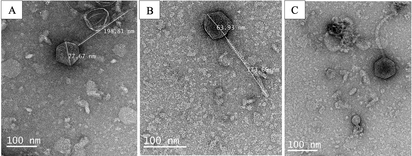
TEM analysis show that all three phages exhibited icosahedral capsids and long, flexible, non-contractile tails, consistent with the sipho morphotype (Figure 1).28
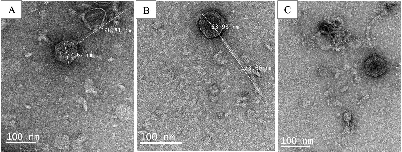
Phage P18 was characterized by an exceptionally long tail (198.81 nm), whereas P24 and P25 exhibited more symmetrical dimensions (Table 2).
The dimensions of P18 (198.81 nm tail) are notably large compared to previously described Vibrio phages with sipho morphology such as ValSw3-329 or RH2G (142 nm).24 This variation in tail length is likely governed by the specific amino acid sequence of the Tape Measure Protein (TMP), which dictates the physical scale of the tail assembly.24 The structural robustness observed in these images correlates with the high viral titers maintained during the purification process.24
Physicochemical stability of bacteriophages
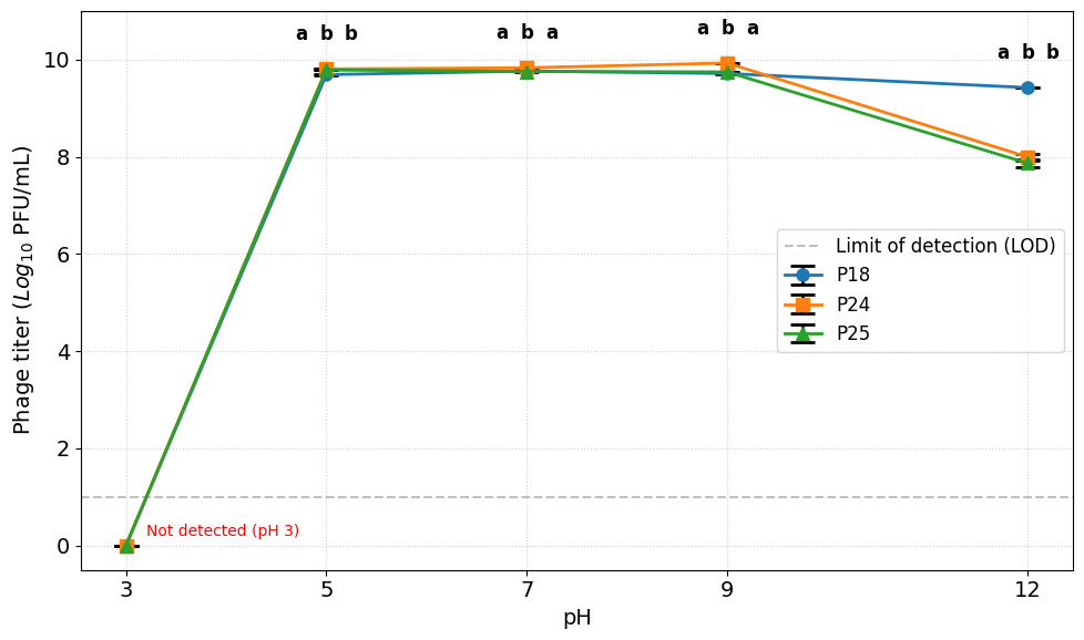
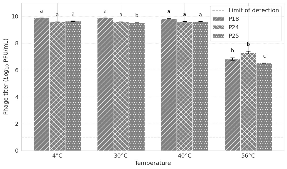
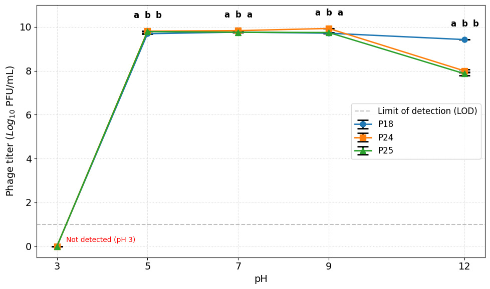
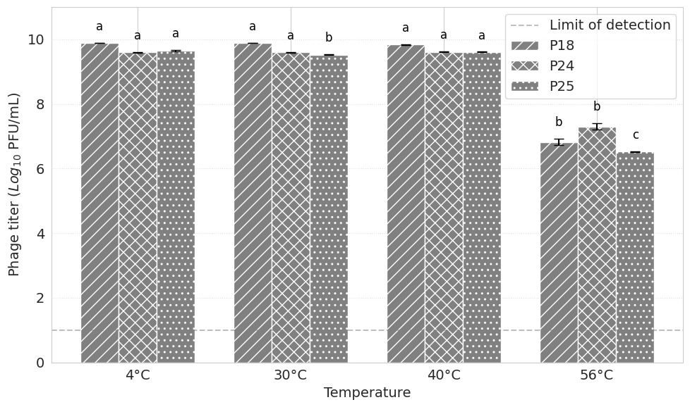
The practical application of phages in aquaculture requires stability under fluctuating environmental conditions. The stability profiles for pH and temperature are illustrated in Figures 2 and 3, respectively.
The infectivity of the phages P18, P24, and P25 was assessed over a broad pH range from 3 to 12. As illustrated in Figure 2, all three phages exhibited a similar stability pattern. They maintained high viral titers (≈108 PFU/mL) within the circumneutral range of pH 5 to pH 9, with optimal activity observed between pH 7 and 9. This range is well within the typical pH range of brackish water in shrimp ponds (7.5–8.5). Notably, extreme acidity (pH 3) completely inactivated all phages.
At highly alkaline conditions (pH 12.0), all three phages exhibited remarkable resilience by maintaining high viable concentrations (>108 PFU/mL), though distinct susceptibility profiles were observed. Phage P18 demonstrated significantly superior stability (p<0.05), retaining a titer of (2.66±0.37)×109 PFU/mL, which corresponds to 45.62% survival and a minor reduction of 0.34 log10 units compared to its neutral control at pH 7.0 ((5.83±0.83)×109 PFU/mL). In contrast, phages P24 and P25 experienced more pronounced reductions (p<0.05). The survival titer of P24 dropped to (9.90±1.44)×108 PFU/mL (representing 14.58% survival and a reduction of 0.84 log10 units compared to its control at pH 7.0), while the titer of P25 was reduced to 7.37±1.48)×108 PFU/mL (representing 12.86% survival and a reduction of 0.89 log10 units compared to its control at pH 7.0).
The thermal tolerance of the isolated phages was evaluated to determine their viability under storage and operational temperatures. As depicted in Figure 3, all three phages (P18, P24, P25) demonstrated high stability at 4°C, 30°C, and 40°C, maintaining titers of 109 PFU/mL. There was no statistically significant difference in infectivity between 40°C and 30°C (p>0.05), indicating suitability for application in tropical aquaculture environments where water temperatures can rise. However, a critical thermal threshold was observed at 56°C. After one hour of incubation at this temperature, the titer of all phages dropped precipitously by approximately 2 to 4 log units (from ∼109 to ∼105-107 PFU/mL), indicating that they are mesophilic and cannot withstand pasteurization temperatures.
In vitro lysis kinetics and comparative efficacy
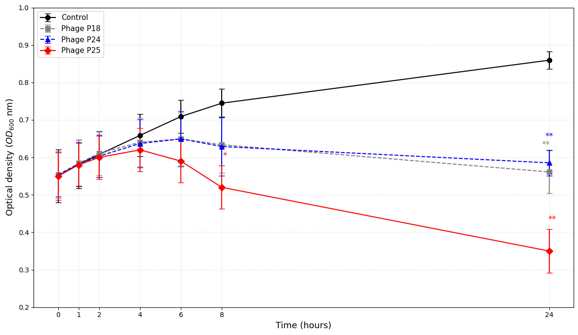
The phages’ lytic activity was monitored over 24 hours. The dynamic changes in bacterial density are presented in Figure 4.
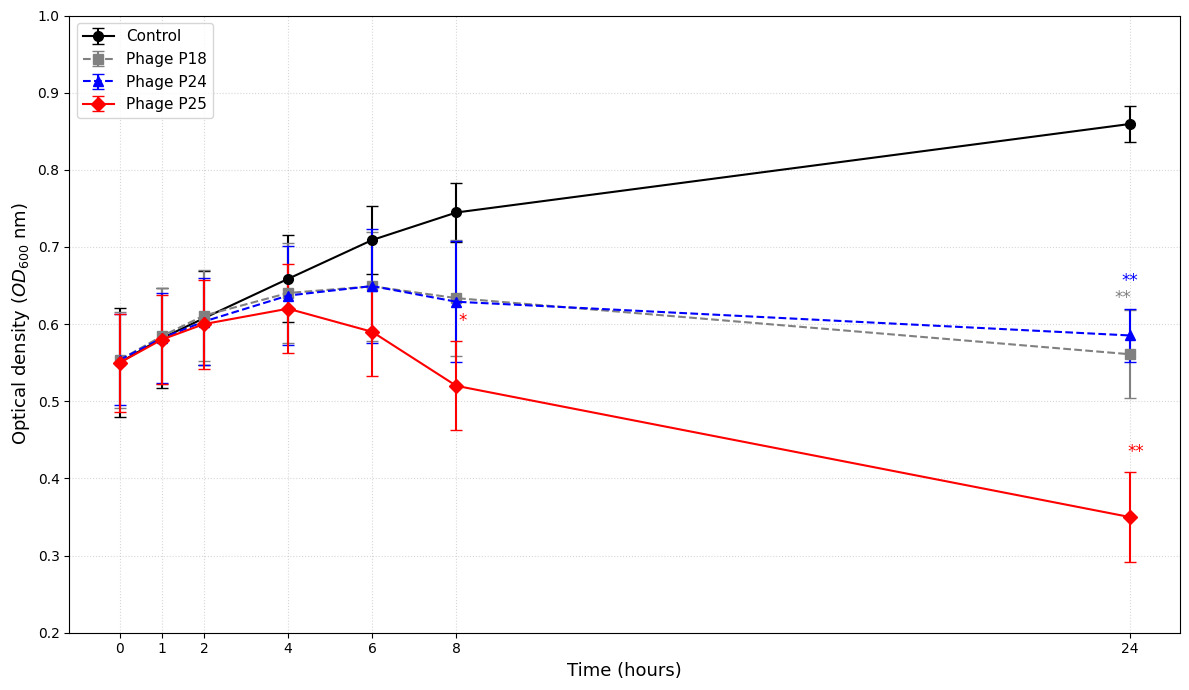
The uninfected control followed a standard logistic growth curve, reaching a peak OD₆₀₀ of 0.856±0.036 at 24 hours. In contrast, Phage P25 exhibited the most aggressive and sustained lytic activity. Following an initial rise during the latent period, the OD declined significantly after 4 hours. Unlike the other treatments, P25 continued to suppress the bacterial population throughout the experiment, reaching a minimum OD₆₀₀ of 0.343±0.101 at 24 hours. While phages P18 and P24 also inhibited growth compared to the control, they displayed a distinct “bacteriostatic-like” profile in the later stages. These two phages reached final ODs between 0.56 and 0.58 but failed to reduce turbidity below the initial inoculum level. Furthermore, the growth curves for P18 and P24 eventually plateaued and showed signs of slight recovery, suggesting either the saturation of lysis or the outgrowth of resistant sub-populations – a stark difference from the continuous suppression observed with P25.
Plating assay insights
To verify the OD findings, viable cell counts (CFU) were determined at the 24-hour endpoint. This revealed a critical divergence in efficacy (Table 3).
Phage P25 achieved a >1.5 log reduction in viable bacterial counts compared to the control (1.6×10⁷ vs 5.78×10⁸ CFU/mL). In contrast, bacterial loads in P18 and P24 treatments remained high (>1.5×10⁸ CFU/mL). Crucially, the surviving colonies in the P18 and P24 groups exhibited a distinct “small opaque” morphology, characteristic of small colony variants (SCVs), a phenotypic hallmark of fitness trade-offs associated with phage resistance.
Discussion
Accurate identification of bacterial pathogens is the prerequisite for specific biocontrol. In this study, the identity of strain V79 was established using a dual-target molecular strategy. While the collagenase gene PCR confirmed the species as V. alginolyticus based on virulence factors,14,29 the amplification of the 700 bp fragment of the 16S rRNA gene using the primers 63f (5’-CAGGCCTAACACATGCAAGTC-3’) and 763r (5’-GCATCTGAGTGTCAGTATCTGTCC-3’)26 provided definitive genus-level confirmation. The protocol by Montieri et al. (2010)30 is particularly valuable in marine microbiology because it utilizes Vibrio-specific variable regions of the 16S gene, thereby avoiding the amplification of non-target marine bacteria that often confound universal 16S primers. This rigorous identification ensures that the isolated phages are indeed Vibrio-specific, a critical factor for their potential application in complex microbial ecosystems such as shrimp ponds.
Resistance phenotype and antibiotic panel completeness
The isolation of V. alginolyticus V79 with a MAR index of 0.9 is a stark indicator of the “post-antibiotic” reality facing modern aquaculture. A MAR index of 0.2 is the standard threshold for distinguishing high-risk contamination sources; a value of 0.9 suggests that V79 has evolved in an environment saturated with diverse antimicrobial agents.6,11 The observed resistance to fluoroquinolones (ciprofloxacin) and tetracyclines is particularly concerning, as these classes represent the backbone of current therapeutic interventions. This resistance profile likely involves multiple mechanisms, including efflux pumps, enzymatic inactivation (e.g., beta-lactamases), and target site mutations (e.g., gyrA mutations for quinolone resistance). The presence of such a strain poses a dual threat: direct pathogenicity to shrimp stocks and the potential for horizontal gene transfer (HGT) of resistance determinants to human pathogens present in the same aquatic environment.11
A limitation of the current resistance profile is that the 10-antibiotic panel did not include chloramphenicol or trimethoprim-sulfamethoxazole, which are heavily used in local Vietnamese farming practices. Future surveillance must expand this panel to completely map local resistance. Nevertheless, demonstrating that phage P25 can suppress an environmental isolate resistant to nine therapeutic antibiotics highlights its value as a candidate.
Environmental stability and morphological parameters
The stability results indicate that P18, P24, and P25 are well-adapted to the physicochemical landscape of aquaculture ponds. Their ability to persist at pH 9 is vital, as phytoplankton photosynthesis often drives pond pH to alkaline levels during the day.31 Thermal stability up to 40°C ensures viability during tropical heatwaves.
The ability of phages P18, P24, and P25 to remain highly active at pH 12.0 is a highly notable biological finding, which is consistent with other alkaline-tolerant marine phages such as the Vibrio siphovirus vB_Va_Val-yong3 and myovirus vB_CoeS_P1 that retain high viability up to pH 12.0.32,33 In intensive penaeid shrimp ponds, daytime carbon dioxide depletion driven by the rapid photosynthetic activities of dense microalgal blooms typically pushes the water pH to alkaline extremes, sometimes reaching 9.5 or even 10.0. The broad alkaline tolerance of these three phages ensures they remain stable and active under these fluctuating diurnal pH conditions. Crucially, the structural assembly of Phage P18 showed a distinct advantage, maintaining a high titer of (2.66±0.37)×109 PFU/mL at pH 12.0. This exceptional stability suggests that the head capsid proteins and tail structures of P18 possess strong molecular resistance to basic denaturation, which may be linked to its unique structural proteins or capsid symmetry observed in our TEM analysis. This finding supports the potential use of P18 as a highly durable component in a therapeutic phage cocktail, ensuring long-term persistence in alkaline aquaculture environments.
The morphological findings provide a structural basis for these phages’ performance. The long, flexible tails observed in the TEM images (up to 198.81 nm) observed in the TEM images are consistent with the sipho morphotype. Variation in tail length may reflect differences in tail structural proteins; however, morphology alone does not establish taxonomic family or receptor usage.28 The variation in tail lengths among the three phages suggests they likely target different host receptors, which is a critical feature for the development of phage cocktails.24 Recent research indicates that combining phages with distinct receptor targets significantly reduces the likelihood that the host will develop simultaneous resistance.34
Mechanisms of phage resistance
A central finding of this study is the differential emergence of resistance among the three phages. While P25 maintained suppression, P18 and P24 treatments were rapidly overtaken by resistant bacterial populations. The “small opaque” morphology of the survivors in P18 and P24 treatments is highly indicative of receptor modification. Phage infection typically begins with adsorption to specific cell surface receptors, such as lipopolysaccharides (LPS) or outer membrane proteins (e.g., OmpK).35–37 Bacteria can evolve resistance by mutating or downregulating these receptors. However, these surface structures often play vital physiological roles in nutrient uptake and environmental interaction. Their loss or alteration incurs a metabolic fitness cost, resulting in slower growth rates and smaller colony sizes – hence the SCVs phenotype observed.38
Resistance trade-offs known as a “fitness trade-off,” are a critical concept in phage therapy. Although the bacteria survive phage attack, they may become less fit for survival in the competitive pond environment or less virulent to the host (e.g., if the phage receptor is also a virulence factor like the LPS O-antigen).12
It is hypothesized that Vibrio species may utilize density-dependent quorum sensing (QS) systems to regulate phage defense mechanisms, potentially upregulating intracellular restriction-modification or CRISPR-Cas systems at high cell densities.37 While no direct QS assays were performed, this density-dependent model remains a compelling speculative hypothesis to explain the non-linear regrowth kinetics in the late stages of high-density killing assays.
A standout finding in this study is the superior lytic efficacy of phage P25 and its sustained suppression of the host population over 24 hours. The continuous decline in optical density (OD600) points to two compelling biological hypotheses: either P25 targets a highly conserved, physiologically essential receptor (such as the flagellar motor or the core component of the lipopolysaccharide—LPS) that the bacterium cannot mutate or downregulate without incurring a severe metabolic fitness cost, or P25 possesses a distinct kinetic advantage (e.g., a shorter latent period and a larger burst size) that outpaces host bacterial replication.24,39 Although P25 did not achieve complete sterilization, leaving a surviving viable count of 1.6×10⁷ CFU/mL, this significant reduction in bacterial load positions it as a promising candidate for therapeutic phage cocktail formulations. By combining P25 with complementary phages (such as P18 or newly isolated candidates targeting alternative receptors), the probability of simultaneous multi-receptor resistance development can be minimized, effectively preventing the regrowth kinetics observed under single-phage selection pressures.
Generalized transduction and biosafety considerations
Prior to any environmental application of bacteriophages, safety evaluation is a critical requirement. A major biological risk associated with using unsequenced phages against extensively drug-resistant (XDR) hosts is generalized or specialized transduction. Temperate phages, as well as some lytic phages, can package host genomic segments (including plasmid-borne antibiotic resistance genes or chromosomal virulence factors) and transfer them to naive environmental bacterial populations.40
Given the high MAR index of strain V79, the use of a phage that mediates high-frequency transduction could accelerate the spread of antibiotic resistance in aquaculture systems. Therefore, complete Whole-Genome Sequencing (WGS) is an absolute prerequisite to verify the absence of integrases, excisionases, recombinases, toxins, and antibiotic resistance genes before these phages can be considered safe for therapeutic or commercial development.40
Limitations and future directions
While this study establishes baseline physical and biological parameters for three novel Vibrio phages, several critical limitations must be addressed before therapeutic application:
(1) Absence of Whole-Genome Sequencing (WGS): The lack of genomic data prevents the screening of virulence genes and the confirmation of a strictly lytic lifecycle.
(2) Lack of One-Step Growth Curves: The latent period and burst size of these phages have not been experimentally determined, limiting the modeling of replication kinetics.
(3) Single-Host Range Panel: The phages were tested only against the single sentinel strain V79. Testing across a broader panel of V. alginolyticus strains and non-target species is required to evaluate host specificity and ecological safety.
(4) No In vivo validation: All lytic assays were performed in vitro. The efficacy of phage P25 in reducing mortality must be validated using shrimp challenge models (e.g., L. vannamei trials).
Conclusions
This study isolated three phages targeting a highly resistant V. alginolyticus strain (MAR index 0.9). While all phages showed good environmental stability, phage P25 was unequivocally identified as the superior therapeutic candidate. In vitro, P25 achieved a significant reduction in bacterial load (>1.5 log) and sustained population suppression over 24 hours. Conversely, Phages P18 and P24 were hampered by the rapid emergence of resistant, small-colony variants. These results advocate for the development of P25-based biological control products, potentially as the core component of a phage cocktail, to combat MDR Vibriosis in sustainable aquaculture.
Acknowledgments
The authors are especially grateful to the Institute of Food and Biotechnology, Can Tho University, for making facilities available for this research. Van-Thanh Vo was funded by the PhD Scholarship Programme of Vingroup Innovation Foundation (VINIF), VinUniversity, code VINIF.2025.TS44. The funder had no role in the study design, data collection, analysis, interpretation, manuscript preparation, or the decision to submit the work for publication.
Authors’ Contribution
Conceptualization: Van-Thanh Vo (Equal), Bui Thanh Liem (Equal), Truong Thi Bich Van (Equal). Methodology: Van-Thanh Vo (Equal), Nguyen Chi Thom (Equal), Truong Thi Bich Van (Equal). Investigation: Van-Thanh Vo (Equal), Van-Thanh Vo (Equal), Nguyen Chi Thom (Equal), Truong Thi Bich Van (Equal). Formal Analysis: Van-Thanh Vo (Equal), Nguyen Chi Thom (Equal), Truong Thi Bich Van (Equal). Writing – original draft: Van-Thanh Vo (Lead). Writing – review & editing: Van-Thanh Vo (Equal), Nguyen Chi Thom (Equal), Bui Thanh Liem (Equal), Truong Thi Bich Van (Equal). Supervision: Bui Thanh Liem (Equal), Truong Thi Bich Van (Equal).
Competing of Interest – COPE
The authors declare no competing financial or professional interests.
Ethical Conduct Approval – IACUC
Invertebrates (such as penaeid shrimp) are exempt from formal institutional IACUC approval under Vietnam’s animal welfare guidelines. However, diseased shrimp samples were collected from the commercial aquaculture facility with the owner’s explicit permission, and all sampling protocols complied with local agricultural biosecurity guidelines.
Informed Consent Statement
All authors and institutions have confirmed this manuscript for publication.
Data Availability Statement
All are available upon reasonable request.