
1. INTRODUCTION
In recent years, the global aquaculture industry has experienced rapid growth, generating significant economic benefits for society. However, the environmental degradation caused by the continuous expansion of intensive aquaculture operations has become increasingly prominent. The rearing environment is closely linked to the survival and health of crustaceans.1 Parameters such as temperature,2 pH,3 salinity,4 ammonia nitrogen,5 nitrite,6 dissolved oxygen,7 and stocking density8 are key factors affecting crustacean survival and health. Among these, hypoxia stress stands as a major challenge in crustacean aquaculture, negatively impacting growth and development, energy metabolism, immune defense, and antioxidant capacity.9
Some crustaceans respond to hypoxic stress through complex behavioral and physiological strategies.10 The physiological response of crustaceans to hypoxia involves biochemical and molecular changes in energy metabolism, antioxidant defense, and immune response, as well as regulation of apoptosis and cell cycle.11,12 Hypoxia affects the metabolic patterns of aquatic animals.13 At the physiological level, crustaceans exposed to low dissolved oxygen initiate the oxygen-carrying protein (hemocyanin) synthesis mechanism to improve oxygen transport efficiency,14 and activate anaerobic metabolism, manifested as increased lactate production15 and tissue-specific induction of glycolytic enzymes (Hexokinase, lactate dehydrogenase, and phosphofructokinase).16–18 Hypoxia may also stimulate lipolysis.19 At the cellular level, metabolic adaptation of crustaceans is reflected by regulating energy metabolism, lipid and carbon metabolism, and nutrient acquisition.20 At the molecular level, short-term hypoxia induced the up-regulated expression of glyceraldehyde-3-phosphate dehydrogenase (GAPDH) gene in the gills of Litopenaeus vannamei in response to stress21 ; Eriocheir sinensis up-regulated HIF-1α during hypoxia, further activated the NF-κB-ALFs pathway, and played an immune defense role in response to hypoxia stress22 ; HIF-1α regulates key genes in glycolysis and TCA cycle under hypoxic conditions to achieve energy programming.23 At present, research on the mechanisms of hypoxia stress in crustaceans is deepening and progressing from basic physiological phenotypes to deeper molecular mechanisms.
Procambarus clarkii, commonly known as crayfish, is one of the most important crustacean aquaculture species in the global freshwater economy and plays a vital role in the world economy.24 According to the ‘China Crayfish Industry Development Report (2025)’, in 2024, crayfish aquaculture production in China will reach 3.4476 million tons, ranking fourth among freshwater aquaculture varieties. P. clarkii is vulnerable to hypoxia stress in aquaculture and commercial transportation, and hypoxia can significantly affect the expression profile and metabolic response of related genes. Previous studies have shown that P. clarkii can increase the concentration of lactic acid and glucose in hemolymph under low dissolved oxygen conditions,25 regulate the dynamic changes of antioxidant enzymes in hepatopancreas, reduce oxidative stress, and up-regulate the expression of key enzymes in glycolysis to activate anaerobic metabolism,26 experiencing changes in the abundance of metabolites related to energy metabolism.27 Zeng et al.28 found that hypoxic stress significantly affected the activities of antioxidant enzymes (SOD, CAT) and anaerobic metabolic enzymes (LDH), as well as the expression of antioxidant and immune-related genes, in the hepatopancreas of juvenile and subadult crayfish. The higher the degree of hypoxia stress, the higher the gene expression level. This indicates that the regulation of the expression of specific enzymes required for metabolic adaptation to hypoxia is achieved through differential transcription. Zhang et al.29 showed that hypoxia significantly affected glucose metabolism (glycolysis/gluconeogenesis, AMPK, and insulin signaling pathways) and oxygen transport (HIF-1, MAPK, and PI3K-Akt signaling pathways) in a transcriptomic analysis of the hepatopancreas of Procambarus clarkii. These metabolic changes are regulated by genes related to glucose metabolism and oxygen transport. The gill is an important organ for crustaceans to breathe, excrete, and regulate osmotic pressure, and is extremely sensitive to changes in dissolved oxygen in water. Because the gills of crustaceans are in direct contact with the external environment, hypoxia stress can directly damage their gill tissues.30,31 At present, the molecular mechanism of metabolic regulation of key genes and pathways of acute hypoxia stress in the gills of Procambarus clarkii is still poorly understood. Therefore, it is of great significance to study the metabolic pattern of the gill tissue of crustaceans in a hypoxic environment.
In a large number of studies on hypoxic environments, aquatic animals do not rely on carbohydrates as the main source but initiate metabolic compensation mechanisms that potentially enhance the metabolism of amino acids, lipids, and other nutrients to maintain energy demand.19,32 In the post-genomic era, the widespread use of transcriptomics, metabolomics, and proteomics analyses has brought new insights to biology.33 In recent years, multi-omics analysis methods have been employed to investigate stress responses in aquatic animals.34 However, the mechanisms and molecular regulatory networks underlying P. clarkii gill responses to acute hypoxia stress have not been clearly elucidated using transcriptomic and metabolomic analyses, an important approach for understanding gene expression and metabolite differences in P. clarkii gills under low dissolved oxygen conditions.
Therefore, it was hypothesized that acute hypoxia triggered a comprehensive metabolic reprogramming in the gills of P. clarkii to restore energy homeostasis and resist oxidative stress. The purpose of this study was to identify differentially expressed genes and metabolites in the gill tissues of P. clarkii under acute hypoxia conditions. Through combined transcriptomic and metabolomic analysis, key pathways and regulatory networks were revealed, and the molecular mechanisms of energy homeostasis and oxidative stress resistance in the gills during hypoxia adaptation were further elucidated. This integrated multi-omics study will deepen our understanding of the hypoxia adaptation strategies of crustaceans and provide a theoretical reference for the sustainable development of the crustacean aquaculture industry.
2. MATERIALS AND METHODS
2.1. Experimental animals and feeding management
The subadult shrimp (Procambarus clarkii) used in this experiment was obtainted from the breeding laboratory of Yancheng Institute of Technology, with an average weight of 7.5±0.5 g. The crayfish were domesticated in a laboratory indoor aquarium (55 cm×25 cm×35 cm) for 7 days. During the domestication, the dissolved oxygen in the aquaculture water was maintained at 7.5±0.2 mg/L, the water temperature was 24±2°C, the pH value was 7.0±0.2, and the ammonia nitrogen content was maintained less than 0.02 mg/L. The photoperiod followed the natural light-dark cycle. The crayfish were fed with special compound feed at 8:00 a.m. and 6:00 p.m., respectively. The feeding rate was 3%-5% of the body weight of crayfish. Residual food and excreta were siphoned daily, and one-third of the water was replaced every two days to maintain water quality and ensure a healthy environment.
2.2. Hypoxia treatment and sample collection
After the end of the acclimation period, the hypoxic environment was induced. The exposure time is set to 24 hours, which can induce a stable physiological metabolic response in P. clarkii, with no deaths, and demonstrates hypoxia adaptability.28,35 Sixty healthy and lively crayfish (unseparated male and female were in the intermolt period) with normal body color were randomly placed in 6 aquariums (55 cm×25 cm×35 cm), 10 crayfish in each aquarium (aquaria were arranged in a randomized block design to minimize potential positional effects within the laboratory). During the experiment, the breeding environment was fully consistent with the domestication period, except for dissolved oxygen conditions. In this experiment, low dissolved oxygen group (Low, 2.0±0.2 mg/L) and normoxic control group (CK, 7.5±0.2 mg/L) were set up, with 3 replicates in each group, all of which were treated for 24 h. Low dissolved oxygen treatment involves applying low oxygen stress while maintaining normal dissolved oxygen levels. The hypoxia experiment in this study was performed using the Wang et al.12 method. In short, the air in the CK group was continuously pumped into the water to maintain a normoxic state of about 7.5 mg/L. The low-oxygen treatment maintained a dissolved oxygen level of about 2.0 mg/L by injecting nitrogen and air into the water and controlling the flow rate. A pen dissolved oxygen meter (AR8210, XIMA) was used to monitor the dissolved oxygen concentration of the water in real time throughout the test. Gill tissue samples of P. clarkii treated with two dissolved oxygen concentrations were collected after the end of hypoxia treatment. Transcription experiments and metabolic experiments were set up with 3 and 6 parallels for the construction of transcriptome libraries and the analysis of metabolite profiles, respectively. After the shrimp were dissected, the tissue was frozen in liquid nitrogen and stored at −80°C in a refrigerator for further RNA extraction and metabolite assays.
2.3. Transcriptome analysis
2.3.1. RNA extraction, library construction and sequencing
According to the manufacturer 's protocol, total RNA was extracted from the gill tissue of P. clarkii using magnetic beads connected with Oligo (dT), and the total RNA amount of each sample was 2 μg. RNA purity was measured using a kaiao K5500® spectrophotometer (Kaiao, Beijing, China), and RNA integrity and concentration were assessed using an Agilent 2100 bioanalyzer system. The construction and sequencing of cDNA and RNA Seq libraries were carried out in Wuhan Benard Technology Co., Ltd.in accordance with the manufacturer 's specifications. The library fragments were purified using Ampure XP magnetic beads, and library quality was evaluated using the Agilent Bioanalyzer 2100. Subsequently, these libraries were further amplified with phi29 polymerase to prepare DNA nanospheres (DNB), which were then loaded onto the patterned nanoarrays and generated 150 bp double-ended sequencing reads on the T7 platform of Wuhan Benard Technology Co., Ltd.
2.3.2. Quality control, mapping and assembly
The original data were first processed using FastQC,36 and the joints and low-quality sequences were filtered from the original sequence (raw reads) to obtain high-quality effective sequences (clean reads). The Trinity software (v2.14) was used to generate unigenes.37 Then, the effective sequences were aligned to the reference genome using HISAT (Procambarus clarkii GCA_040958095.1.genome.fa). The RSEM (v1.3.1) software package was used to calculate gene expression levels for each sample,38 expressed as per-million alignment fragments per thousand base transcripts (FPKM), with a threshold of FPKM > 0.5. The gene expression levels of the hypoxia stress samples were compared with those of the control samples to identify differentially expressed genes (DEGs).
2.3.3. DEGs screening and functional prediction
DEseq2 (v1.34.0)39 was used to screen differentially expressed genes (DEGs) of Low vs CK, and the screening criteria was |log2FoldChange|≥2 and P<0.05. ClusterProfiler (v3.8)40 was used to perform gene ontology (GO) and Kyoto Encyclopedia of Genes and Genomes (KEGG) enrichment analysis, and P<0.05 was used as the standard.
2.4. Non-targeted metabolomics analysis
2.4.1. Metabolite extraction
25 mg gill tissue samples were weighed and thawed at low temperature, and then 500 μL -40°C pre-cooled extract [methanol:acetonitrile:water=2:2:1(v/v/v)] was added. The extract contained an isotope-labeled internal standard; steel balls were added, and the mixture was homogenized in a homogenizer (35 Hz, 4 min), then transferred to an ice-water bath for ultrasound for 5min. This step was repeated three times. Then it was allowed to stand at −40°C for 1 h, and 300 μL was added to a 96-well filter plate. The 96-well filter plate-collection plate combination was placed in a positive-pressure device and slowly pressurized to 6 psi for 3 min. The filter plate-collection plate combination was removed from the positive pressure device, and the same amount of supernatant was mixed to prepare a quality control (QC) sample. The treatment and analysis methods of QC samples are the same as those of test samples.
2.4.2. LC-MS analysis
LC-MS analysis of samples was performed using a Vanquish (Thermo Fisher Scientific) ultra-high-performance liquid chromatograph. Chromatographic separation of target compounds was achieved via a Waters ACQUITY UPLC BEH Amide column (2.1 mm×50 mm, 1.7 μm). LC mobile phase A was an aqueous phase containing 25 mmol/L ammonium acetate and 25 mmol/L ammonium hydroxide, while phase B was acetonitrile. The sample tray temperature was maintained at 4°C, with an injection volume of 2 μL.
2.4.3. Data analysis
In this study, raw data were converted to mzXML format using ProteoWizard and processed with an internally developed program in R, based on XCMS, for peak detection, extraction, alignment, and integration. The R package and BiotreeDB (V3.0) were used to identify metabolites. Principal component analysis (PCA) and orthogonal partial least squares discriminant analysis (OPLS-DA) were performed on the normalized data using SIMCA software (v18.0.1). The OPLS-DA replacement test model was used to identify and screen for differential metabolites between the control and hypoxia groups. Combined with t-test analysis (P1) and the difference fold of metabolites |log2FoldChange|≥2, they were screened as potential biomarkers and differential metabolites. In addition, the differential metabolites were compared, annotated and analyzed by KEGG (http://www.genome.jp/kegg/) database.40
2.5. Integration analysis between transcriptome and metabolome
Differential genes (DEGs, P<0.05 and |log2FoldChange|>2) and differential metabolites (DEMs, VIP>1 and P<0.05) were used for the integrated analysis of Low vs CK. The Pearson correlation coefficient method was used to integrate correlation coefficient analysis of metabolic groups and transcriptome data. The heat map intuitively shows the relationship between genes and metabolites. R software (R Version 3.5.1) was used to combine the information of each group, KEGG pathway enrichment analysis was performed, and matrix heat map was constructed according to Pearson correlation coefficient. CytoScape software (CytoScape 3.5.1) was used to generate a gene and metabolite interaction network.
2.6. Statistical analysis
All analyses and charts are produced using R software (v3.5.1). DEGs were identified using DEseq2 (v1.34.0), and GO term annotation and KEGG pathway enrichment were performed using hypergeometric test. After FDR adjustment, adjusted P<0.05 indicated statistical significance.
3. Results
3.1. Transcriptome analysis
The sequencing results of 6 libraries are shown in Table 1. The number of Raw reads and Clean reads was stable between 40.02 Mb and 40.06 Mb. Q20 and Q30 were greater than 97.52 % and 93.58 %, respectively. The GC content of all samples was greater than 36.95 %.
3.1.1. Identification and annotation of DEGs
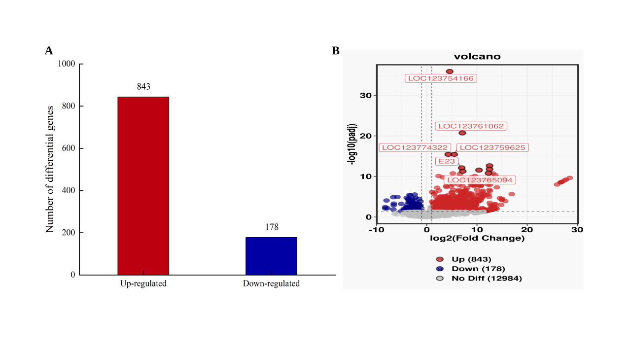
The number of differentially expressed genes of P. clarkii under two different dissolved oxygen levels is shown in Fig.1. Compared with the CK group, a total of 1021 differentially expressed genes were identified in the Low group (843 up-regulated and 178 down-regulated).
__volcano_.svg)
3.1.2. GO and KEGG enrichment analysis of DEGs
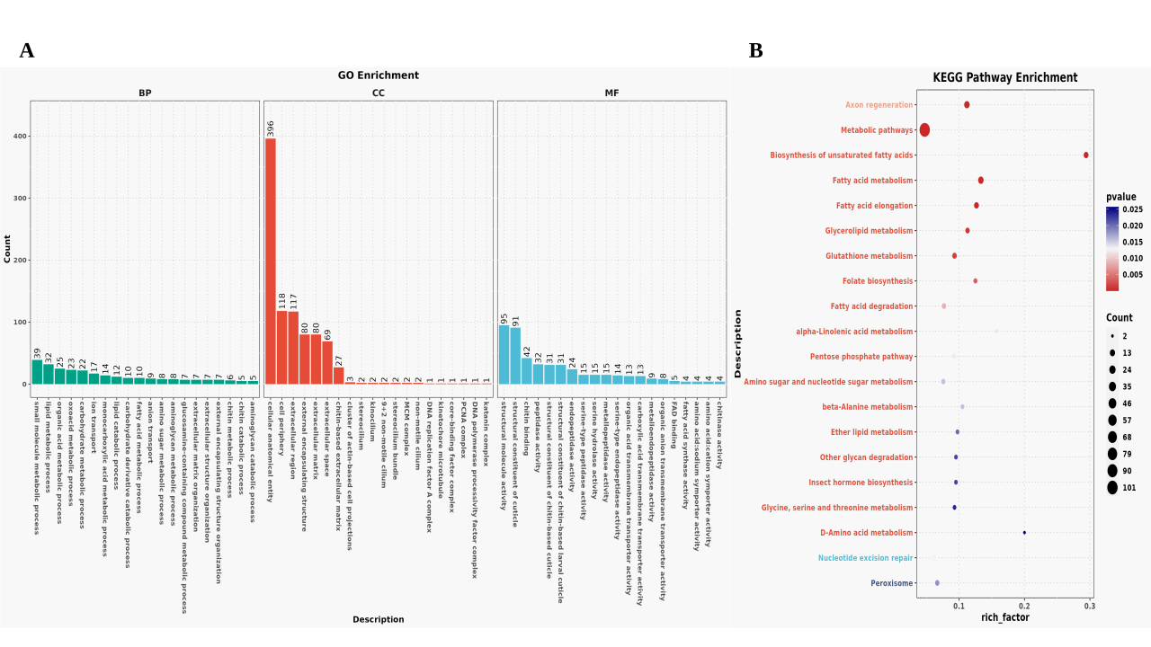
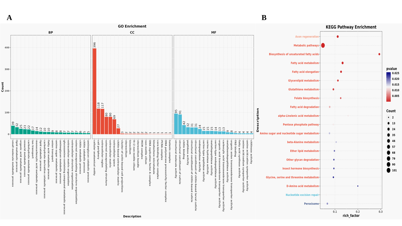
In order to further explore the biological functions of differentially expressed genes in the gill transcriptome under hypoxia stress, GO and KEGG enrichment analysis were performed on the differentially expressed genes detected under two different dissolved oxygen levels. In terms of GO enrichment analysis, most of the differentially expressed genes between Low group and CK group were mainly enriched in cellular anatomical entity (396), cell periphery (118) and extracellular region (117). Structural molecule activity (95), structural component of cuticle (91), and so on were enriched in molecular function. In the biological process, small molecule metabolic process (39), lipid metabolic process (32) and organic acid metabolic process (25) were enriched, such as Fig 2A.
In order to identify the biological pathways activated or inhibited in the response of gill tissue of Procambarus clarkii to hypoxia stress, DEGs were annotated into the KEGG database. KEGG enrichment analysis showed that 226 DEGs in Low vs CK were enriched into 116 KEGG pathways. Fig 2B showed the first 20 significantly enriched pathways in the gills, and were significantly enriched in multiple metabolic pathways (P<0.05). It includes unsaturated fatty acid biosynthesis (map01040), fatty acid metabolism (map01212), fatty acid elongation (map00062), glycerolipid metabolism (map00561), glutathione metabolism (map00480), folic acid biosynthesis (map00790), fatty acid degradation (map00071), α-linolenic acid metabolism (map00592), pentose phosphate pathway (map00030), D-amino acid metabolism (map00470) and other lipid metabolism, other amino acid metabolism, cofactor and vitamin metabolism and carbohydrate metabolism pathways. However, we cannot completely prove that these physiological processes occur in organisms. Therefore, we further tested the metabolome to identify metabolic pathways or metabolites related to the transcriptomic results, thereby confirming that hypoxia stress induces changes in physiological activity and energy metabolism in the gill tissue.
3.2. Metabolome analysis
3.2.1. Overall changes in metabolites
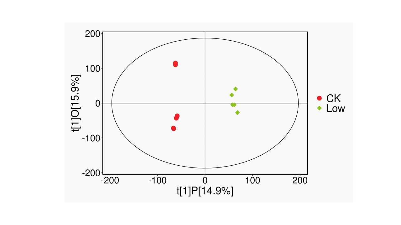
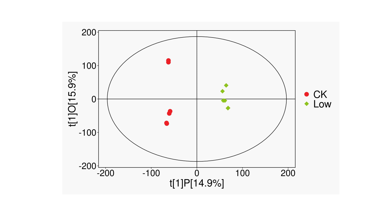
To detect changes in metabolites in Procambarus clarkii under low dissolved oxygen stress, LC-MS was used for non-targeted metabolomics analysis. Based on orthogonal partial least squares discriminant analysis (OPLS-DA), R2Y≥0.99, and Q2>0.84 (Table 2), indicating that the model stability is better; according to the OPLS-DA score scatter plot in Fig 3, it can be seen that there is a significant difference between the two groups, which can be used for subsequent analysis.
3.2.2. Screening of DEMs
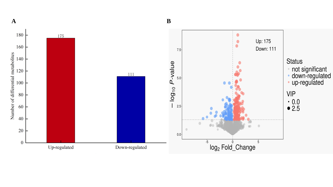
The metabolic changes of gill tissue of Procambarus clarkii under low dissolved oxygen stress were studied by non-targeted metabolomics analysis. According to the criteria of VIP>1.0 and P<0.05, a total of 286 DEMs were identified between the Low group and the CK group, of which 175 increased and 111 decreased (Fig 4).
__differentially_expressed_metabolite_volcano_pl.svg)
3.2.3. Hierarchical Cluster Analysis of DEMs
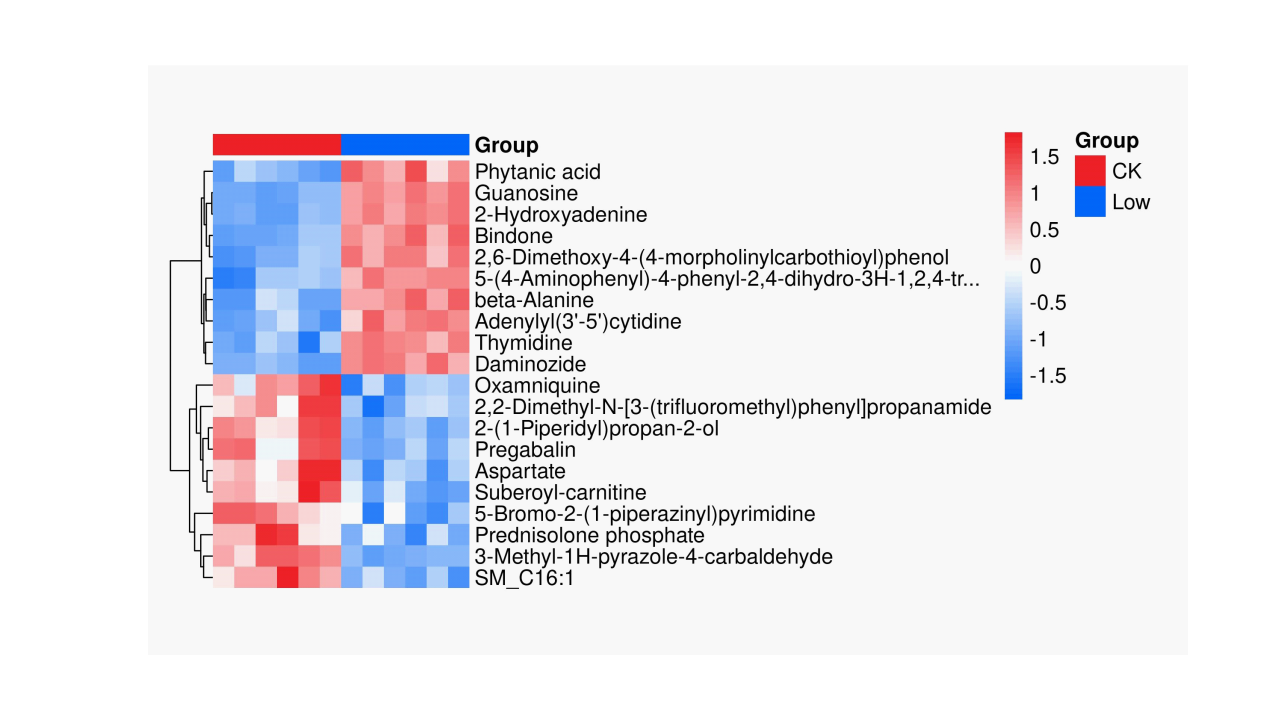
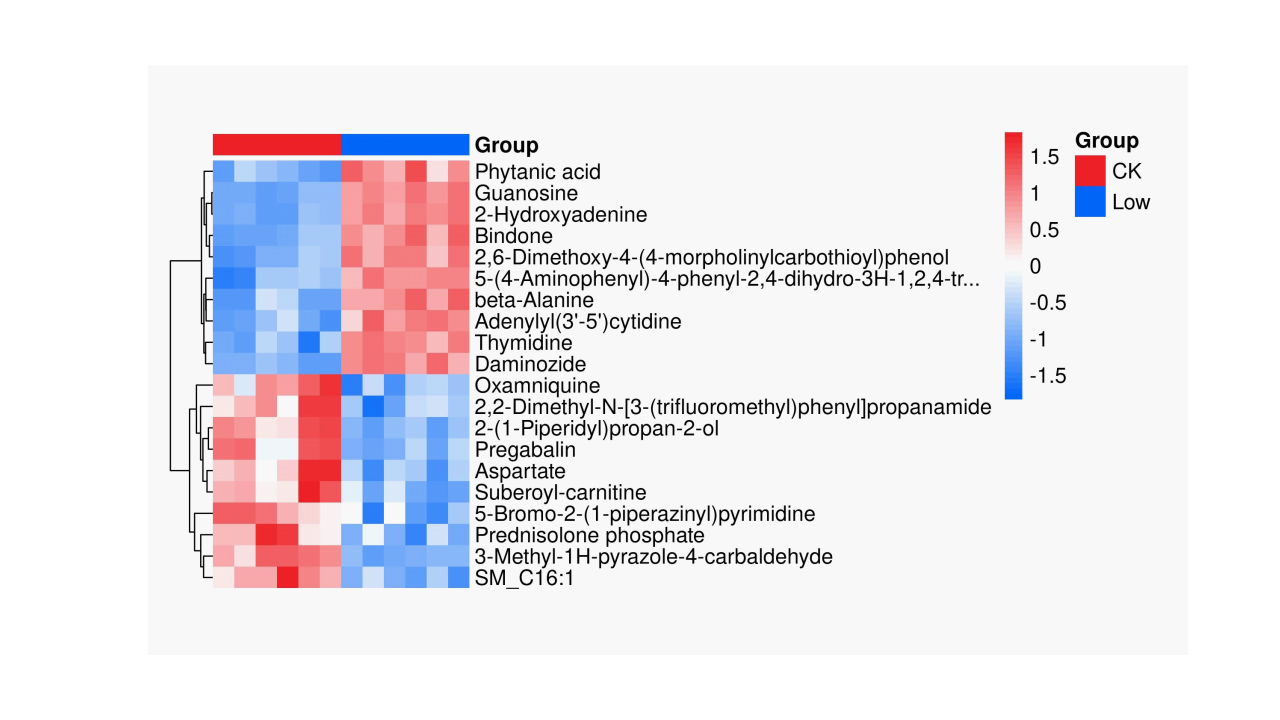
Fig 5 shows the heat map of Low vs CK based on the top 20 DEMs in abundance. Metabolites with similar expression patterns are usually functionally related, further underscoring the differences in expression patterns between the two groups. According to the cluster heat map, 20 DEMs can be distributed into 7 clusters. Beta-Alanine and Aspartate showed different expression trends in the CK and Low groups, both of which belonged to organic acids and their derivatives. In addition, the samples in the Low group and the CK group showed similar colors, but the color difference between the two groups was significant; that is, greater intra-group similarity was observed between the groups.
3.2.4. Functional enrichment analysis of DEMs related to hypoxia stress
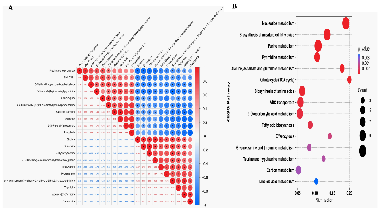
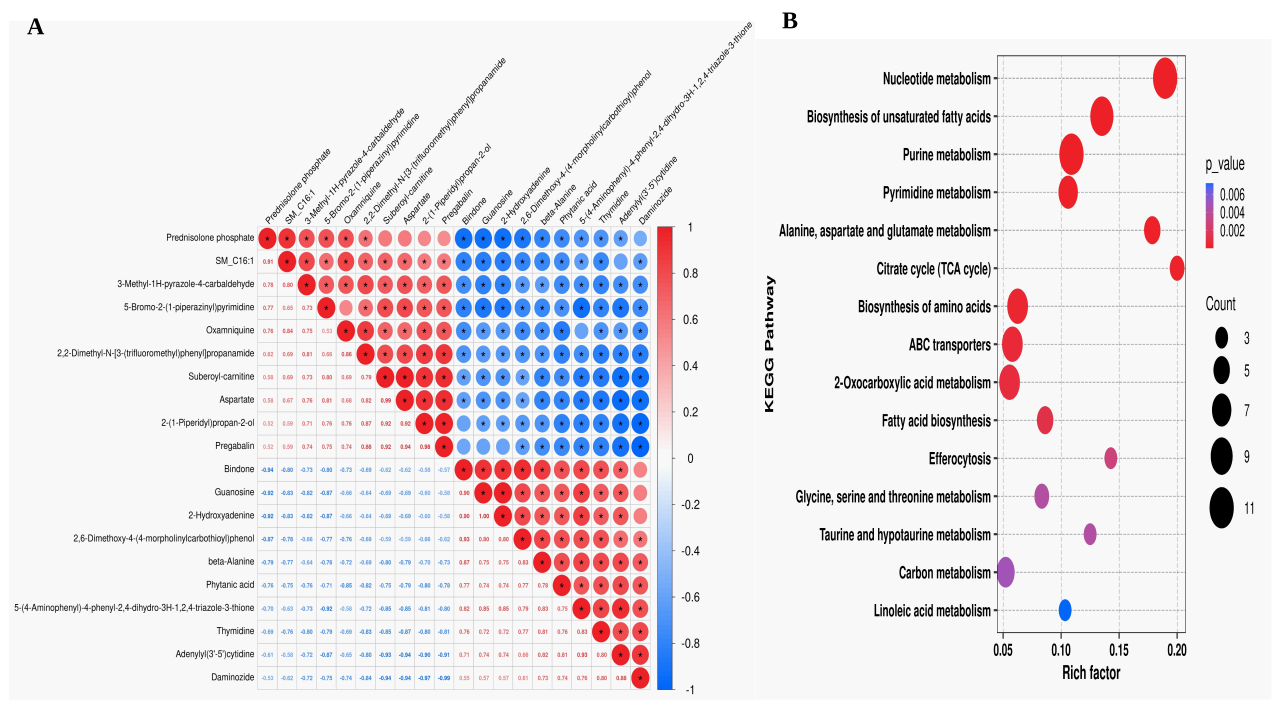
This study found that the abundance of differential metabolites identified in the Low vs CK group showed a correlation, and a variety of metabolites showed a significant positive correlation (P<0.05, Fig 6A). The KEGG pathway enrichment analysis of differential metabolites showed that the significantly enriched pathways in the Low vs CK group were mainly: Nucleotide metabolism; purine metabolism; biosynthesis of unsaturated fatty acids; ABC transporters; alanine, aspartate and glutamate metabolism; 2-Oxocarboxylic acid metabolism; citric acid cycle. Among them, there are differences in the degree of enrichment of different pathways (Fig 6B).
3.3. Conjoint analysis of transcriptome and metabolome
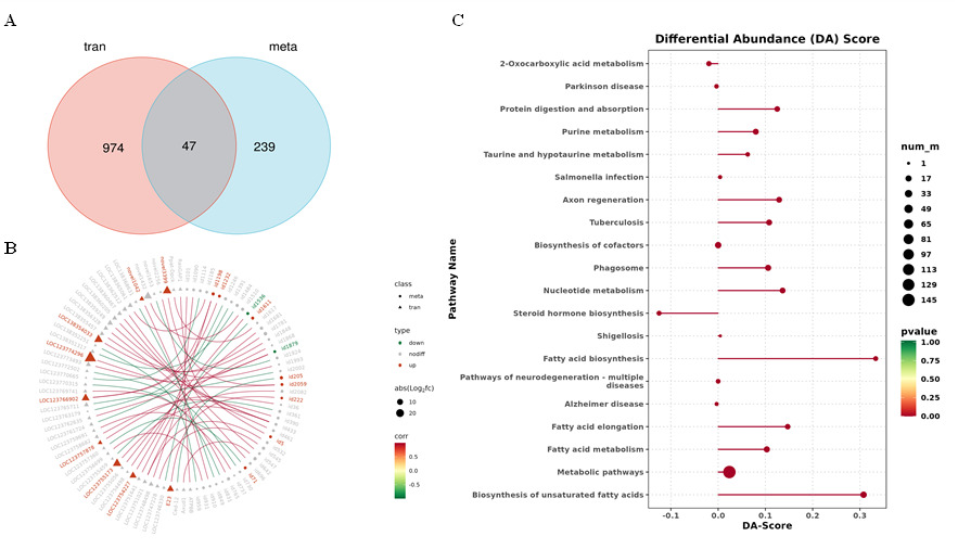
To further understand the association between DEGs and DEMs, conjoint analysis of transcriptome and metabolome was performed. Fig 7A shows that a total of 47 genes and 47 metabolites were associated between the 1021 differentially expressed genes identified by transcriptome and the 286 differentially expressed metabolites identified by metabolomics. The circular diagram of the association between differentially expressed genes and metabolites showed that the ABCG2 gene (E23) in the ABC transporter membrane family was associated with three types of metabolites (lipids, organic nitrogen-containing compounds, and organic heterocyclic compounds), synergistically regulating the energy metabolism of P. clarkii (Fig.7B). The KEGG pathway of transcription and metabolic association analysis was mainly enriched in lipid, nucleotide, carbohydrate and amino acid metabolic pathways (Table 3). In terms of lipid metabolism, the fatty acid biosynthesis (map00061), fatty acid elongation (map00062) and fatty acid degradation (map00071) pathways were significantly enriched and up-regulated, and the steroid hormone biosynthesis (map00140) pathway was significantly enriched and down-regulated (Fig 7C); in terms of nucleotide metabolism, the pathways of purine metabolism (map00230) and pyrimidine metabolism (map00240) were significantly enriched and up-regulated (Fig 7C). In carbohydrate metabolism, the mutual conversion of pentose and glucuronate (map00040) and the pentose phosphate pathway (map00030) were significantly enriched; in terms of amino acid metabolism, alanine, aspartic acid and glutamic acid metabolism (map00250) and glycine, serine and threonine metabolism (map00260) pathways were significantly enriched; in addition, it is also enriched in biosynthetic pathways of secondary metabolites, such as caffeine metabolism (map00232).
__correlation_loop_diagram_(b)__da-sc.png)
4. DISCUSSION
The research on the stress response of P. clarkii to hypoxia primarily focuses on physiological and biochemical aspects, as well as single transcriptomic analyses related to energy metabolism and immune function, but research on the hypoxia response of key gene functions and regulatory networks remains limited. In particular, the combined transcriptomic and metabolomic analysis of the effects of hypoxia stress on P. clarkii remains unclear. In this study, the combined analysis of transcriptional and metabolic KEGG enrichment pathways showed that hypoxia significantly affected lipid, carbohydrate, nucleotide, and amino acid metabolism in P. clarkii. This is similar to the metabolic changes of Takifugu Obscurus gills after acute hypoxia 6 h stress studied by Zhang et al.41 It was found that energy synthesis and metabolism-related pathways such as lipid metabolism, amino acid metabolism, and purine metabolism were significantly enriched. However, compared with fish, crustaceans have an open circulatory system and gill tissue is directly exposed to the water environment. Their metabolic reprogramming may entail more urgent physiological needs to resist oxidative stress and maintain ion exchange function. The results of this study will provide a scientific basis for the characteristics of hypoxia adaptation metabolic reprogramming in crustacean gill tissue.
4.1. Hypoxia stress affects lipid metabolism of Procambarus clarkii
Under hypoxic conditions, organisms must adjust their energy requirements to maintain basic physiological processes. Lipids, as important energy sources and precursors of signaling molecules, play key roles in maintaining membrane structure, regulating energy metabolism, and controlling signal transduction.42 Therefore, lipid homeostasis is essential for maintaining health.43 More and more studies show that the compensatory mechanism of aquatic animals under hypoxic stress involves enhanced lipid metabolism.13 Nguyen et al.44 found that metabolites, such as fatty acids and amino acids, in the hemolymph of Litopenaeus vannamei changed significantly after hypoxia exposure by GC-MS metabolomics analysis, suggesting that hypoxia stress led to an energy metabolism disorder and a disturbance of amino acid metabolism. The gills of crustaceans are important sites for lipid storage and metabolism.45 This study found that under hypoxic conditions, the contents of most fatty acyl and glycerophospholipid metabolites in the significantly enriched differential metabolites in the gills of P. clarkii were significantly increased, and the expression of fatty acid biosynthesis-related genes, such as fatty acid synthase (fasn), long-chain fatty acid-CoA ligase (acsbg), and fatty acid degradation-related gene carnitine palmitoyltransferase 1 (cpt-1A), was up-regulated. Among the significantly enriched metabolic pathways, four were directly related to lipid metabolism, indicating that fatty acid synthesis in the gills of Procambarus clarkii was significantly enhanced under hypoxia stress. At the same time, polyunsaturated fatty acids (PUFAs) also changed significantly in the gills of P. clarkii under hypoxic conditions; metabolites such as arachidonic acid, linolenic acid, palmitic acid, arachidonic acid, and oleic acid were significantly accumulated after 24 h of hypoxia. Fasn is a key enzyme involved in the synthesis of endogenous fatty acids. cpt-1 is a key enzyme that limits the rate of fatty acid β-oxidation and can transport fatty acids to mitochondria. The fasn and cpt-1 genes in this study were up-regulated simultaneously, indicating that the crayfish initiated the synthesis and degradation of fatty acids in the gills under hypoxic conditions. This “dynamic lipid turnover” strategy has important physiological significance: on the one hand, the enhancement of fatty acid synthesis can provide raw materials for cell membrane repair and signal molecule generation; on the other hand, moderately accumulated unsaturated fatty acids can participate in cell stress protection through esterification and reduce lipid peroxidation damage, thereby maintaining the integrity of gill epithelial cells. In addition, crustacean molting is a biological process that requires substantial energy.46,47 Enhancing lipid catabolism under hypoxic conditions may also help conserve protein reserves and allocate limited amino acids to more important physiological processes, such as molting-related structural reconstruction. The above results indicate that lipid metabolism is the primary energy-metabolism adjustment pathway in gill tissue to cope with acute hypoxia.
4.2. Hypoxia stress affects carbohydrate metabolism of Procambarus clarkii
The metabolic adaptation of crustaceans to hypoxic stress means a shift from aerobic metabolism to anaerobic metabolism.48 In the study on Litopenaeus vannamei, HIF-1 signaling pathway has been confirmed to transcriptionally regulate the expression of glycolytic enzymes such as GAPDH in gill tissue under hypoxic conditions, and mediate the activation of anaerobic glycolysis.49 Carbohydrate metabolism is an important pathway of energy metabolism, serving as a hypoxic metabolic compensation strategy to maintain the balance between oxidation and anaerobic metabolism and energy supply.50 Studies have shown that during hypoxia, the expression of phosphofructokinase (pfk) and fructose 1,6-bisphosphatase (fbp) in the hepatopancreas of Penaeus vannamei is up-regulated, which activates glycolysis and gluconeogenesis pathways.18 This study showed that hypoxia stress activated the pentose phosphate pathway in the gill tissue of P. clarkii, increased the final metabolite pyruvate in the glycolytic process, and down-regulated the expression of fructose-1,6-bisphosphatase 1 (fbp), a key rate-limiting enzyme in the gluconeogenesis pathway, indicating that acute hypoxia may enhance glycolysis and inhibit gluconeogenesis. This may be due to the fact that, under hypoxic conditions, the glycolytic product pyruvate cannot directly enter the mitochondria and is recycled via the TCA cycle, resulting in decreased ATP production. The inhibition of fbp leads to the closure of gluconeogenesis, and the accumulation of glycolytic intermediate glucose-6-phosphate (G6P), which in turn promotes the up-regulation of glucose-1-dehydrogenase (GLD) gene expression related to the pentose phosphate pathway (PPP). The carbon flow eventually enters the pentose phosphate pathway, providing a large amount of NADPH for cell energy metabolism. It is worth noting that the pentose phosphate pathway is not only the main source of NADPH,51 but also provides raw materials for the synthesis of fatty acids, ribonucleic acid, and cholesterol. NADPH, as a reducing agent, can maintain glutathione in its reduced state and effectively remove reactive oxygen species, thereby reducing oxidative damage caused by hypoxia stress.52 In this study, the synergistic activation of lipid anabolism and the pentose phosphate pathway under hypoxia provided sufficient energy to support P. clarkii’s metabolic demands and prevent oxidative damage to gill tissue. This is similar to the multi-omics analysis of tilapia (GIFT, Oreochromis niloticus) under hypoxia stress, which is characterized by activation of the pentose phosphate pathway and upregulation of unsaturated fatty acid biosynthesis and fatty acid metabolism.53 Similar to the results of this study, Sun et al.54 found that glycolysis was enhanced and gluconeogenesis was inhibited in largemouth bass under acute hypoxia conditions. In this study, similar metabolic patterns were observed in the gill tissues of crustaceans, but their open circulatory system exposed them to greater direct oxidative stress. Therefore, the activation of the PPP pathway may have more urgent physiological significance in protecting the function of gill epithelial cells.
4.3. Hypoxia stress affects other metabolisms of Procambarus clarkii
Most of the 286 differential metabolites identified under hypoxia stress were amino acids, peptides, and analogs, and were significantly enriched in alanine, aspartic acid, and glutamic acid metabolism (map00250) and glycine, serine, and threonine metabolism (map00260) pathways. Huo et al.55 showed that amino acid metabolism is an important process for sea cucumbers to adapt to hypoxia stress, and may play a role in osmotic regulation and energy regulation. Nguyen et al.44 also found that hypoxia could disrupt amino acid metabolism in Litopenaeus vannamei, further supporting the universality of amino acid metabolism in the hypoxia response of crustaceans. In this study, the contents of alanine, β-alanine and glutamic acid increased under hypoxia stress, and the KEGG pathway of transcriptome and metabolome combined analysis was significantly enriched in amino acid metabolic pathway. These results indicated that P. clarkii initiated amino acid metabolism to improve its immune response and anti-stress capacity under hypoxic stress.56
The final product of purine metabolism is uric acid, which is an important antioxidant that helps maintain stable blood pressure and reduce oxidative stress.57 Among them, xanthine dehydrogenase (XDH) is the rate-limiting enzyme for hypoxanthine to produce uric acid.58 This study showed that hypoxia down-regulated the expression of XDH gene and up-regulated the expression of adenosine kinase (ADK) gene, which synergistically regulated the purine metabolism of P. clarkii to cope with hypoxia stress. This finding is consistent with the view of synergistic regulation of multiple signaling pathways in crustaceans under hypoxia, as reported in the review by Zheng et al.,9 suggesting that purine metabolism may play an important role in crustacean antioxidant defense.
ABC transporters are a family of membrane proteins widely distributed in prokaryotes and eukaryotes. The energy generated by ATP hydrolysis is used to transport a variety of substrates (inorganic and organic small molecules and larger organic compounds) across the membrane in an inverse concentration gradient. Kropf et al.59 demonstrated the existence, diversity and functional activity of ATP-binding cassette (ABC) transporters in the gills of rainbow trout, and may be involved in the active transport of endogenous substances and exogenous compounds through basal side transport. In this study, metabolomic results showed that the ABC transporter pathway was significantly enriched, indicating that the ABC transporter in the gill of P. clarkii under hypoxic stress may transport small molecules, such as nucleosides and organic acids, across the membrane to support energy metabolism and maintain normal life activities. In view of the multiple functions of crustacean gill tissue, such as respiration, ion regulation, and excretion, the activation of ABC transporters may play a key compensatory role in maintaining ion homeostasis and metabolite exchange of gill epithelial cells under hypoxic conditions. Therefore, hypoxia stress may lead to metabolic reprogramming of P. clarkii. Likewise, short-term temperature stress has been documented to significantly alter key metabolic enzymes in P. clarkii, indicating metabolic adjustment/reprogramming under stress conditions.60
5. CONCLUSSION
In summary, this study examined the response of P. clarkii to low dissolved oxygen stress by analyzing gill transcriptional and metabolic levels and identified the key regulatory pathways involved in the hypoxic stress response, along with differentially expressed genes and differentially abundant metabolites. This work identified 1021 DEGs and 286 DEMs. The combined analysis of transcriptome and metabolome showed that fasn and cpt-1A genes related to fatty acid synthesis showed a strong correlation with DEMs under hypoxia stress, and were involved in improving the hypoxia tolerance of P. clarkii. Hypoxia stress significantly affected the metabolism of lipids, amino acids, nucleotides, and carbohydrates in P. clarkii. Lipid metabolism is the primary pathway underlying the response of P. clarkii to acute hypoxia stress. Under hypoxia, P. clarkii improves hypoxia tolerance by enhancing the synthesis of unsaturated fatty acids and activating the pentose phosphate pathway to provide a large amount of NADPH to respond to hypoxia stress and defend cells and cell membranes from oxidative damage, while remodeling energy metabolism to meet its own needs. In addition, the functional initiation of ABC transporters and the adjustment of amino acid and nucleotide metabolism also provide conditions for metabolic reprogramming of Procambarus clarkii in hypoxic environment.
ACKNOWLEDGEMENTS
This work was supported by the transfer and transformation project of scientific and technological achievements of the Science and Technology Department of Xinjiang Uygur Autonomous Region (ZYYD2025CG03) and the special fund project of Jiangsu modern agricultural industry technology system construction (JATS[2023]471).
AUTHORS’CONTRIBUTION
Conceptualization: Yuting Xie (Equal). Data curation: Yuting Xie (Lead). Formal Analysis: Yuting Xie (Equal), Yuan Liu (Equal). Validation: Yuting Xie (Equal), Yongjun Dai (Equal). Writing – original draft: Yuting Xie (Lead). Writing – review & editing: Yuting Xie (Equal). Methodology: Dingxiang Xu (Equal), Siqin Ouyang (Equal). Software: Fei Liu (Equal), Yebing Yu (Equal). Investigation: Wenping Yang (Equal), Hongyan Tian (Equal). Resources: Wuxiao Zhang (Equal), Silei Xia (Equal). Supervision: Gongneng Feng (Equal), Aimin Wang (Equal). Funding acquisition: Xiaoping Guan (Equal), Aimin Wang (Equal). Project administration: Aimin Wang (Lead).
COMPETING OF INTEREST – COPE
No competing interests were disclosed.
ETHICAL CONDUCT APPROVAL – IACUC
The article adheres to the Convention on Biological Diversity and the Convention on Trade in Endangered Species of Wild Fauna and Flora Research.
INFORMED CONSENT STATEMENT
All authors and institutions have confirmed this manuscript for publication.
DATA AVAILABILITY STATEMENT
Data are available upon reasonable request.