
Introduction
Aeromonas veronii belongs to the order Aeromonadales, family Aeromonadaceae, and genus Aeromonas. It is a Gram-negative, facultatively anaerobic, motile, mesophilic bacterium commonly found in freshwater and estuarine environments.1–4 Numerous studies have shown that this bacterium can infect a wide range of aquatic animals as well as mammals,5–8 including humans, causing diseases such as gastroenteritis, peritonitis, meningitis, septicemia, and wound infections.9–12 For instance, Tian et al. reported that A. veronii strains isolated in Guizhou province caused symptoms in Ictalurus punctatus including body surface ulceration, fin-base hemorrhage, gill erosion, and intestinal hyperemia.13 Gao et al. documented that A. veronii isolates from Guizhou exhibited strong pathogenicity toward Megalobrama amblycephala, Carassius auratus, and Aristichthys nobilis.14 It was also shown that A. veronii strains isolated in Sichuan could induce ascites, ulceration, and high mortality in fish.15–18 Collectively, these findings indicate that A. veronii has significant pathogenic potential, posing a serious threat to both aquaculture and public health. Therefore, investigating the genetic diversity of A. veronii, clarifying phylogenetic relationships and evolutionary patterns among strains from different sources, and conducting in-depth identification and typing studies are essential.
Currently, most studies on A. veronii have focused on isolation, identification, and preliminary characterization from single host species or limited geographic regions. However, comprehensive analyses of genetic diversity, virulence gene profiles, and antimicrobial resistance patterns among A. veronii isolates from representative cultured aquatic species in Guangxi remain limited. Traditional identification methods—such as morphological observation, physiological and biochemical tests, and 16S rRNA gene sequence analysis—are susceptible to environmental interference and often fail to distinguish closely related species.19–25 In comparison, the gyrB gene evolves faster, with interspecies divergence reaching up to 15.2%, which partly overcomes the limitations of the 16S rRNA gene for identifying closely related taxa.26 Multilocus sequence typing (MLST), which analyzes sequence variations in multiple housekeeping genes, can reveal genetic relationships and evolutionary patterns among bacterial strains with high resolution and excellent reproducibility, and has been widely applied in bacterial molecular epidemiological studies.27 For example, Wei et al. used MLST to identify dominant sequence types in three Escherichia coli strains,28 and Wu et al. applied MLST to the epidemiological study of Aeromonas hydrophila.29 Robinson and Johnson used MLST to elucidate the evolutionary pathways of MRSA and the correlations between isolates from different periods, respectively.30,31
Virulence genes are key determinants of A. veronii pathogenicity. It is known that A. veronii carries various virulence factors, including the cytotoxic enterotoxin (act), aerolysin (aerA), and collagenase gene (acg), among others.32 Furthermore, studies by Geng et al. indicate that this bacterium exhibits multidrug resistance, suggesting that conventional antibiotic therapies may have limited efficacy against it.33,34 In Guangxi, Micropterus salmoides, Ctenopharyngodon idella, and Procambarus clarkii are important aquaculture species that have been frequently affected by A. veronii infections in recent years. Although some progress has been made in the study of A. veronii domestically and internationally, systematic research on the genetic diversity, virulence gene profiles, and antimicrobial resistance characteristics of A. veronii strains from specific cultured species in Guangxi remains insufficient, making it difficult to provide precise guidance for disease prevention and control in local aquaculture. Therefore, this study focused on A. veronii strains isolated from cultured species in Guangxi, including M. salmoides, C. idella, and P. clarkii. The isolates were identified through morphological observation, physiological and biochemical tests, and molecular methods. MLST was used to analyze genetic relationships and evolutionary patterns among the strains. Meanwhile, virulence gene profiles and antimicrobial resistance characteristics were examined to elucidate the genetic diversity, distribution of virulence genes, and resistance features of A. veronii from different cultured species in Guangxi, thereby providing a scientific basis for disease prevention and control in local aquaculture.
Materials and Methods
2.1. Materials
The samples of diseased C. idella and M. salmoides were collected from the same fish farm in Guangxi, while the samples of diseased P. clarkii were from another different fish farm in Guangxi. All the diseased animals showed typical clinical symptoms of bacterial infection at the time of sampling. C. idella and M. salmoides presented with external bleeding, abdominal distension, and ascites. The P. clarkii showed loss of appetite, listlessness, and deep spots on the local carapace, with some edges ulcerated. Healthy control samples of the same species were collected from the freshwater aquaculture base of the Guangxi Academy of Fishery Sciences. Blood nutrient agar plates used in the experiments were purchased from Guangdong Huankai Microbial Science and Technology Co., Ltd. Antimicrobial susceptibility test discs were obtained from Beijing Tiantan Pharmaceutical Technology Development Company. PCR reagents, including 2×Taq PCR Master Mix, bacterial DNA extraction kits, and agarose gel recovery kits, were all sourced from Beijing Aidlab Biotechnologies Co., Ltd.35 The animal experiments involved in this study were approved by the Animal Ethics Committee of Guangxi Academy of Fishery Sciences (Approval No. 2026-GAFS-08).
2.2. Experimental Methods
2.2.1. Isolation and Purification of Pathogenic Bacteria
Under aseptic conditions, tissues, including the hepatopancreas and kidney, were collected from experimental samples of M. salmoides, C. idella, and P. clarkii. These tissues were inoculated onto both nutrient agar and blood nutrient agar plates, then incubated at 30 °C for 24 h in a constant-temperature incubator. Single colonies were selected and repeatedly streaked for purification over three cycles. This procedure yielded seven purified pathogenic bacterial isolates, which were designated as GX-PC-22041102, GX-MS-22040201, GX-MS-24110416, GX-CI-22041401, GX-CI-22041402, GX-CI-24110403, and GX-CI-24110404. The purified strains were stored at -80 °C for subsequent use.28
2.2.2. Morphological Observation and Phenotypic Identification of Pathogens
The isolates were streaked on agar plates for cultivation to observe colonial morphological characteristics. Physiological and biochemical properties of the strains were determined using micro-bacterial biochemical identification tubes.36
2.2.3. Experimental infection
To determine the pathogenicity of the isolated bacterial strains, artificial challenge experiments were conducted on healthy M. salmoides, C. idella, and P. clarkii. Purified strains GX-PC-22041102, GX-MS-22040201, GX-MS-24110416, GX-CI-22041401, GX-CI-22041402, GX-CI-24110403, and GX-CI-24110404 were inoculated onto nutrient agar plates and incubated at 30 °C for 18 h. Bacterial lawns were then washed off with sterile physiological saline to prepare bacterial suspensions. Bacterial concentrations were adjusted to three serial dilutions: 9×10⁸, 9×10⁷, and 9×10⁶ CFU/mL using the plate counting method.
A total of 720 healthy animals were selected after a two-week acclimatization period. This included 120 P. clarkii (body weight 25 ± 2 g), randomly divided into 4 groups (3 challenge groups and 1 control group); 210 M. salmoides (body weight 13 ± 2 g), divided into 7 groups (6 challenge groups and 1 control group); and 390 C. idella (body weight 30 ± 5 g), divided into 13 groups (12 challenge groups and 1 control group). Each animal received an intraperitoneal injection of 0.1 mL of bacterial suspension for the challenge groups or 0.1 mL of sterile saline for the control group. Disease onset and mortality in each group were observed and recorded daily for 15 consecutive days. Dead individuals were dissected, and liver and spleen samples were collected for bacterial re-isolation and identification. The median lethal dose (LD₅₀) for each strain was calculated using the modified Karber’s method.35 The specific LD₅₀ calculation steps are illustrated with P. clarkii as an example. 120 healthy P. clarkii were randomly divided into 4 groups (3 dose groups and 1 control group), with 30 individuals in each group. The experimental groups were injected with doses of 9×10⁷, 9×10⁶ and 9×10⁵ CFU/individual respectively, while the control group was injected with the same volume of sterile physiological saline. The observation period was 15 days, and the cumulative mortality rate of each group was recorded. The injection doses and mortality rates of each experimental group are shown in Table 4. The LD₅₀ was calculated using the formula: log10(LD50)=Xm−i(∑p−0.5). In this formula, LD₅₀ represents the median lethal dose, with the unit of CFU/individual; Xm represents the logarithmic dose of the highest dose group, that is, log10 of the highest group dose; i represents the difference in logarithmic doses between adjacent groups, and it is required that each dose group forms a geometric sequence; p represents the mortality rate of each group, expressed as a decimal; ∑p represents the sum of the mortality rates of all groups; 0.5 is a constant for formula correction. The specific calculation steps are as follows: First, convert the injection dose per individual of each group into logarithmic form X=log10 dose; Second, determine Xm and the difference in logarithmic doses between adjacent groups i; Third, calculate the sum of the mortality rates of all groups ∑p; Fourth, substitute the above parameters into the formula to calculate log10(LD50); Fifth, obtain the LD₅₀ value by taking the antilogarithm 10log10(LD50).
2.2.4. 16S rRNA and gyrB Gene Identification and Phylogenetic Analysis
Bacterial genomic DNA was extracted using a bacterial DNA extraction kit (Beijing Aidlab Biotechnology Co., Ltd.). PCR primers were synthesized by TaKaRa Bio (Dalian, China). The primers used for amplifying the bacterial 16S rRNA gene were 5’-AGAGTTTGATCCTGGCTCAG-3’ (forward) and 5’-ACGGCTACCTTGTTACGACTT-3’ (reverse), yielding a product of approximately 1441 bp. The primers for the gyrB gene were UP1 (5’-GAAGTCATCATGACCGTTCTGCAYGCNGGNGGNAARTTYGA-3’) and UP2 (5’-AGCAGGGTACGGATGTGCGAGCCRTCNACRTCNGCRTCNGTCAT-3’), yielding a product of approximately 1143 bp.4 The PCR amplification reaction was performed in a 50 μL system containing 25 μL of 2×F8 FastLong PCR MasterMix, 2 μL each of forward and reverse primers, 5 μL of template DNA, and 18 μL of nuclease-free water. The PCR conditions were as follows: initial denaturation at 94 °C for 3 min; 35 cycles of denaturation at 94 °C for 10 s, annealing at 55 °C for 15 s, and extension at 72 °C for 10 s; followed by a final extension at 72 °C for 5 min. The PCR products were then analyzed by 2% agarose gel electrophoresis.35 Specific bands were excised and sent to TaKaRa Bio (Dalian) for sequencing. The obtained sequences were subjected to BLAST homology analysis on the NCBI website. Phylogenetic trees based on the 16S rRNA and gyrB genes were constructed using the neighbor-joining method with 1,000 bootstrap replicates in MEGA 5 software.
2.2.5. Antimicrobial Susceptibility Testing
Bacterial suspensions were adjusted to a turbidity of 0.5 McFarland standard and uniformly spread onto the surface of nutrient agar plates. Antimicrobial susceptibility test discs were then aseptically placed onto the inoculated plates. After incubation at 30 °C for 24 h, the diameters of the inhibition zones were measured. The results were interpreted and categorized as susceptible (S), intermediately susceptible (I), or resistant (R) according to the standards established by the Clinical and Laboratory Standards Institute (CLSI).
2.2.6. Detection of Virulence Genes
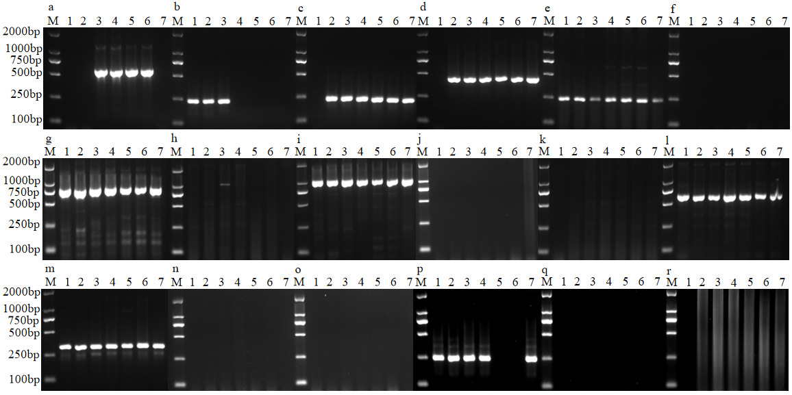
Eighteen virulence genes in Aeromonas veronii were detected by PCR. These included the genes for the ADP-ribosylating toxin (AexT), the type III secretion system inner membrane component (Ascv), glycerophospholipid-cholesterol acyltransferase (GcaT), cytotoxic enterotoxin (Act), the extracellular vir ulence factor aerolysin (aerA), collagenase (Acg), serine protease (Ahp), hemolysin (Hly), elastase (AhyB), lipase (Lip), heat-stable enterotoxin (Ast), heat-labile enterotoxin (Alt), flagellin (LafA), temperature sensitive protease (EprCAI), outer membrane protein (OmpAI), flagella (Fla), nuclease (Exu), and serine protease (Ser). The sequences of the corresponding primers are detailed in Table 1. The PCR reaction system and conditions were the same as those described in section 2.2.4.35
2.2.7. MLST of Strains
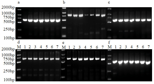
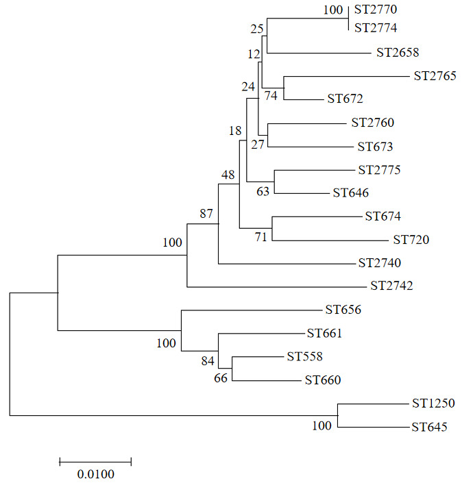
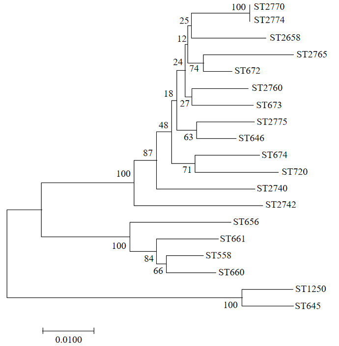
In this study, six housekeeping genes (gyrB, groL, gltA, metG, ppsA, and recA) were selected for MLST analysis. The sequences of the corresponding primers are detailed in Table 1. The PCR reaction system and conditions were the same as those described in section 2.2.4. The sequences of the six obtained housekeeping genes were subjected to BLAST homology analysis on the NCBI website. These sequences were then submitted to the PubMLST database for comparison and analysis to determine the allele number for each gene and ultimately define the novel sequence type (ST). Additionally, 12 Aeromonas strains from the PubMLST Aeromonas database were selected as reference strains (Table 5). A phylogenetic tree based on the different STs was constructed using the neighbor-joining method with 1,000 bootstrap replicates in MEGA 5 software. The sequences have been deposited in the National Genomics Data Center, Beijing Institute of Genomics, Chinese Academy of Sciences, and GenBase accession numbers were obtained (Table 2).
Results
3.1. Isolation and Identification of Pathogenic Bacteria
3.1.1. Morphological Characteristics of Isolates
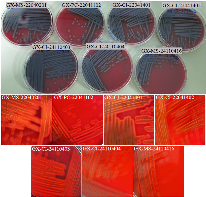
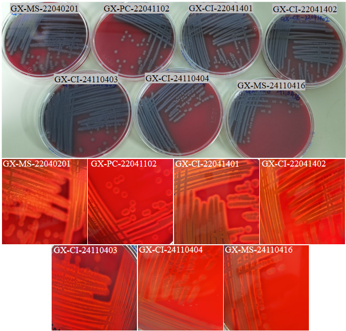
Seven bacterial strains were isolated from diseased M.salmoides, C.idella, and P.clarkii. The isolates were designated as GX-PC-22041102, GX-MS-22040201, GX-MS-24110416, GX-CI-22041401, GX-CI-22041402, GX-CI-24110403, and GX-CI-24110404. On blood agar plates, all seven isolates formed grayish-white, round, raised colonies with smooth margins, uniform texture, and a moist, glossy surface. They exhibited β-hemolysis, which is consistent with the typical colony characteristics of A. veronii (Figure 1).
3.1.2. Physiological and Biochemical Identification Results
Seven isolated strains were identified using the VITEK 2 compact fully automatic microbial identification system. As shown in Table 3, the seven isolated strains were consistent with the physiological and biochemical characteristics of A.veronii. The identification results indicated that they were A. veronii, with a probability of 97%.
3.1.3. Molecular Biological Identification Results
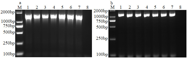
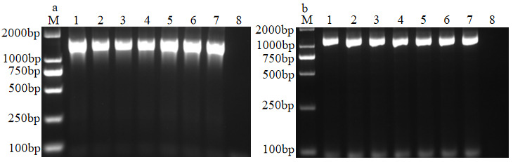
The amplification products of the 16S rRNA and gyrB genes from the seven isolated strains were analyzed by 2% agarose gel electrophoresis. The results showed specific bands for both the 16S rRNA and gyrB gene amplification products of all seven isolates, approximately 1441 bp and 1143 bp in size, respectively, which was consistent with the expected outcomes (Figure 2).
3.1.4. Phylogenetic Analysis
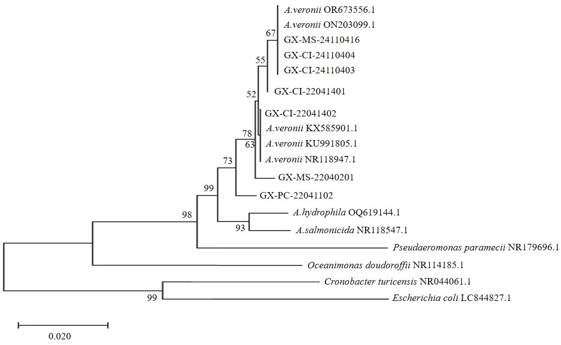
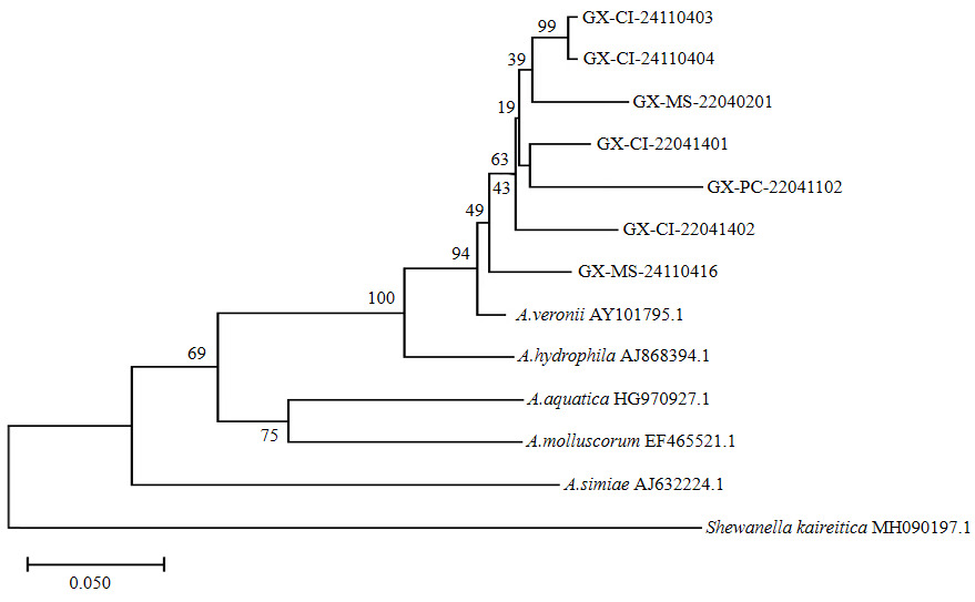
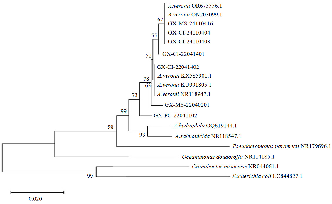
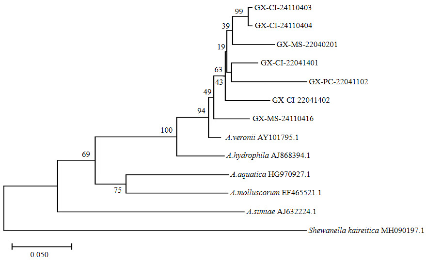
BLAST homology analysis of the 16S rRNA and gyrB gene sequences from the seven isolates revealed high homology with A.veronii. Phylogenetic trees constructed using sequences from different bacterial strains demonstrated that all seven isolates clustered within a distinct clade together with A. veronii (Figures 3, 4), confirming their identification as A. veronii.
3.1.5. Results of Artificial Challenge Infection Assay
The artificial challenge infection assay demonstrated that M. salmoides, C. idella, and P. clarkii injected with different concentrations of A. veronii suspensions exhibited symptoms similar to those observed in natural infections. These symptoms included hemorrhage, skin ulceration, abdominal swelling, and ascites in C. idella and M. salmoides. Infected P. clarkii showed reduced appetite and lethargy, with post-mortem examination revealing hepatopancreatic atrophy, softening, and pallor. Mortality increased with higher bacterial injection concentrations. Deaths began to occur from the third day post-challenge, with the mortality rates for each group detailed in Table 4.The calculated LD₅₀ values for strains GX-PC-22041102, GX-MS-22040201, GX-MS-24110416, GX-CI-22041401, GX-CI-22041402, GX-CI-24110403, and GX-CI-24110404 were 3.58×10⁶, 1.71×10⁶, 1.94×10⁶, 1.66×10⁶, 1.43×10⁶, 1.43×10⁶ and 1.66×10⁶ CFU/individual, respectively(Table 4). Bacteriological re-isolation and identification from the M. salmoides, C. idella, and P. clarkii subjected to artificial challenge yielded a single dominant bacterium with identical morphological characteristics to the original challenge strains GX-PC-22041102, GX-MS-22040201, GX-MS-24110416, GX-CI-22041401, GX-CI-22041402, GX-CI-24110403, and GX-CI-24110404.
3.2. Antimicrobial Susceptibility Testing Results
The antimicrobial susceptibility results of the seven isolated strains against 19 antibiotics are presented in Table 5. All seven isolates were susceptible to ceftriaxone, cefepime, fosfomycin, ceftazidime, ceftizoxime, and cefuroxime sodium. In contrast, all strains exhibited resistance to clavulanic acid, amoxicillin, teicoplanin, and sulbactam. These results indicate that the seven A. veronii isolates exhibited multidrug resistance.
3.3. Virulence Gene Detection Results
The detection results of 18 virulence genes in the seven isolated strains are shown in Figure 5. The GcaT, OmpAI, Hly, Fla, and Exu genes were detected in all isolates. In contrast, the Acg, Ast, Alt, Ser, AhyB, lafA, and eprCAI genes were not detected.
_dl2000dn.png)
3.4. MLST Typing
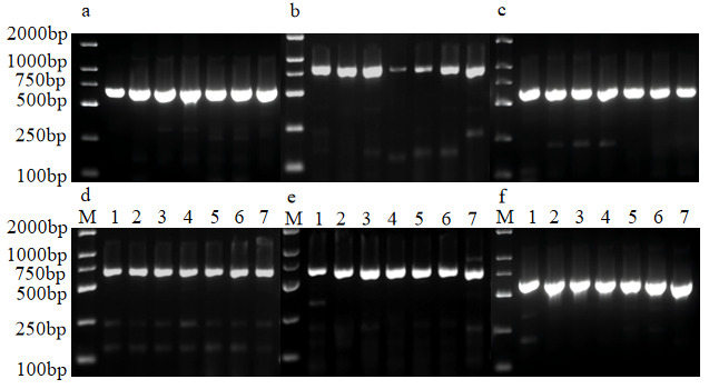
The PCR detection results for the six housekeeping genes of the seven isolated strains are shown in Figure 6. All six housekeeping genes from the seven strains amplified specific bands at the expected positions, with consistent fragment lengths.
The gene sequences of 7 isolated bacterial strains were uploaded to the Pubmlst database, resulting in 42 allele numbers and 7 new ST types (Table 6). Using the 7 new ST types and 12 ST types from the Aeromonas MLST database to construct a phylogenetic tree, the results showed that all the new STs clustered with A.veronii. Among them, ST2770 (GX-CI-24110403) and ST2774 (GX-CI-24110404) formed a single branch, with the closest genetic relationship; ST2765 (GX-CI-22041401), ST2760 (GX-CI-22041402), and ST2775 (GX-MS-24110416) formed adjacent branches, with a secondary genetic relationship; ST2742 (GX-MS-22040201) was relatively distant. Among them, ST2770 and ST2774 were more closely related to A.veronii from freshwater in China; ST2765 was more closely related to A.veronii from Polish Trachemys scripta elegans; ST2760 was more closely related to A. veronii from Polish T. scripta elegans; ST2775 was more closely related to A. veronii from Chinese P. clarkii. The 7 newly discovered sequence types showed relatively distant genetic relationships with Cavia porcellus and aquatic Aeromonas, and the correlation was not significant (Figure 7). The results indicated that the internal typing of the 7 isolated A.veronii strains GX-MS-22040201, GX-PC-22041102, GX-CI-22041401, GX-CI-22041402, GX-CI-24110403, GX-CI-24110404, and GX-MS-24110416 was rich, further confirming the genetic diversity and complexity of A.veronii in Guangxi region.
Discussion
A. veronii is an important zoonotic pathogen that infects fish, crustaceans, and humans, posing substantial risks to global aquaculture and public health.37–43 In this study, seven bacterial strains isolated from diseased P. clarkii, M. salmoides, and C. idella in Guangxi were conclusively identified as A. veronii using phenotypic and molecular methods. Artificial challenge trials confirmed that these isolates could reproduce typical clinical signs similar to natural outbreaks, with mortality rates increasing in a dose-dependent manner. The LD₅₀ values ranged from 1.43×10⁶ to 3.58×10⁶ CFU/individual, indicating moderate to high pathogenicity. These results are consistent with previous studies showing that the pathogenicity of A. veronii is closely related to infection dose.13–15,35
Studies have shown that the pathogenicity of pathogenic bacteria is closely associated with the virulence factors they carry, with different factors playing distinct roles during the infection process. For example, aerolysin (aerA), enterotoxins (Act/Ast/Alt), and enzymes such as GcaT, Lip, and Exu can disrupt host cell membranes, nucleic acids, and normal physiological functions, thereby enhancing bacterial adhesion and facilitating attachment and invasion.44 In this study, the carriage rates of the GcaT, OmpAI, Hly, Fla, and Exu genes among the seven isolated A. veronii strains were all 100%. The carriage rates of the Act and aerA genes were both 85.71%, while those of the Lip and Acsv genes were 71.43% and 57.14%, respectively. In contrast, the Acg, Ast, Alt, Ser, AhyB, lafA, and eprCAI genes were not detected (0%). These findings differ from those reported by other researchers. For instance, among the 119 A.veronii strains isolated by Liu et al., the carriage rates of GcaT, Fla, and Exu genes were 82.35%, 76.47%, and 92.44% respectively.45 Wu et al. found that the carriage rates of Act and Lip genes in the isolated A. veronii strains were 35.0% and 5.0% respectively.12 Similarly, Kingom et al. found that among 350 A. veronii isolates, the carriage rates of the aerA, Act, and Hly genes were 65%, whereas Sen et al. reported a carriage rate of 70% for the aerA, Act, and Hly genes in their isolates.46,47 Analyzing the results of virulence gene carriage in A. veronii isolates from other regions, it was found that the five virulence genes, GcaT, OmpAI, Hly, Fla, and Exu, were not universally present in all A. veronii strains, but rather showed differences among the strains. In this study, the 100% carriage rates of virulence genes such as GcaT, OmpAI, and Hly might be due to all strains being isolated from Guangxi or to the relatively small sample size. Among all the strains, only the GX-MS-22040201 strain isolated from largemouth bass did not carry Act and aerA. The LD₅₀ of this strain was 1.71×10⁶ CFU/individual, while the LD₅₀ of the other six strains ranged from 1.43×10⁶ to 3.58×10⁶ CFU/individual. There was no significant difference in either LD₅₀ values or host species. This phenomenon might be related to the relatively small sample size of the isolated strains in this study. Such discrepancies in virulence gene profiles may reflect the complexity and regional characteristics of A. veronii pathogenicity. Strains from different geographical or host origins may adapt to specific environments and exert pathogenic effects through distinct combinations of virulence genes. This variation may be attributed to multiple interacting factors, including environmental growth conditions, host species, horizontal gene transfer, evolutionary history, ecological niche differentiation, and laboratory cultivation conditions, all of which could contribute to the significant differences in virulence gene carriage among A. veronii strains from diverse sources.48 Detection of virulence genes can help predict the pathogenic potential of bacterial strains, thereby providing a theoretical basis for formulating targeted prevention and control measures.
Currently, antibiotic therapy remains the primary approach to treating bacterial diseases, making the analysis of antibiotic resistance in A. veronii particularly important. The seven A. veronii strains isolated in this study exhibited multidrug resistance, which aligns with the multidrug resistance phenomenon reported by Fadel et al.43 However, some strains showed differential responses to the same antibiotic. For instance, strain GX-CI-24110403 was sensitive to ampicillin, whereas the remaining six strains were resistant. This indicates variability in resistance phenotypes among the strains, which may be associated with differences in genetic background, history of antibiotic exposure, or expression of multidrug resistance genes.49
MLST typing provides a high-resolution tool for epidemiological studies of bacteria and offers critical insights into bacterial population structure, evolutionary history, and transmission patterns.50 In this study, MLST typing revealed that the seven isolated A. veronii strains represented seven novel STs, indicating a high degree of genetic diversity among A. veronii populations in the Guangxi region. This considerable genetic diversity may be associated with the complex ecological environment, diverse host species, and genetic recombination among strains in this area. The novel STs were closely related to STs originating from freshwater environments in China and from species such as red-eared sliders in Poland, whereas they were more distantly related to STs derived from other hosts (e.g., guinea pigs) or different species (e.g., A. hydrophila). This suggests that strains from different geographic and host origins may exhibit both genetic similarities and differences, which may be attributed to variations in local ecological conditions, host species, and the evolutionary histories of the strains.51
Acknowledgments
This research was funded by the Guangxi Key Research and Development Program (Guike Nong AB2506910047), The Guangxi Natural Science Foundation (Grant No. 2026GXNSFBA00640361), the Guangxi County Characteristic Modern Agricultural Industry Science and Technology Strengthening Chain Project (Guike Nong AA2512260008), and the Guangxi Special Freshwater Fish Innovation Team Project of the National Modern Agricultural Industry Technology System (nycytxgxcxtd-2021-08-02).
Authors’ Contribution
Conceptualization: Meizhen Su (Equal), Honglian Tan (Equal). Methodology: Meizhen Su (Lead). Writing – original draft: Meizhen Su (Lead). Formal Analysis: Honglian Tan (Equal), Shikun Huang (Equal). Investigation: Honglian Tan (Equal), Shikun Huang (Equal). Data curation: Shikun Huang (Lead). Resources: Jiankun Wu (Equal), Mingwei Yang (Equal). Supervision: Fuyan Chen (Equal), Jiancheng Wei (Equal). Visualization: Liping Li (Lead). Validation: Jing Chen (Lead). Software: Aiying Lei (Lead). Writing – review & editing: Xinxian Wei (Equal), Guixiang Tong (Equal). Funding acquisition: Xinxian Wei (Equal), Guixiang Tong (Equal). Project administration: Xinxian Wei (Lead).
Competing of Interest – COPE
The authors declare no conflicts of interest.
Ethical Conduct Approval – IACUC
All procedures involving animals were approved by the relevant ethics committee of Guangxi Academy of Fishery Sciences on 01-05-2026 (Approval No.2026-GAFS-08) and conducted in accordance with applicable animal welfare guidelines.
Informed Consent Statement
All authors and institutions have confirmed this manuscript for publication.
Data Availability Statement
All are available upon reasonable request.